ムービー
ムービー コントローラー


+ データを開く

- 基本情報
基本情報
| 登録情報 | データベース: EMDB / ID: EMD-5937 | |||||||||
|---|---|---|---|---|---|---|---|---|---|---|
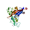
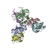
| タイトル | Electron cryo-microscopy of the Moloney murine leukemia virus furin precursor Env in its isomerization arrested state (IAS) form | |||||||||
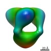
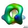
 マップデータ マップデータ | Reconstruction of the furin precursor of the Moloney murine leukemia virus at isomerization arrested intermediate stage (IAS) | |||||||||
 試料 試料 |
| |||||||||
 キーワード キーワード | Furin precursor / Moloney murine leukemia virus / Env maturation / intermediate form | |||||||||
| 生物種 |  Moloney murine leukemia virus (ウイルス) Moloney murine leukemia virus (ウイルス) | |||||||||
| 手法 | 単粒子再構成法 / クライオ電子顕微鏡法 / 解像度: 23.0 Å | |||||||||
 データ登録者 データ登録者 | Sjoberg M / Wu SR / Loving R / Rantalainen K / Lindqvist B / Garoff H | |||||||||
 引用 引用 | ジャーナル: Proc Natl Acad Sci U S A / 年: 2014 タイトル: Furin cleavage of the Moloney murine leukemia virus Env precursor reorganizes the spike structure. 著者: Mathilda Sjöberg / Shang-Rung Wu / Robin Löving / Kimmo Rantalainen / Birgitta Lindqvist / Henrik Garoff /  要旨: The trimeric Moloney murine leukemia virus Env protein matures by two proteolytic cleavages. First, furin cleaves the Env precursor into the surface (SU) and transmembrane (TM) subunits in the cell ...The trimeric Moloney murine leukemia virus Env protein matures by two proteolytic cleavages. First, furin cleaves the Env precursor into the surface (SU) and transmembrane (TM) subunits in the cell and then the viral protease cleaves the R-peptide from TM in new virus. Here we analyzed the structure of the furin precursor, by cryoelectron microscopy. We transfected 293T cells with a furin cleavage site provirus mutant, R466G/K468G, and produced the virus in the presence of amprenavir to also inhibit the R-peptide cleavage. Although Env incorporation into particles was inhibited, enough precursor could be isolated and analyzed by cryoelectron microscopy to yield a 3D structure at 22 Å resolution. This showed an open cage-like structure like that of the R-peptide precursor and the mature Env described before. However, the middle protrusion of the protomeric unit, so prominently pointing out from the side of the more mature forms of the Env, was absent. Instead, there was extra density in the top protrusion. This suggested that the C-terminal SU domain was associated alongside the receptor binding N-terminal SU domain in the furin precursor. This was supported by mapping with a SU C-terminal domain-specific antigen binding fragment. We concluded that furin cleavage not only separates the subunits and liberates the fusion peptide at the end of TM but also allows the C-terminal domain to relocate into a peripheral position. This conformational change might explain how the C-terminal domain of SU gains the potential to undergo disulfide isomerization, an event that facilitates membrane fusion. | |||||||||
| 履歴 |
|
- 構造の表示
構造の表示
| ムービー |
 ムービービューア ムービービューア |
|---|---|
| 構造ビューア | EMマップ: SurfViewMolmilJmol/JSmol |
| 添付画像 |

- ダウンロードとリンク
-EMDBアーカイブ
| マップデータ | emd_5937.map.gz | 329.9 KB | EMDBマップデータ形式 | |
|---|---|---|---|---|
| ヘッダ (付随情報) | emd-5937-v30.xmlemd-5937.xml | 11.7 KB 11.7 KB | 表示 表示 | EMDBヘッダ |
| 画像 |  400_5937.gif 400_5937.gif 80_5937.gif 80_5937.gif | 22.2 KB 2.5 KB | ||
| アーカイブディレクトリ |  http://ftp.pdbj.org/pub/emdb/structures/EMD-5937ftp://ftp.pdbj.org/pub/emdb/structures/EMD-5937 http://ftp.pdbj.org/pub/emdb/structures/EMD-5937ftp://ftp.pdbj.org/pub/emdb/structures/EMD-5937 | HTTPS FTP |
-検証レポート
| 文書・要旨 | emd_5937_validation.pdf.gz | 79.3 KB | 表示 | EMDB検証レポート |
|---|---|---|---|---|
| 文書・詳細版 | emd_5937_full_validation.pdf.gz | 78.4 KB | 表示 | |
| XML形式データ | emd_5937_validation.xml.gz | 495 B | 表示 | |
| アーカイブディレクトリ | https://ftp.pdbj.org/pub/emdb/validation_reports/EMD-5937ftp://ftp.pdbj.org/pub/emdb/validation_reports/EMD-5937 | HTTPS FTP |
-関連構造データ


-リンク
| EMDBのページ | EMDB (EBI/PDBe) / EMDataResource |
|---|
-マップ
| ファイル | ダウンロード / ファイル: emd_5937.map.gz / 形式: CCP4 / 大きさ: 422.9 KB / タイプ: IMAGE STORED AS FLOATING POINT NUMBER (4 BYTES) | ||||||||||||||||||||||||||||||||||||||||||||||||||||||||||||||||||||
|---|---|---|---|---|---|---|---|---|---|---|---|---|---|---|---|---|---|---|---|---|---|---|---|---|---|---|---|---|---|---|---|---|---|---|---|---|---|---|---|---|---|---|---|---|---|---|---|---|---|---|---|---|---|---|---|---|---|---|---|---|---|---|---|---|---|---|---|---|---|
| 注釈 | Reconstruction of the furin precursor of the Moloney murine leukemia virus at isomerization arrested intermediate stage (IAS) | ||||||||||||||||||||||||||||||||||||||||||||||||||||||||||||||||||||
| 投影像・断面図 | 画像のコントロール
画像は Spider により作成 | ||||||||||||||||||||||||||||||||||||||||||||||||||||||||||||||||||||
| ボクセルのサイズ | X=Y=Z: 3.5 Å | ||||||||||||||||||||||||||||||||||||||||||||||||||||||||||||||||||||
| 密度 |
| ||||||||||||||||||||||||||||||||||||||||||||||||||||||||||||||||||||
| 対称性 | 空間群: 1 | ||||||||||||||||||||||||||||||||||||||||||||||||||||||||||||||||||||
| 詳細 | EMDB XML:
CCP4マップ ヘッダ情報:
| ||||||||||||||||||||||||||||||||||||||||||||||||||||||||||||||||||||
 Z (Sec.)
 Y (Row.)
 X (Col.)


-添付データ
- 試料の構成要素
-全体 : Isomerization arrested stage (IAS) of the furin cleavage deficien...
| 全体 | 名称: Isomerization arrested stage (IAS) of the furin cleavage deficient mutant (R466G/K468G) Env of Moloney murine leukemia virus |
|---|---|
| 要素 |
|
-超分子 #1000: Isomerization arrested stage (IAS) of the furin cleavage deficien...
| 超分子 | 名称: Isomerization arrested stage (IAS) of the furin cleavage deficient mutant (R466G/K468G) Env of Moloney murine leukemia virus タイプ: sample / ID: 1000 詳細: Affinity purified, gradient separated protein in 0.05% Triton X-100 集合状態: trimer / Number unique components: 1 |
|---|---|
| 分子量 | 実験値: 500 KDa / 理論値: 270 KDa / 手法: Estimated from Blue native PAGE |
-分子 #1: gp90
| 分子 | 名称: gp90 / タイプ: protein_or_peptide / ID: 1 / Name.synonym: Furin precursor / コピー数: 3 / 集合状態: Trimer / 組換発現: No / データベース: NCBI |
|---|---|
| 由来(天然) | 生物種: Moloney murine leukemia virus (ウイルス) |
| 分子量 | 実験値: 500 KDa / 理論値: 270 KDa |
-実験情報
-構造解析
| 手法 | クライオ電子顕微鏡法 |
|---|---|
 解析 解析 | 単粒子再構成法 |
| 試料の集合状態 | particle |
-試料調製
| 濃度 | 0.1 mg/mL |
|---|---|
| 緩衝液 | pH: 7.4 / 詳細: 50 mM HEPES, 100 mM NaCl, 1.8 mM CaCl2, pH 7.4 |
| グリッド | 詳細: 400 mesh holey carbon grid, glow discharged |
| 凍結 | 凍結剤: ETHANE / チャンバー内湿度: 90 % / チャンバー内温度: 77 K / 装置: FEI VITROBOT MARK II 手法: Blotted for 3 seconds before plunging in liquid ethane, followed by transfer into liquid nitrogen. |
- 電子顕微鏡法
| 顕微鏡 | JEOL 2100F |
|---|---|
| 温度 | 最低: 93 K / 最高: 96 K / 平均: 95 K |
| アライメント法 | Legacy - 非点収差: Objective lens astigmatism was corrected using online FFT. |
| 日付 | 2013年1月25日 |
| 撮影 | カテゴリ: CCD フィルム・検出器のモデル: TVIPS TEMCAM-F416 (4k x 4k) デジタル化 - サンプリング間隔: 3.5 µm / 実像数: 456 / 平均電子線量: 9 e/Å2 / ビット/ピクセル: 14 |
| 電子線 | 加速電圧: 200 kV / 電子線源:  FIELD EMISSION GUN FIELD EMISSION GUN |
| 電子光学系 | 倍率(補正後): 43200 / 照射モード: SPOT SCAN / 撮影モード: BRIGHT FIELD / Cs: 2.0 mm / 最大 デフォーカス(公称値): 4.0 µm / 最小 デフォーカス(公称値): 2.5 µm / 倍率(公称値): 43200 |
| 試料ステージ | 試料ホルダーモデル: GATAN LIQUID NITROGEN |
+画像解析
-原子モデル構築 1
| 初期モデル | PDB ID: |
|---|---|
| ソフトウェア | 名称: Chimera |
| 詳細 | The atomic model for the Mo-RBD was obtained using the atomic structure of the highly homologous F-RBD (Fass et al, 1997) (PDB ID 1AOL) and the SWISS-MODEL protein structure homology-modeling server (accessible through the ExPASy web server). |
| 精密化 | 空間: REAL / プロトコル: RIGID BODY FIT |