 Movie
Movie Controller

[English] 日本語
 Yorodumi
- PDB-5id4: Crystal structure of Proteus mirabilis ScsC in an extended confor... -
+ Open data

- Basic information
| Entry | Database: PDB / ID: 5id4 | ||||||
|---|---|---|---|---|---|---|---|
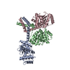
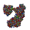
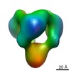
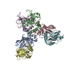
| Title | Crystal structure of Proteus mirabilis ScsC in an extended conformation | ||||||
 Components Components | DsbA-like protein | ||||||
 Keywords Keywords | ISOMERASE / thioredoxin fold / disulfide isomerase / trimer / copper resistance | ||||||
| Function / homology | :  Function and homology information Function and homology information | ||||||
| Biological species |  Proteus mirabilis ATCC 29906 (bacteria) Proteus mirabilis ATCC 29906 (bacteria) | ||||||
| Method |  X-RAY DIFFRACTION / SYNCHROTRON / MOLECULAR REPLACEMENT / Resolution: 2.921 Å X-RAY DIFFRACTION / SYNCHROTRON / MOLECULAR REPLACEMENT / Resolution: 2.921 Å | ||||||
 Authors Authors | Furlong, E.J. / Kurth, F. / Choudhury, H.G. / Martin, J.L. | ||||||
| Funding support |  Australia, 1items Australia, 1items
| ||||||
 Citation Citation | Journal: Nat Commun / Year: 2017 Title: Proteus mirabilis ScsC is a highly dynamic, novel trimeric protein disulfide isomerase Authors: Kurth, F. / Furlong, E.J. / Lo, A.W. / Premkumar, L. / Totsika, M. / Whitten, A.E. / Achard, M.E.S. / Halili, M.A. / Heras, B. / Choudhury, H.G. / Schembri, M.A. / Martin, J.L. | ||||||
| History |
|
- Structure visualization
| Structure viewer | Molecule: MolmilJmol/JSmol |
|---|
- Downloads & links
-Download
| PDBx/mmCIF format | 5id4.cif.gz | 138.5 KB | Display | PDBx/mmCIF format |
|---|---|---|---|---|
| PDB format | pdb5id4.ent.gz | 113.7 KB | Display | PDB format |
| PDBx/mmJSON format | 5id4.json.gz | Tree view | PDBx/mmJSON format | |
| Others |  Other downloads Other downloads |
-Validation report
| Arichive directory | https://data.pdbj.org/pub/pdb/validation_reports/id/5id4ftp://data.pdbj.org/pub/pdb/validation_reports/id/5id4 | HTTPS FTP |
|---|
-Related structure data
| Related structure data |  4yx8 S: Starting model for refinement |
|---|---|
| Similar structure data |

-Links
 PDBj
PDBj- Assembly
| Deposited unit | 
| ||||||||
|---|---|---|---|---|---|---|---|---|---|
| 1 | 
| ||||||||
| Unit cell |
|
-Components
| #1: Protein | Mass: 24800.443 Da / Num. of mol.: 1 / Fragment: UNP residues 22-243 Source method: isolated from a genetically manipulated source Source: (gene. exp.) Proteus mirabilis ATCC 29906 (bacteria)Gene: HMPREF0693_3732 / Plasmid: pMCSG7 / Production host: |
|---|---|
| Has protein modification | Y |
-Experimental details
-Experiment
| Experiment | Method: X-RAY DIFFRACTION / Number of used crystals: 1 |
|---|
- Sample preparation
| Crystal | Density Matthews: 4.83 Å3/Da / Density % sol: 74.53 % |
|---|---|
| Crystal grow | Temperature: 293.15 K / Method: vapor diffusion, hanging drop Details: 32% Jeffamine M-600 pH 7, 0.1 M HEPES pH 8, 2.5 mM Copper(II) chloride PH range: 7-8 |
-Data collection
| Diffraction | Mean temperature: 100 K | ||||||||||||||||||||||||||||||
|---|---|---|---|---|---|---|---|---|---|---|---|---|---|---|---|---|---|---|---|---|---|---|---|---|---|---|---|---|---|---|---|
| Diffraction source | Source: SYNCHROTRON / Site: Australian Synchrotron / Beamline: MX2 / Wavelength: 0.9537 Å | ||||||||||||||||||||||||||||||
| Detector | Type: ADSC QUANTUM 315r / Detector: CCD / Date: Dec 10, 2015 | ||||||||||||||||||||||||||||||
| Radiation | Protocol: SINGLE WAVELENGTH / Monochromatic (M) / Laue (L): M / Scattering type: x-ray | ||||||||||||||||||||||||||||||
| Radiation wavelength | Wavelength: 0.9537 Å / Relative weight: 1 | ||||||||||||||||||||||||||||||
| Reflection | Resolution: 2.92→110.29 Å / Num. obs: 10755 / % possible obs: 99.4 % / Redundancy: 4.1 % / Biso Wilson estimate: 85.97 Å2 / CC1/2: 0.998 / Rmerge(I) obs: 0.059 / Rpim(I) all: 0.033 / Rrim(I) all: 0.068 / Net I/σ(I): 14.2 / Num. measured all: 44513 | ||||||||||||||||||||||||||||||
| Reflection shell | Diffraction-ID: 1 / Rejects: _
|
- Processing
| Software |
| |||||||||||||||||||||||||||||||||||||||||||||||||||||||||||||||||||||||||||
|---|---|---|---|---|---|---|---|---|---|---|---|---|---|---|---|---|---|---|---|---|---|---|---|---|---|---|---|---|---|---|---|---|---|---|---|---|---|---|---|---|---|---|---|---|---|---|---|---|---|---|---|---|---|---|---|---|---|---|---|---|---|---|---|---|---|---|---|---|---|---|---|---|---|---|---|---|
| Refinement | Method to determine structure: MOLECULAR REPLACEMENT Starting model: 4YX8 4yx8 Resolution: 2.921→40.363 Å / SU ML: 0.41 / Cross valid method: FREE R-VALUE / σ(F): 1.34 / Phase error: 35.3 / Stereochemistry target values: ML
| |||||||||||||||||||||||||||||||||||||||||||||||||||||||||||||||||||||||||||
| Solvent computation | Shrinkage radii: 0.9 Å / VDW probe radii: 1.11 Å / Solvent model: FLAT BULK SOLVENT MODEL | |||||||||||||||||||||||||||||||||||||||||||||||||||||||||||||||||||||||||||
| Displacement parameters | Biso max: 214.71 Å2 / Biso mean: 108.8807 Å2 / Biso min: 9.16 Å2 | |||||||||||||||||||||||||||||||||||||||||||||||||||||||||||||||||||||||||||
| Refinement step | Cycle: final / Resolution: 2.921→40.363 Å
| |||||||||||||||||||||||||||||||||||||||||||||||||||||||||||||||||||||||||||
| Refine LS restraints |
| |||||||||||||||||||||||||||||||||||||||||||||||||||||||||||||||||||||||||||
| LS refinement shell | Refine-ID: X-RAY DIFFRACTION / Total num. of bins used: 4
| |||||||||||||||||||||||||||||||||||||||||||||||||||||||||||||||||||||||||||
| Refinement TLS params. | Method: refined / Refine-ID: X-RAY DIFFRACTION
| |||||||||||||||||||||||||||||||||||||||||||||||||||||||||||||||||||||||||||
| Refinement TLS group |
|
