 Movie
Movie Controller


+ Open data

- Basic information
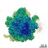
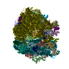
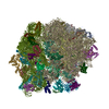
| Entry | Database: EMDB / ID: EMD-5889 | |||||||||
|---|---|---|---|---|---|---|---|---|---|---|
| Title | Cryo-electron microscopy of human 80S ribosome | |||||||||
 Map data Map data | human 80S ribosome | |||||||||
 Sample Sample |
| |||||||||
| Biological species |  Homo sapiens (human) Homo sapiens (human) | |||||||||
| Method | single particle reconstruction / cryo EM / Resolution: 3.85 Å | |||||||||
 Authors Authors | Penczek PA / Fang J / Li X / Cheng Y / Loerke J / Spahn CMT | |||||||||
 Citation Citation | Journal: Ultramicroscopy / Year: 2014 Title: CTER-rapid estimation of CTF parameters with error assessment. Authors: Pawel A Penczek / Jia Fang / Xueming Li / Yifan Cheng / Justus Loerke / Christian M T Spahn /   Abstract: In structural electron microscopy, the accurate estimation of the Contrast Transfer Function (CTF) parameters, particularly defocus and astigmatism, is of utmost importance for both initial ...In structural electron microscopy, the accurate estimation of the Contrast Transfer Function (CTF) parameters, particularly defocus and astigmatism, is of utmost importance for both initial evaluation of micrograph quality and for subsequent structure determination. Due to increases in the rate of data collection on modern microscopes equipped with new generation cameras, it is also important that the CTF estimation can be done rapidly and with minimal user intervention. Finally, in order to minimize the necessity for manual screening of the micrographs by a user it is necessary to provide an assessment of the errors of fitted parameters values. In this work we introduce CTER, a CTF parameters estimation method distinguished by its computational efficiency. The efficiency of the method makes it suitable for high-throughput EM data collection, and enables the use of a statistical resampling technique, bootstrap, that yields standard deviations of estimated defocus and astigmatism amplitude and angle, thus facilitating the automation of the process of screening out inferior micrograph data. Furthermore, CTER also outputs the spatial frequency limit imposed by reciprocal space aliasing of the discrete form of the CTF and the finite window size. We demonstrate the efficiency and accuracy of CTER using a data set collected on a 300kV Tecnai Polara (FEI) using the K2 Summit DED camera in super-resolution counting mode. Using CTER we obtained a structure of the 80S ribosome whose large subunit had a resolution of 4.03Å without, and 3.85Å with, inclusion of astigmatism parameters. | |||||||||
| History |
|
- Structure visualization
| Movie |
 Movie viewer Movie viewer |
|---|---|
| Structure viewer | EM map: SurfViewMolmilJmol/JSmol |
| Supplemental images |

- Downloads & links
-EMDB archive
| Map data | emd_5889.map.gz | 19.2 MB | EMDB map data format | |
|---|---|---|---|---|
| Header (meta data) | emd-5889-v30.xmlemd-5889.xml | 8.3 KB 8.3 KB | Display Display | EMDB header |
| Images | 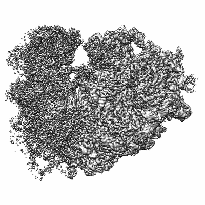 400_5889.gif 400_5889.gif 80_5889.gif 80_5889.gif | 59.6 KB 4.1 KB | ||
| Archive directory |  http://ftp.pdbj.org/pub/emdb/structures/EMD-5889ftp://ftp.pdbj.org/pub/emdb/structures/EMD-5889 http://ftp.pdbj.org/pub/emdb/structures/EMD-5889ftp://ftp.pdbj.org/pub/emdb/structures/EMD-5889 | HTTPS FTP |
-Related structure data
| Similar structure data |
|---|


-Links
| EMDB pages | EMDB (EBI/PDBe) / EMDataResource |
|---|---|
| Related items in Molecule of the Month |

-Map
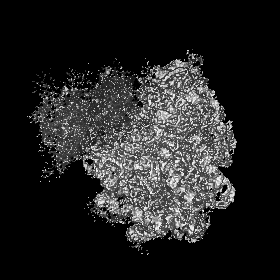
| File | Download / File: emd_5889.map.gz / Format: CCP4 / Size: 81.8 MB / Type: IMAGE STORED AS FLOATING POINT NUMBER (4 BYTES) | ||||||||||||||||||||||||||||||||||||||||||||||||||||||||||||||||||||
|---|---|---|---|---|---|---|---|---|---|---|---|---|---|---|---|---|---|---|---|---|---|---|---|---|---|---|---|---|---|---|---|---|---|---|---|---|---|---|---|---|---|---|---|---|---|---|---|---|---|---|---|---|---|---|---|---|---|---|---|---|---|---|---|---|---|---|---|---|---|
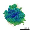
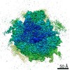
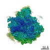
| Annotation | human 80S ribosome | ||||||||||||||||||||||||||||||||||||||||||||||||||||||||||||||||||||
| Projections & slices | Image control
Images are generated by Spider. | ||||||||||||||||||||||||||||||||||||||||||||||||||||||||||||||||||||
| Voxel size | X=Y=Z: 1.264 Å | ||||||||||||||||||||||||||||||||||||||||||||||||||||||||||||||||||||
| Density |
| ||||||||||||||||||||||||||||||||||||||||||||||||||||||||||||||||||||
| Symmetry | Space group: 1 | ||||||||||||||||||||||||||||||||||||||||||||||||||||||||||||||||||||
| Details | EMDB XML:
CCP4 map header:
| ||||||||||||||||||||||||||||||||||||||||||||||||||||||||||||||||||||
 Z (Sec.)
 Y (Row.)
 X (Col.)


-Supplemental data
- Sample components
-Entire : human 80S ribosome
| Entire | Name: human 80S ribosome |
|---|---|
| Components |
|
-Supramolecule #1000: human 80S ribosome
| Supramolecule | Name: human 80S ribosome / type: sample / ID: 1000 / Details: sample was monodisperse / Oligomeric state: 1 / Number unique components: 1 |
|---|---|
| Molecular weight | Theoretical: 4.2 MDa |
-Supramolecule #1: 80S ribosome
| Supramolecule | Name: 80S ribosome / type: complex / ID: 1 / Recombinant expression: No / Database: NCBI / Ribosome-details: ribosome-eukaryote: ALL |
|---|---|
| Source (natural) | Organism: Homo sapiens (human) / synonym: human / Tissue: kidney / Cell: HEK293 / Organelle: cytosol |
| Molecular weight | Theoretical: 4.2 MDa |
-Experimental details
-Structure determination
| Method | cryo EM |
|---|---|
 Processing Processing | single particle reconstruction |
| Aggregation state | particle |
-Sample preparation
| Buffer | pH: 7.5 Details: 20 mM HEPES, pH 7.5, 100 mM KCl, 1.5 mM MgCl2, 0.5 mM spermidine, 0.04 mM spermine, 1 mM DTT |
|---|---|
| Grid | Details: Quantifoil 2/4 Cu/Re with custom continuous carbon film |
| Vitrification | Cryogen name: ETHANE / Instrument: OTHER |
- Electron microscopy
| Microscope | FEI TECNAI F30 |
|---|---|
| Date | Feb 12, 2013 |
| Image recording | Category: CCD / Film or detector model: GATAN K2 (4k x 4k) / Number real images: 893 Details: The data was collected in movie mode at 39,000x magnification. Frames 3 to 20 were decimated twice, motion corrected with UCSHImage4, and averaged. |
| Electron beam | Acceleration voltage: 300 kV / Electron source:  FIELD EMISSION GUN FIELD EMISSION GUN |
| Electron optics | Illumination mode: FLOOD BEAM / Imaging mode: BRIGHT FIELD / Cs: 2 mm |
| Sample stage | Specimen holder model: GATAN LIQUID NITROGEN |
| Experimental equipment |  Model: Tecnai F30 / Image courtesy: FEI Company |
-Image processing
| CTF correction | Details: Per-particle |
|---|---|
| Final reconstruction | Algorithm: OTHER / Resolution.type: BY AUTHOR / Resolution: 3.85 Å / Resolution method: OTHER / Software - Name: SPARX Details: The resolution reported is for the large subunit (60S) only. Number images used: 35198 |
