 Movie
Movie Controller

[English] 日本語
 Yorodumi
- EMDB-3624: Hibernating ribosome from Staphylococcus aureus (Unrotated state) -
+ Open data

- Basic information
| Entry | Database: EMDB / ID: EMD-3624 | ||||||||||||||||||
|---|---|---|---|---|---|---|---|---|---|---|---|---|---|---|---|---|---|---|---|
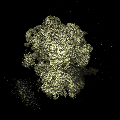
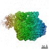
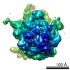
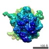
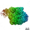
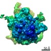
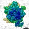
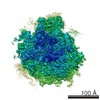
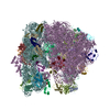
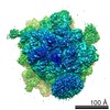
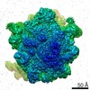
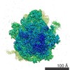
| Title | Hibernating ribosome from Staphylococcus aureus (Unrotated state) | ||||||||||||||||||
 Map data Map data | |||||||||||||||||||
 Sample Sample |
| ||||||||||||||||||
 Keywords Keywords | S.aureus / HPF / hibernation / 100S ribosome / ribosome | ||||||||||||||||||
| Function / homology |  Function and homology information Function and homology informationnegative regulation of translational elongation / ribosomal small subunit binding / large ribosomal subunit / transferase activity / ribosome biogenesis / ribosomal small subunit biogenesis / 5S rRNA binding / ribosomal large subunit assembly / small ribosomal subunit / small ribosomal subunit rRNA binding ...negative regulation of translational elongation / ribosomal small subunit binding / large ribosomal subunit / transferase activity / ribosome biogenesis / ribosomal small subunit biogenesis / 5S rRNA binding / ribosomal large subunit assembly / small ribosomal subunit / small ribosomal subunit rRNA binding / large ribosomal subunit rRNA binding / cytosolic small ribosomal subunit / cytosolic large ribosomal subunit / cytoplasmic translation / tRNA binding / negative regulation of translation / rRNA binding / structural constituent of ribosome / ribosome / translation / ribonucleoprotein complex / mRNA binding / RNA binding / zinc ion binding / cytoplasm / cytosol Similarity search - Function | ||||||||||||||||||
| Biological species |  Staphylococcus aureus (strain NCTC 8325) (bacteria) / Staphylococcus aureus subsp. aureus NCTC 8325 (bacteria) Staphylococcus aureus (strain NCTC 8325) (bacteria) / Staphylococcus aureus subsp. aureus NCTC 8325 (bacteria) | ||||||||||||||||||
| Method | single particle reconstruction / cryo EM / Resolution: 3.7 Å | ||||||||||||||||||
 Authors Authors | Khusainov I / Vicens Q | ||||||||||||||||||
| Funding support |  France, France,  Russian Federation, 5 items Russian Federation, 5 items
| ||||||||||||||||||
 Citation Citation | Journal: EMBO J / Year: 2017 Title: Structures and dynamics of hibernating ribosomes from mediated by intermolecular interactions of HPF. Authors: Iskander Khusainov / Quentin Vicens / Rustam Ayupov / Konstantin Usachev / Alexander Myasnikov / Angelita Simonetti / Shamil Validov / Bruno Kieffer / Gulnara Yusupova / Marat Yusupov / Yaser Hashem / Abstract: In bacteria, ribosomal hibernation shuts down translation as a response to stress, through reversible binding of stress-induced proteins to ribosomes. This process typically involves the formation of ...In bacteria, ribosomal hibernation shuts down translation as a response to stress, through reversible binding of stress-induced proteins to ribosomes. This process typically involves the formation of 100S ribosome dimers. Here, we present the structures of hibernating ribosomes from human pathogen containing a long variant of the hibernation-promoting factor (SaHPF) that we solved using cryo-electron microscopy. Our reconstructions reveal that the N-terminal domain (NTD) of SaHPF binds to the 30S subunit as observed for shorter variants of HPF in other species. The C-terminal domain (CTD) of SaHPF protrudes out of each ribosome in order to mediate dimerization. Using NMR, we characterized the interactions at the CTD-dimer interface. Secondary interactions are provided by helix 26 of the 16S ribosomal RNA We also show that ribosomes in the 100S particle adopt both rotated and unrotated conformations. Overall, our work illustrates a specific mode of ribosome dimerization by long HPF, a finding that may help improve the selectivity of antimicrobials. | ||||||||||||||||||
| History |
|
- Structure visualization
| Movie |
Movie viewer |
|---|---|
| Structure viewer | EM map: SurfViewMolmilJmol/JSmol |
| Supplemental images |
- Downloads & links
-EMDB archive
| Map data | emd_3624.map.gz | 140.4 MB | EMDB map data format | |
|---|---|---|---|---|
| Header (meta data) | emd-3624-v30.xmlemd-3624.xml | 74.8 KB 74.8 KB | Display Display | EMDB header |
| Images |  emd_3624.png emd_3624.png | 149.7 KB | ||
| Filedesc metadata | emd-3624.cif.gz | 13.7 KB | ||
| Archive directory |  http://ftp.pdbj.org/pub/emdb/structures/EMD-3624ftp://ftp.pdbj.org/pub/emdb/structures/EMD-3624 http://ftp.pdbj.org/pub/emdb/structures/EMD-3624ftp://ftp.pdbj.org/pub/emdb/structures/EMD-3624 | HTTPS FTP |
-Related structure data
| Related structure data | 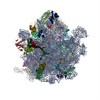 5nd8MC 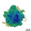 3625C 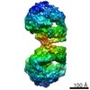 3638C  3639C 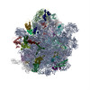 5nd9C  5nkoC M: atomic model generated by this map C: citing same article ( |
|---|---|
| Similar structure data |
-Links
| EMDB pages | EMDB (EBI/PDBe) / EMDataResource |
|---|---|
| Related items in Molecule of the Month |


-Map
| File | Download / File: emd_3624.map.gz / Format: CCP4 / Size: 149.9 MB / Type: IMAGE STORED AS FLOATING POINT NUMBER (4 BYTES) | ||||||||||||||||||||||||||||||||||||||||||||||||||||||||||||
|---|---|---|---|---|---|---|---|---|---|---|---|---|---|---|---|---|---|---|---|---|---|---|---|---|---|---|---|---|---|---|---|---|---|---|---|---|---|---|---|---|---|---|---|---|---|---|---|---|---|---|---|---|---|---|---|---|---|---|---|---|---|
| Projections & slices | Image control
Images are generated by Spider. | ||||||||||||||||||||||||||||||||||||||||||||||||||||||||||||
| Voxel size | X=Y=Z: 1.1 Å | ||||||||||||||||||||||||||||||||||||||||||||||||||||||||||||
| Density |
| ||||||||||||||||||||||||||||||||||||||||||||||||||||||||||||
| Symmetry | Space group: 1 | ||||||||||||||||||||||||||||||||||||||||||||||||||||||||||||
| Details | EMDB XML:
CCP4 map header:
| ||||||||||||||||||||||||||||||||||||||||||||||||||||||||||||
 Z (Sec.)
 Y (Row.)
 X (Col.)


-Supplemental data
- Sample components
+Entire : Hibernating ribosome from Staphylococcus aureus (Unrotated state)
+Supramolecule #1: Hibernating ribosome from Staphylococcus aureus (Unrotated state)
+Supramolecule #2: Hibernating ribosome from Staphylococcus aureus (Unrotated state)
+Supramolecule #3: Ribosome hibernation promotion factor
+Macromolecule #1: 16S ribosomal RNA
+Macromolecule #23: 23S ribosomal RNA
+Macromolecule #24: 5S ribosomal RNA
+Macromolecule #2: 30S ribosomal protein S2
+Macromolecule #3: 30S ribosomal protein S3
+Macromolecule #4: 30S ribosomal protein S4
+Macromolecule #5: 30S ribosomal protein S5
+Macromolecule #6: 30S ribosomal protein S6
+Macromolecule #7: 30S ribosomal protein S7
+Macromolecule #8: 30S ribosomal protein S8
+Macromolecule #9: 30S ribosomal protein S9
+Macromolecule #10: 30S ribosomal protein S10
+Macromolecule #11: 30S ribosomal protein S11
+Macromolecule #12: 30S ribosomal protein S12
+Macromolecule #13: 30S ribosomal protein S13
+Macromolecule #14: 30S ribosomal protein S14 type Z
+Macromolecule #15: 30S ribosomal protein S15
+Macromolecule #16: 30S ribosomal protein S16
+Macromolecule #17: 30S ribosomal protein S17
+Macromolecule #18: 30S ribosomal protein S18
+Macromolecule #19: 30S ribosomal protein S19
+Macromolecule #20: 30S ribosomal protein S20
+Macromolecule #21: 30S ribosomal protein S21
+Macromolecule #22: Ribosome hibernation promotion factor
+Macromolecule #25: 50S ribosomal protein L2
+Macromolecule #26: 50S ribosomal protein L3
+Macromolecule #27: 50S ribosomal protein L4
+Macromolecule #28: 50S ribosomal protein L5
+Macromolecule #29: 50S ribosomal protein L6
+Macromolecule #30: 50S ribosomal protein L13
+Macromolecule #31: 50S ribosomal protein L14
+Macromolecule #32: 50S ribosomal protein L15
+Macromolecule #33: 50S ribosomal protein L16
+Macromolecule #34: 50S ribosomal protein L17
+Macromolecule #35: 50S ribosomal protein L18
+Macromolecule #36: 50S ribosomal protein L19
+Macromolecule #37: 50S ribosomal protein L20
+Macromolecule #38: 50S ribosomal protein L21
+Macromolecule #39: 50S ribosomal protein L22
+Macromolecule #40: 50S ribosomal protein L23
+Macromolecule #41: 50S ribosomal protein L24
+Macromolecule #42: 50S ribosomal protein L25
+Macromolecule #43: 50S ribosomal protein L27
+Macromolecule #44: 50S ribosomal protein L28
+Macromolecule #45: 50S ribosomal protein L29
+Macromolecule #46: 50S ribosomal protein L30
+Macromolecule #47: 50S ribosomal protein L31 type B
+Macromolecule #48: 50S ribosomal protein L32
+Macromolecule #49: 50S ribosomal protein L33 2
+Macromolecule #50: 50S ribosomal protein L34
+Macromolecule #51: 50S ribosomal protein L35
+Macromolecule #52: 50S ribosomal protein L36
-Experimental details
-Structure determination
| Method | cryo EM |
|---|---|
 Processing Processing | single particle reconstruction |
| Aggregation state | particle |
-Sample preparation
| Buffer | pH: 7.5 Details: 5 mM Hepes-KOH pH 7.5 50 mM KCl 10 mM NH4Cl 10 mM Mg(OAc)2 1 mM DTT |
|---|---|
| Grid | Material: COPPER / Pretreatment - Type: GLOW DISCHARGE / Pretreatment - Time: 20 sec. |
| Vitrification | Cryogen name: ETHANE / Chamber humidity: 100 % / Chamber temperature: 277 K / Instrument: FEI VITROBOT MARK IV / Details: blot force 5, blot waiting time 30 s. |
- Electron microscopy
| Microscope | FEI TITAN KRIOS |
|---|---|
| Image recording | Film or detector model: FEI FALCON II (4k x 4k) / Detector mode: COUNTING / Digitization - Frames/image: 2-8 / Number grids imaged: 1 / Average exposure time: 1.0 sec. / Average electron dose: 60.0 e/Å2 |
| Electron beam | Acceleration voltage: 300 kV / Electron source:  FIELD EMISSION GUN FIELD EMISSION GUN |
| Electron optics | Illumination mode: FLOOD BEAM / Imaging mode: DARK FIELD |
| Experimental equipment |  Model: Titan Krios / Image courtesy: FEI Company |
+Image processing
-Atomic model buiding 1
| Refinement | Space: REAL / Protocol: RIGID BODY FIT |
|---|---|
| Output model | PDB-5nd8: |
