ムービー
ムービー コントローラー


+ データを開く

- 基本情報
基本情報
| 登録情報 | データベース: EMDB / ID: EMD-5710 | |||||||||
|---|---|---|---|---|---|---|---|---|---|---|
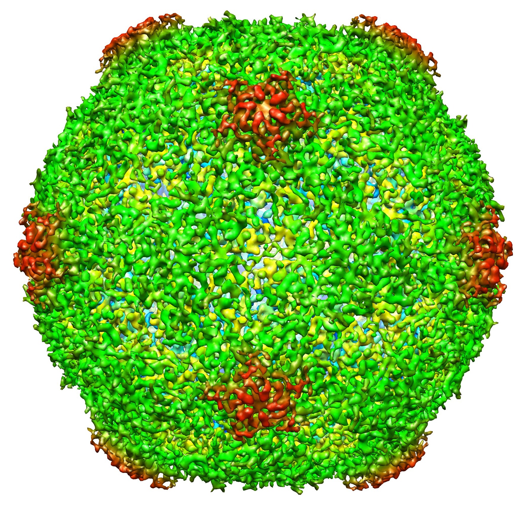
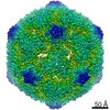
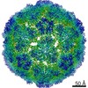
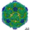
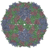
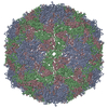
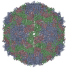
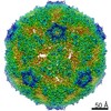
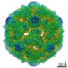
| タイトル | Cryo-EM structure of Poliovirus 135S particles | |||||||||
 マップデータ マップデータ | Poliovirus 135S particle | |||||||||
 試料 試料 |
| |||||||||
 キーワード キーワード | cell entry / cryo-electron microscopy / poliovirus / single particle analysis | |||||||||
| 機能・相同性 |  機能・相同性情報 機能・相同性情報symbiont-mediated suppression of host translation initiation / symbiont-mediated suppression of host cytoplasmic pattern recognition receptor signaling pathway via inhibition of RIG-I activity / symbiont-mediated suppression of host cytoplasmic pattern recognition receptor signaling pathway via inhibition of MDA-5 activity / symbiont-mediated suppression of host cytoplasmic pattern recognition receptor signaling pathway via inhibition of MAVS activity / ribonucleoside triphosphate phosphatase activity / picornain 2A / symbiont-mediated suppression of host mRNA export from nucleus / symbiont genome entry into host cell via pore formation in plasma membrane / picornain 3C / T=pseudo3 icosahedral viral capsid ...symbiont-mediated suppression of host translation initiation / symbiont-mediated suppression of host cytoplasmic pattern recognition receptor signaling pathway via inhibition of RIG-I activity / symbiont-mediated suppression of host cytoplasmic pattern recognition receptor signaling pathway via inhibition of MDA-5 activity / symbiont-mediated suppression of host cytoplasmic pattern recognition receptor signaling pathway via inhibition of MAVS activity / ribonucleoside triphosphate phosphatase activity / picornain 2A / symbiont-mediated suppression of host mRNA export from nucleus / symbiont genome entry into host cell via pore formation in plasma membrane / picornain 3C / T=pseudo3 icosahedral viral capsid / host cell cytoplasmic vesicle membrane / nucleoside-triphosphate phosphatase / channel activity / monoatomic ion transmembrane transport / RNA helicase activity / endocytosis involved in viral entry into host cell / symbiont-mediated activation of host autophagy / RNA-directed RNA polymerase / cysteine-type endopeptidase activity / viral RNA genome replication / RNA-directed RNA polymerase activity / DNA-templated transcription / virion attachment to host cell / host cell nucleus / structural molecule activity / proteolysis / RNA binding / zinc ion binding / ATP binding / membrane 類似検索 - 分子機能 | |||||||||
| 生物種 |   Human poliovirus 1 Mahoney (ポリオウイルス) Human poliovirus 1 Mahoney (ポリオウイルス) | |||||||||
| 手法 | 単粒子再構成法 / クライオ電子顕微鏡法 / 解像度: 5.5 Å | |||||||||
 データ登録者 データ登録者 | Butan C / Filman DJ / Hogle JM | |||||||||
 引用 引用 | ジャーナル: J Virol / 年: 2014 タイトル: Cryo-electron microscopy reconstruction shows poliovirus 135S particles poised for membrane interaction and RNA release. 著者: Carmen Butan / David J Filman / James M Hogle /  要旨: During infection, binding of mature poliovirus to cell surface receptors induces an irreversible expansion of the capsid, to form an infectious cell-entry intermediate particle that sediments at 135S. ...During infection, binding of mature poliovirus to cell surface receptors induces an irreversible expansion of the capsid, to form an infectious cell-entry intermediate particle that sediments at 135S. In these expanded virions, the major capsid proteins (VP1 to VP3) adopt an altered icosahedral arrangement to open holes in the capsid at 2-fold and quasi-3-fold axes, and internal polypeptides VP4 and the N terminus of VP1, which can bind membranes, become externalized. Cryo-electron microscopy images for 117,330 particles were collected using Leginon and reconstructed using FREALIGN. Improved rigid-body positioning of major capsid proteins established reliably which polypeptide segments become disordered or rearranged. The virus-to-135S transition includes expansion of 4%, rearrangements of the GH loops of VP3 and VP1, and disordering of C-terminal extensions of VP1 and VP2. The N terminus of VP1 rearranges to become externalized near its quasi-3-fold exit, binds to rearranged GH loops of VP3 and VP1, and attaches to the top surface of VP2. These details improve our understanding of subsequent stages of infection, including endocytosis and RNA transfer into the cytoplasm. | |||||||||
| 履歴 |
|
- 構造の表示
構造の表示
| ムービー |
ムービービューア |
|---|---|
| 構造ビューア | EMマップ: SurfViewMolmilJmol/JSmol |
| 添付画像 |
- ダウンロードとリンク
-EMDBアーカイブ
| マップデータ | emd_5710.map.gz | 225.1 MB | EMDBマップデータ形式 | |
|---|---|---|---|---|
| ヘッダ (付随情報) | emd-5710-v30.xmlemd-5710.xml | 12 KB 12 KB | 表示 表示 | EMDBヘッダ |
| 画像 | emd_5710.tif | 8.9 MB | ||
| アーカイブディレクトリ |  http://ftp.pdbj.org/pub/emdb/structures/EMD-5710ftp://ftp.pdbj.org/pub/emdb/structures/EMD-5710 http://ftp.pdbj.org/pub/emdb/structures/EMD-5710ftp://ftp.pdbj.org/pub/emdb/structures/EMD-5710 | HTTPS FTP |
-検証レポート
| 文書・要旨 | emd_5710_validation.pdf.gz | 377.9 KB | 表示 | EMDB検証レポート |
|---|---|---|---|---|
| 文書・詳細版 | emd_5710_full_validation.pdf.gz | 377.5 KB | 表示 | |
| XML形式データ | emd_5710_validation.xml.gz | 7.1 KB | 表示 | |
| アーカイブディレクトリ | https://ftp.pdbj.org/pub/emdb/validation_reports/EMD-5710ftp://ftp.pdbj.org/pub/emdb/validation_reports/EMD-5710 | HTTPS FTP |
-関連構造データ
-リンク
| EMDBのページ | EMDB (EBI/PDBe) / EMDataResource |
|---|---|
| 「今月の分子」の関連する項目 |


-マップ
| ファイル | ダウンロード / ファイル: emd_5710.map.gz / 形式: CCP4 / 大きさ: 238.4 MB / タイプ: IMAGE STORED AS FLOATING POINT NUMBER (4 BYTES) | ||||||||||||||||||||||||||||||||||||||||||||||||||||||||||||||||||||
|---|---|---|---|---|---|---|---|---|---|---|---|---|---|---|---|---|---|---|---|---|---|---|---|---|---|---|---|---|---|---|---|---|---|---|---|---|---|---|---|---|---|---|---|---|---|---|---|---|---|---|---|---|---|---|---|---|---|---|---|---|---|---|---|---|---|---|---|---|---|
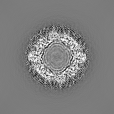
| 注釈 | Poliovirus 135S particle | ||||||||||||||||||||||||||||||||||||||||||||||||||||||||||||||||||||
| 投影像・断面図 | 画像のコントロール
画像は Spider により作成 | ||||||||||||||||||||||||||||||||||||||||||||||||||||||||||||||||||||
| ボクセルのサイズ | X=Y=Z: 1.37 Å | ||||||||||||||||||||||||||||||||||||||||||||||||||||||||||||||||||||
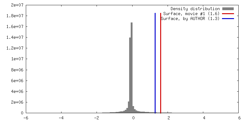
| 密度 |
| ||||||||||||||||||||||||||||||||||||||||||||||||||||||||||||||||||||
| 対称性 | 空間群: 1 | ||||||||||||||||||||||||||||||||||||||||||||||||||||||||||||||||||||
| 詳細 | EMDB XML:
CCP4マップ ヘッダ情報:
| ||||||||||||||||||||||||||||||||||||||||||||||||||||||||||||||||||||
 Z (Sec.)
 Y (Row.)
 X (Col.)


-添付データ
- 試料の構成要素
-全体 : Poliovirus 135S particle
| 全体 | 名称: Poliovirus 135S particle |
|---|---|
| 要素 |
|
-超分子 #1000: Poliovirus 135S particle
| 超分子 | 名称: Poliovirus 135S particle / タイプ: sample / ID: 1000 詳細: Native virus 160S is converted by heat treatment to 135S. 集合状態: icosahedrally ordered capsid: 60 copies of VP1, VP2, VP3 Number unique components: 1 |
|---|---|
| 分子量 | 実験値: 8.6 MDa / 理論値: 9 MDa |
-超分子 #1: Human poliovirus 1 Mahoney
| 超分子 | 名称: Human poliovirus 1 Mahoney / タイプ: virus / ID: 1 / Name.synonym: Poliovirus type 1 (strain Mahoney) / NCBI-ID: 12081 / 生物種: Human poliovirus 1 Mahoney / Sci species strain: Mahoney / ウイルスタイプ: VIRION / ウイルス・単離状態: STRAIN / ウイルス・エンベロープ: No / ウイルス・中空状態: No / Syn species name: Poliovirus type 1 (strain Mahoney) |
|---|---|
| 宿主 | 生物種:  Homo sapiens (ヒト) / 別称: VERTEBRATES Homo sapiens (ヒト) / 別称: VERTEBRATES |
| Host system | 生物種: Homo sapiens (ヒト) / 組換細胞: HeLa |
| 分子量 | 実験値: 8.6 MDa / 理論値: 9 MDa |
| ウイルス殻 | Shell ID: 1 / 直径: 308 Å / T番号(三角分割数): 1 |
-実験情報
-構造解析
| 手法 | クライオ電子顕微鏡法 |
|---|---|
 解析 解析 | 単粒子再構成法 |
| 試料の集合状態 | particle |
-試料調製
| 濃度 | 0.3 mg/mL |
|---|---|
| 緩衝液 | pH: 7.4 / 詳細: 2 mM CaCl2, 20 mM HEPES |
| グリッド | 詳細: glow-discharged holey carbon-grids (200 mesh C-flat grids) |
| 凍結 | 凍結剤: ETHANE / チャンバー内温度: 90 K / 装置: HOMEMADE PLUNGER 詳細: Vitrification carried out in ambient atmosphere. Ethane cooled by liquid nitrogen. 手法: Blotted manually before plunging into liquid ethane |
- 電子顕微鏡法
| 顕微鏡 | FEI TECNAI F20 |
|---|---|
| 温度 | 最低: 90 K / 最高: 93 K / 平均: 90 K |
| アライメント法 | Legacy - 非点収差: Objective lens astigmatism was corrected. |
| 日付 | 2011年2月28日 |
| 撮影 | カテゴリ: CCD フィルム・検出器のモデル: TVIPS TEMCAM-F415 (4k x 4k) 実像数: 1020 / 平均電子線量: 15 e/Å2 |
| Tilt angle min | 0 |
| Tilt angle max | 0 |
| 電子線 | 加速電圧: 200 kV / 電子線源:  FIELD EMISSION GUN FIELD EMISSION GUN |
| 電子光学系 | 照射モード: FLOOD BEAM / 撮影モード: BRIGHT FIELD / Cs: 2 mm / 最大 デフォーカス(公称値): 2.0 µm / 最小 デフォーカス(公称値): 0.98 µm / 倍率(公称値): 62000 |
| 試料ステージ | 試料ホルダー: Side entry liquid nitrogen-cooled cryo specimen holder 試料ホルダーモデル: GATAN LIQUID NITROGEN |
| 実験機器 |  モデル: Tecnai F20 / 画像提供: FEI Company |
-画像解析
| 詳細 | The particles were selected using an automatic selection program. |
|---|---|
| CTF補正 | 詳細: Each micrograph |
| 最終 再構成 | アルゴリズム: OTHER / 解像度のタイプ: BY AUTHOR / 解像度: 5.5 Å / 解像度の算出法: FSC 0.143 CUT-OFF / ソフトウェア - 名称: FREALIGN / 使用した粒子像数: 117330 |
-原子モデル構築 1
| 初期モデル | PDB ID: Chain - #0 - Chain ID: 0 / Chain - #1 - Chain ID: 1 / Chain - #2 - Chain ID: 3 |
|---|---|
| ソフトウェア | 名称: COOT, REFMAC |
| 詳細 | Most of the model was docked, with specific areas of discrepancy fitted. The fitting was rigid body with flexible fitting or deletion of selected polypeptide segments. Rigid bodies for VP1, VP2, VP3, and the VP3 beta tube were defined to include beta barrels and non-covalently attached polypeptides. Each rigid body was repeatedly fitted manually and then refined. Disordered polypeptide segments were removed. Several rearranged segments were included as approximate backbone traces and refined. |
| 精密化 | 空間: RECIPROCAL / プロトコル: RIGID BODY FIT 当てはまり具合の基準: mean amplitude-weighted cosine of the phase difference |
| 得られたモデル | 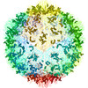 PDB-3j48: |