cell envelope Sec protein transport complex / protein transport by the Sec complex / intracellular protein transmembrane transport / protein-transporting ATPase activity / SRP-dependent cotranslational protein targeting to membrane, translocation / signal sequence receptor activity / Secretion of toxins / transmembrane protein transporter activity / protein secretion / intracellular protein transport ...cell envelope Sec protein transport complex / protein transport by the Sec complex / intracellular protein transmembrane transport / protein-transporting ATPase activity / SRP-dependent cotranslational protein targeting to membrane, translocation / signal sequence receptor activity / Secretion of toxins / transmembrane protein transporter activity / protein secretion / intracellular protein transport / ribosomal large subunit assembly / large ribosomal subunit rRNA binding / cytosolic large ribosomal subunit / cytoplasmic translation / rRNA binding / structural constituent of ribosome / translation / membrane / plasma membrane / cytoplasm / cytosol Similarity search - Function
Preprotein translocase SecG subunit / Preprotein translocase SecG subunit / SecE subunit of protein translocation complex, bacterial-like / SecE superfamily / Protein translocase subunit SecY / Protein secE/sec61-gamma signature. / Protein secY signature 1. / Protein secY signature 2. / SecE/Sec61-gamma subunits of protein translocation complex / Protein translocase complex, SecE/Sec61-gamma subunit ...Preprotein translocase SecG subunit / Preprotein translocase SecG subunit / SecE subunit of protein translocation complex, bacterial-like / SecE superfamily / Protein translocase subunit SecY / Protein secE/sec61-gamma signature. / Protein secY signature 1. / Protein secY signature 2. / SecE/Sec61-gamma subunits of protein translocation complex / Protein translocase complex, SecE/Sec61-gamma subunit / SecY/SEC61-alpha family / SecY domain superfamily / SecY conserved site / SecY / : / : / Large ribosomal subunit protein uL24, C-terminal domain / Ribosomal protein L24 / Ribosomal protein L23/L25, conserved site / Ribosomal protein L23 signature. / Ribosomal protein L29, conserved site / Ribosomal protein L29 signature. / Ribosomal L29 protein / Ribosomal protein L29/L35 / Ribosomal protein L29/L35 superfamily / Ribosomal protein L24 signature. / Ribosomal protein L24/L26, conserved site / KOW (Kyprides, Ouzounis, Woese) motif. / Ribosomal protein L23 / Ribosomal protein L25/L23 / Ribosomal protein L26/L24, KOW domain / Ribosomal protein L23/L15e core domain superfamily / Translation protein SH3-like domain superfamily / KOW motif / KOW / Ribosomal protein L2, domain 2 / Nucleotide-binding alpha-beta plait domain superfamily Similarity search - Domain/homology
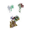
Large ribosomal subunit protein uL29 / Large ribosomal subunit protein uL23 / Protein translocase subunit SecE / Protein-export membrane protein SecG / Protein translocase subunit SecY / Large ribosomal subunit protein uL24 Similarity search - Component
Biological species
Escherichia coli (E. coli)
Method
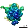
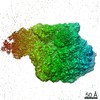
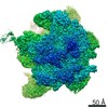
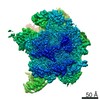
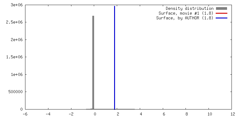
single particle reconstruction / cryo EM / Resolution: 9.5 Å
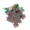
Journal: Nature / Year: 2014 Title: Structure of the SecY channel during initiation of protein translocation. Authors: Eunyong Park / Jean-François Ménétret / James C Gumbart / Steven J Ludtke / Weikai Li / Andrew Whynot / Tom A Rapoport / Christopher W Akey / Abstract: Many secretory proteins are targeted by signal sequences to a protein-conducting channel, formed by prokaryotic SecY or eukaryotic Sec61 complexes, and are translocated across the membrane during ...Many secretory proteins are targeted by signal sequences to a protein-conducting channel, formed by prokaryotic SecY or eukaryotic Sec61 complexes, and are translocated across the membrane during their synthesis. Crystal structures of the inactive channel show that the SecY subunit of the heterotrimeric complex consists of two halves that form an hourglass-shaped pore with a constriction in the middle of the membrane and a lateral gate that faces the lipid phase. The closed channel has an empty cytoplasmic funnel and an extracellular funnel that is filled with a small helical domain, called the plug. During initiation of translocation, a ribosome-nascent chain complex binds to the SecY (or Sec61) complex, resulting in insertion of the nascent chain. However, the mechanism of channel opening during translocation is unclear. Here we have addressed this question by determining structures of inactive and active ribosome-channel complexes with cryo-electron microscopy. Non-translating ribosome-SecY channel complexes derived from Methanocaldococcus jannaschii or Escherichia coli show the channel in its closed state, and indicate that ribosome binding per se causes only minor changes. The structure of an active E. coli ribosome-channel complex demonstrates that the nascent chain opens the channel, causing mostly rigid body movements of the amino- and carboxy-terminal halves of SecY. In this early translocation intermediate, the polypeptide inserts as a loop into the SecY channel with the hydrophobic signal sequence intercalated into the open lateral gate. The nascent chain also forms a loop on the cytoplasmic surface of SecY rather than entering the channel directly.
History
Deposition
Jun 14, 2013
-
Header (metadata) release
Aug 21, 2013
-
Map release
Oct 23, 2013
-
Update
Feb 5, 2014
-
Current status
Feb 5, 2014
Processing site: RCSB / Status: Released
-
Structure visualization
Movie
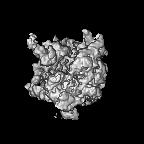
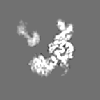
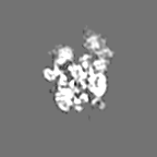
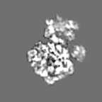
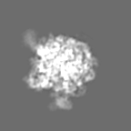
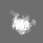
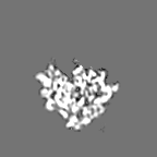
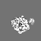
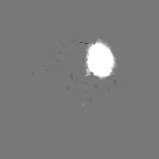
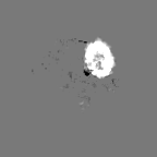
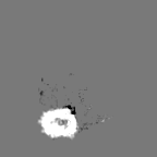
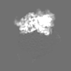
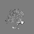
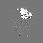
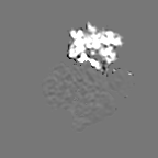
Surface view with section colored by density value
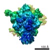
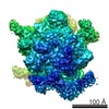
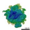
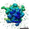
Entire : E. coli 70S ribosome with recombinant E. coli SecYEG
Entire
Name: E. coli 70S ribosome with recombinant E. coli SecYEG
Components
Sample: E. coli 70S ribosome with recombinant E. coli SecYEG
Complex: non-translating 70S ribosome
Protein or peptide: SecYEG
-
Supramolecule #1000: E. coli 70S ribosome with recombinant E. coli SecYEG
Supramolecule
Name: E. coli 70S ribosome with recombinant E. coli SecYEG / type: sample / ID: 1000 Details: Ribosome-SecY complexes were prepared by mixing ribosomes at 4 uM with SecY (32 uM) and incubating them on ice for 30 min before freezing. Oligomeric state: one ribosome and one SecYEG / Number unique components: 2
pH: 7.5 Details: 50 mM HEPES-KOH, 100 mM KOAc, 10 mM Mg(OAc)2, 0.05% DDM
Grid
Details: 400 mesh Cu grids with continuous or holey carbon films
Vitrification
Cryogen name: ETHANE / Chamber humidity: 95 % / Chamber temperature: 90 K / Instrument: HOMEMADE PLUNGER / Method: Blot 1 second before plunging.
-
Electron microscopy
Microscope
FEI TECNAI 20
Temperature
Average: 93 K
Alignment procedure
Legacy - Astigmatism: imaging of carbon film at 175,000 times magnification
Details
low dose imaging with manual data collection
Date
Apr 10, 2006
Image recording
Category: FILM / Film or detector model: KODAK SO-163 FILM / Digitization - Scanner: OTHER / Digitization - Sampling interval: 4.5 µm / Number real images: 360 / Average electron dose: 20 e/Å2 / Od range: 1 / Bits/pixel: 16
Tilt angle min
0
Electron beam
Acceleration voltage: 200 kV / Electron source: FIELD EMISSION GUN
Particles were picked with boxer and CTF-corrected with EMAN1.
CTF correction
Details: per micrograph
Final reconstruction
Algorithm: OTHER / Resolution.type: BY AUTHOR / Resolution: 9.5 Å / Resolution method: OTHER / Software - Name: EMAN1 Details: CTF correction was done on untilted and 30 degree tilted images. Number images used: 39000
In the structure databanks used in Yorodumi, some data are registered as the other names, "COVID-19 virus" and "2019-nCoV". Here are the details of the virus and the list of structure data.
Jan 31, 2019. EMDB accession codes are about to change! (news from PDBe EMDB page)
EMDB accession codes are about to change! (news from PDBe EMDB page)
The allocation of 4 digits for EMDB accession codes will soon come to an end. Whilst these codes will remain in use, new EMDB accession codes will include an additional digit and will expand incrementally as the available range of codes is exhausted. The current 4-digit format prefixed with “EMD-” (i.e. EMD-XXXX) will advance to a 5-digit format (i.e. EMD-XXXXX), and so on. It is currently estimated that the 4-digit codes will be depleted around Spring 2019, at which point the 5-digit format will come into force.
The EM Navigator/Yorodumi systems omit the EMD- prefix.
Related info.:Q: What is EMD? / ID/Accession-code notation in Yorodumi/EM Navigator
Yorodumi is a browser for structure data from EMDB, PDB, SASBDB, etc.
This page is also the successor to EM Navigator detail page, and also detail information page/front-end page for Omokage search.
The word "yorodu" (or yorozu) is an old Japanese word meaning "ten thousand". "mi" (miru) is to see.
Related info.:EMDB / PDB / SASBDB / Comparison of 3 databanks / Yorodumi Search / Aug 31, 2016. New EM Navigator & Yorodumi / Yorodumi Papers / Jmol/JSmol / Function and homology information / Changes in new EM Navigator and Yorodumi
 Movie
Movie Controller


 Open data
 Basic information
 Map data
 Sample
 Keywords
 Function and homology information
 Authors
 Citation
 Structure visualization
 Downloads & links
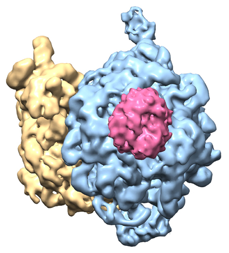 emd_5692.jpg
 http://ftp.pdbj.org/pub/emdb/structures/EMD-5692


 Z (Sec.)
 Y (Row.)
 X (Col.)


 Sample components
 Processing
 Electron microscopy
 FIELD EMISSION GUN
