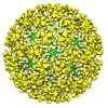
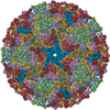
+ Open data
- Basic information
| Entry | Database: EMDB / ID: EMD-5563 | |||||||||
|---|---|---|---|---|---|---|---|---|---|---|
| Title | Electron microscopy of Everglades virus | |||||||||
 Map data Map data | Reconstruction of wild type everglades virus | |||||||||
 Sample Sample |
| |||||||||
 Keywords Keywords | electron microscopy / cryo-electron microscopy / alphavirus / everglades virus | |||||||||
| Biological species |  Everglades virus Everglades virus | |||||||||
| Method | single particle reconstruction / cryo EM / Resolution: 10.0 Å | |||||||||
 Authors Authors | Sherman MB / Trujillo J / Leahy I / Razmus D / DeHate R / Lorcheim P / Czarneski MA / Zimmerman D / Newton JPM / Haddow AM / Weaver SC | |||||||||
 Citation Citation |  Journal: J Struct Biol / Year: 2013 Journal: J Struct Biol / Year: 2013Title: Construction and organization of a BSL-3 cryo-electron microscopy laboratory at UTMB. Authors: Michael B Sherman / Juan Trujillo / Ian Leahy / Dennis Razmus / Robert Dehate / Paul Lorcheim / Mark A Czarneski / Domenica Zimmerman / Je T'aime M Newton / Andrew D Haddow / Scott C Weaver /  Abstract: A unique cryo-electron microscopy facility has been designed and constructed at the University of Texas Medical Branch (UTMB) to study the three-dimensional organization of viruses and bacteria ...A unique cryo-electron microscopy facility has been designed and constructed at the University of Texas Medical Branch (UTMB) to study the three-dimensional organization of viruses and bacteria classified as select agents at biological safety level (BSL)-3, and their interactions with host cells. A 200keV high-end cryo-electron microscope was installed inside a BSL-3 containment laboratory and standard operating procedures were developed and implemented to ensure its safe and efficient operation. We also developed a new microscope decontamination protocol based on chlorine dioxide gas with a continuous flow system, which allowed us to expand the facility capabilities to study bacterial agents including spore-forming species. The new unified protocol does not require agent-specific treatment in contrast to the previously used heat decontamination. To optimize the use of the cryo-electron microscope and to improve safety conditions, it can be remotely controlled from a room outside of containment, or through a computer network world-wide. Automated data collection is provided by using JADAS (single particle imaging) and SerialEM (tomography). The facility has successfully operated for more than a year without an incident and was certified as a select agent facility by the Centers for Disease Control. | |||||||||
| History |
|
- Structure visualization
| Movie |
 Movie viewer Movie viewer |
|---|---|
| Structure viewer | EM map:  SurfView SurfView Molmil Molmil Jmol/JSmol Jmol/JSmol |
| Supplemental images |
- Downloads & links
-EMDB archive
| Map data |  emd_5563.map.gz emd_5563.map.gz | 15.2 MB |  EMDB map data format EMDB map data format | |
|---|---|---|---|---|
| Header (meta data) |  emd-5563-v30.xml emd-5563-v30.xml emd-5563.xml emd-5563.xml | 10.8 KB 10.8 KB | Display Display |  EMDB header EMDB header |
| Images |  emd_5563_1.jpg emd_5563_1.jpg | 266.9 KB | ||
| Archive directory |  http://ftp.pdbj.org/pub/emdb/structures/EMD-5563 http://ftp.pdbj.org/pub/emdb/structures/EMD-5563 ftp://ftp.pdbj.org/pub/emdb/structures/EMD-5563 ftp://ftp.pdbj.org/pub/emdb/structures/EMD-5563 | HTTPS FTP |
-Validation report
| Summary document |  emd_5563_validation.pdf.gz emd_5563_validation.pdf.gz | 78.9 KB | Display |  EMDB validaton report EMDB validaton report |
|---|---|---|---|---|
| Full document |  emd_5563_full_validation.pdf.gz emd_5563_full_validation.pdf.gz | 78 KB | Display | |
| Data in XML |  emd_5563_validation.xml.gz emd_5563_validation.xml.gz | 494 B | Display | |
| Arichive directory |  https://ftp.pdbj.org/pub/emdb/validation_reports/EMD-5563 https://ftp.pdbj.org/pub/emdb/validation_reports/EMD-5563 ftp://ftp.pdbj.org/pub/emdb/validation_reports/EMD-5563 ftp://ftp.pdbj.org/pub/emdb/validation_reports/EMD-5563 | HTTPS FTP |
-Related structure data
| Similar structure data |
|---|
- Links
Links
| EMDB pages |  EMDB (EBI/PDBe) / EMDB (EBI/PDBe) /  EMDataResource EMDataResource |
|---|
- Map
Map
| File |  Download / File: emd_5563.map.gz / Format: CCP4 / Size: 29.8 MB / Type: IMAGE STORED AS FLOATING POINT NUMBER (4 BYTES) Download / File: emd_5563.map.gz / Format: CCP4 / Size: 29.8 MB / Type: IMAGE STORED AS FLOATING POINT NUMBER (4 BYTES) | ||||||||||||||||||||||||||||||||||||||||||||||||||||||||||||||||||||
|---|---|---|---|---|---|---|---|---|---|---|---|---|---|---|---|---|---|---|---|---|---|---|---|---|---|---|---|---|---|---|---|---|---|---|---|---|---|---|---|---|---|---|---|---|---|---|---|---|---|---|---|---|---|---|---|---|---|---|---|---|---|---|---|---|---|---|---|---|---|
| Annotation | Reconstruction of wild type everglades virus | ||||||||||||||||||||||||||||||||||||||||||||||||||||||||||||||||||||
| Projections & slices | Image control
Images are generated by Spider. | ||||||||||||||||||||||||||||||||||||||||||||||||||||||||||||||||||||
| Voxel size | X=Y=Z: 4 Å | ||||||||||||||||||||||||||||||||||||||||||||||||||||||||||||||||||||
| Density |
| ||||||||||||||||||||||||||||||||||||||||||||||||||||||||||||||||||||
| Symmetry | Space group: 1 | ||||||||||||||||||||||||||||||||||||||||||||||||||||||||||||||||||||
| Details | EMDB XML:
CCP4 map header:
| ||||||||||||||||||||||||||||||||||||||||||||||||||||||||||||||||||||
-Supplemental data
- Sample components
-Entire : Everglades virus
| Entire | Name:  Everglades virus Everglades virus |
|---|---|
| Components |
|
-Supramolecule #1000: Everglades virus
| Supramolecule | Name: Everglades virus / type: sample / ID: 1000 / Details: monodisperse sample / Number unique components: 4 |
|---|
-Supramolecule #1: Everglades virus
| Supramolecule | Name: Everglades virus / type: virus / ID: 1 / Sci species name: Everglades virus / Sci species strain: Fe5-47et / Virus type: VIRION / Virus isolate: STRAIN / Virus enveloped: Yes / Virus empty: No |
|---|---|
| Host (natural) | Organism: unidentified (others) / synonym: VERTEBRATES |
| Host system | Organism: unidentified (others) |
| Virus shell | Shell ID: 1 / Name: glycoprotein E1/E2 / Diameter: 700 Å / T number (triangulation number): 4 |
-Experimental details
-Structure determination
| Method | cryo EM |
|---|---|
 Processing Processing | single particle reconstruction |
| Aggregation state | particle |
- Sample preparation
| Concentration | 1 mg/mL |
|---|---|
| Buffer | pH: 7.4 / Details: 0.05 M Tris HCl, pH 7.4, 0.1 M NaCl, 0.001 M EDTA |
| Grid | Details: C-flat 1.2/1.3 holey film |
| Vitrification | Cryogen name: ETHANE / Chamber humidity: 60 % / Chamber temperature: 90 K / Instrument: HOMEMADE PLUNGER / Method: blot for 4 seconds before plunging |
- Electron microscopy
| Microscope | JEOL 2200FS |
|---|---|
| Temperature | Min: 87 K / Max: 90 K / Average: 88 K |
| Alignment procedure | Legacy - Astigmatism: manual correction at 150,000x |
| Specialist optics | Energy filter - Name: omega |
| Date | Feb 9, 2012 |
| Image recording | Category: CCD / Film or detector model: GATAN ULTRASCAN 4000 (4k x 4k) / Digitization - Sampling interval: 15 µm / Number real images: 80 / Average electron dose: 20 e/Å2 / Bits/pixel: 16 |
| Electron beam | Acceleration voltage: 200 kV / Electron source:  FIELD EMISSION GUN FIELD EMISSION GUN |
| Electron optics | Calibrated magnification: 74800 / Illumination mode: FLOOD BEAM / Imaging mode: BRIGHT FIELD / Cs: 2.0 mm / Nominal defocus max: 2.3 µm / Nominal defocus min: 0.3 µm / Nominal magnification: 60000 |
| Sample stage | Specimen holder: Gatan 626 70 degree holder / Specimen holder model: GATAN LIQUID NITROGEN |
- Image processing
| Details | particles picked with EMAN2 boxer, reconstructed using IMAGIC-5 |
|---|---|
| CTF correction | Details: micrograph |
| Final reconstruction | Algorithm: OTHER / Resolution.type: BY AUTHOR / Resolution: 10.0 Å / Resolution method: FSC 0.5 CUT-OFF / Software - Name: IMAGIC-5 Details: final maps were calculated from individual particles Number images used: 1000 |
| Final angle assignment | Details: icosahedral asymmetric unit |
 Movie
Movie Controller


 Z (Sec.)
 Y (Row.)
 X (Col.)


