National Institutes of Health/National Institute Of Allergy and Infectious Diseases (NIH/NIAID)
U54AI170791
United States
Wellcome Trust
CC2058
United Kingdom
Medical Research Council (MRC, United Kingdom)
CC2058
United Kingdom
Cancer Research UK
CC2058
United Kingdom
Citation
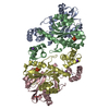
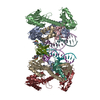
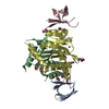
Journal: Nature / Year: 2026 Title: Integrase anchors viral RNA to the HIV-1 capsid interior. Authors: Matthew R Singer / Zhen Li / Juan S Rey / Joshua Hope / Florian Chenavier / Nicola J Cook / Emma Punch / Jamie Smith / Zhiyu Zhou / Sarah Maslen / Laura Masino / Andrea Nans / Mark Skehel / ...Authors: Matthew R Singer / Zhen Li / Juan S Rey / Joshua Hope / Florian Chenavier / Nicola J Cook / Emma Punch / Jamie Smith / Zhiyu Zhou / Sarah Maslen / Laura Masino / Andrea Nans / Mark Skehel / Ian A Taylor / Giulia Zanetti / Peijun Zhang / Juan R Perilla / Alan N Engelman / Peter Cherepanov / Abstract: HIV-1 integrase (IN) promotes encapsulation of viral genomic RNA into mature viral cores, and this function is a target for ongoing antiretroviral drug development efforts. Here we determined the ...HIV-1 integrase (IN) promotes encapsulation of viral genomic RNA into mature viral cores, and this function is a target for ongoing antiretroviral drug development efforts. Here we determined the cryogenic electron microscopy (cryo-EM) structure of a primate lentiviral IN in a complex with RNA, revealing a linear filament made of IN octamer repeat units, each comprising a pair of asymmetric homotetramers. The assembly is stabilized through IN-RNA interactions involving mainly the IN C-terminal domains and RNA backbone. The spacing and orientation of the IN filament repeat units closely matched those of consecutive capsid (CA) hexamers within the mature CA lattice. Using cryo-EM images of native purified HIV-1 cores, we refined the structure of the IN filament as it propagates along the luminal side of the CA lattice. Each IN tetramer within the filament nestled in a CA hexamer, engaging closely with the major homology regions. Substitutions of residues involved in IN-CA contacts yielded eccentric virions with RNA nucleoids located outside of the cores. Collectively, our results establish the structural basis for the HIV-1 IN-RNA interaction and reveal that IN forms an RNA-binding module on the luminal side of the mature CA lattice.
In the structure databanks used in Yorodumi, some data are registered as the other names, "COVID-19 virus" and "2019-nCoV". Here are the details of the virus and the list of structure data.
Jan 31, 2019. EMDB accession codes are about to change! (news from PDBe EMDB page)
EMDB accession codes are about to change! (news from PDBe EMDB page)
The allocation of 4 digits for EMDB accession codes will soon come to an end. Whilst these codes will remain in use, new EMDB accession codes will include an additional digit and will expand incrementally as the available range of codes is exhausted. The current 4-digit format prefixed with “EMD-” (i.e. EMD-XXXX) will advance to a 5-digit format (i.e. EMD-XXXXX), and so on. It is currently estimated that the 4-digit codes will be depleted around Spring 2019, at which point the 5-digit format will come into force.
The EM Navigator/Yorodumi systems omit the EMD- prefix.
Related info.:Q: What is EMD? / ID/Accession-code notation in Yorodumi/EM Navigator
Yorodumi is a browser for structure data from EMDB, PDB, SASBDB, etc.
This page is also the successor to EM Navigator detail page, and also detail information page/front-end page for Omokage search.
The word "yorodu" (or yorozu) is an old Japanese word meaning "ten thousand". "mi" (miru) is to see.
Related info.:EMDB / PDB / SASBDB / Comparison of 3 databanks / Yorodumi Search / Aug 31, 2016. New EM Navigator & Yorodumi / Yorodumi Papers / Jmol/JSmol / Function and homology information / Changes in new EM Navigator and Yorodumi
 Movie
Movie Controller

 Yorodumi
 Open data
 Basic information
 Map data
 Sample
 Keywords
 Function and homology information
 Human immunodeficiency virus 1
 Authors
 United States,
  United Kingdom, 4 items
  Citation
 Structure visualization
 Downloads & links
 emd_54071.png
 http://ftp.pdbj.org/pub/emdb/structures/EMD-54071


 Z (Sec.)
 Y (Row.)
 X (Col.)


 Sample components
 Homo sapiens (human) / Strain: NL4-3
 Processing
 Electron microscopy
 FIELD EMISSION GUN