- EMDB-5352: Structure of a type III secretion needle -
+
データを開く
IDまたはキーワード:
読み込み中...
-
基本情報
登録情報
データベース: EMDB / ID: EMD-5352
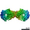
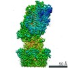
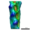
タイトル
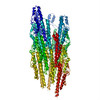
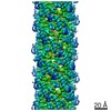
Structure of a type III secretion needle
マップデータ
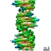
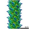
This is an reconstruction of the type III secretion needle
試料
試料: Shigella needle
タンパク質・ペプチド: MxiH
キーワード
type III secretion system / needle / helical filament
機能・相同性
機能・相同性情報
type III protein secretion system complex / protein secretion by the type III secretion system / cell surface / extracellular region / identical protein binding 類似検索 - 分子機能
Type III secretion, needle-protein-like / Type III secretion, needle-protein-like superfamily / Type III secretion needle MxiH, YscF, SsaG, EprI, PscF, EscF / Type III secretion system, needle protein 類似検索 - ドメイン・相同性
Type 3 secretion system needle filament protein / Type 3 secretion system needle filament protein 類似検索 - 構成要素
ジャーナル: Proc Natl Acad Sci U S A / 年: 2012 タイトル: Structure of a type III secretion needle at 7-Å resolution provides insights into its assembly and signaling mechanisms. 著者: Takashi Fujii / Martin Cheung / Amandine Blanco / Takayuki Kato / Ariel J Blocker / Keiichi Namba / 要旨: Type III secretion systems of Gram-negative bacteria form injection devices that deliver effector proteins into eukaryotic cells during infection. They span both bacterial membranes and the ...Type III secretion systems of Gram-negative bacteria form injection devices that deliver effector proteins into eukaryotic cells during infection. They span both bacterial membranes and the extracellular space to connect with the host cell plasma membrane. Their extracellular portion is a needle-like, hollow tube that serves as a secretion conduit for effector proteins. The needle of Shigella flexneri is approximately 50-nm long and 7-nm thick and is made by the helical assembly of one protein, MxiH. We provide a 7-Å resolution 3D image reconstruction of the Shigella needle by electron cryomicroscopy, which resolves α-helices and a β-hairpin that has never been observed in the crystal and solution structures of needle proteins, including MxiH. An atomic model of the needle based on the 3D-density map, in comparison with that of the bacterial-flagellar filament, provides insights into how such a thin tubular structure is stably assembled by intricate intermolecular interactions. The map also illuminates how the needle-length control protein functions as a ruler within the central channel during export of MxiH for assembly at the distal end of the needle, and how the secretion-activation signal may be transduced through a conformational change of the needle upon host-cell contact.
 ムービー
ムービー コントローラー


 データを開く
 基本情報
基本情報 マップデータ
 試料
試料 キーワード
キーワード 機能・相同性情報
 Shigella flexneri (フレクスナー赤痢菌)
 データ登録者
 引用
引用
 構造の表示
構造の表示
 ダウンロードとリンク
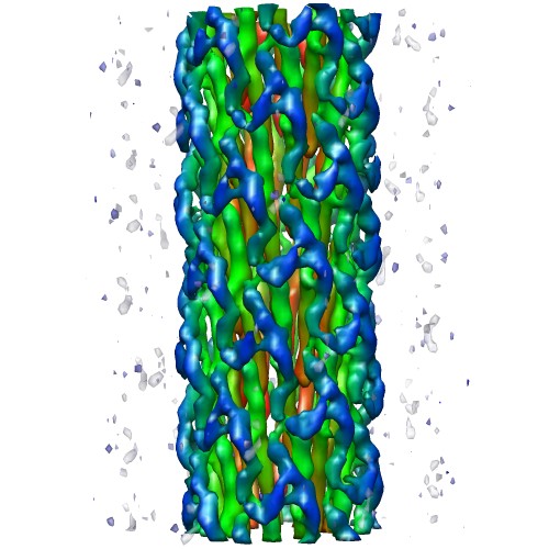 emd_5352_1.jpg
 http://ftp.pdbj.org/pub/emdb/structures/EMD-5352
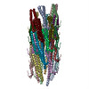
 Z (Sec.)
 Y (Row.)
 X (Col.)


 試料の構成要素
 解析
解析 電子顕微鏡法
 FIELD EMISSION GUN