Movie
Movie Controller

[English] 日本語
 Yorodumi
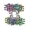
- EMDB-52560: Mycobacterium smegmatis inosine monophosphate dehydrogenase (IMPD... -
+ Open data

- Basic information
| Entry |  | |||||||||
|---|---|---|---|---|---|---|---|---|---|---|
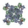
| Title | Mycobacterium smegmatis inosine monophosphate dehydrogenase (IMPDH) E-XMP* intermediate, extended | |||||||||
 Map data Map data | LocScale primary viewing map | |||||||||
 Sample Sample |
| |||||||||
 Keywords Keywords | Octamer / reaction intermediate / Purine metabolism / IMPDH / OXIDOREDUCTASE | |||||||||
| Function / homology |  Function and homology information Function and homology informationIMP dehydrogenase / IMP dehydrogenase activity / GMP biosynthetic process / GTP biosynthetic process / nucleotide binding / metal ion binding Similarity search - Function | |||||||||
| Biological species |  Mycolicibacterium smegmatis MC2 155 (bacteria) Mycolicibacterium smegmatis MC2 155 (bacteria) | |||||||||
| Method | single particle reconstruction / cryo EM / Resolution: 2.39 Å | |||||||||
 Authors Authors | Bulvas O / Kouba T / Pichova I | |||||||||
| Funding support | European Union, 1 items
| |||||||||
 Citation Citation | Journal: J Struct Biol / Year: 2025 Title: Conformational landscape of the mycobacterial inosine 5'-monophosphate dehydrogenase octamerization interface. Authors: Ondřej Bulvas / Zdeněk Knejzlík / Anatolij Filimoněnko / Tomáš Kouba / Iva Pichová /  Abstract: Inosine 5'-monophosphate dehydrogenase (IMPDH), a key enzyme in bacterial purine metabolism, plays an essential role in the biosynthesis of guanine nucleotides and shows promise as a target for ...Inosine 5'-monophosphate dehydrogenase (IMPDH), a key enzyme in bacterial purine metabolism, plays an essential role in the biosynthesis of guanine nucleotides and shows promise as a target for antimicrobial drug development. Despite its significance, the conformational dynamics and substrate-induced structural changes in bacterial IMPDH remain poorly understood, particularly with respect to its octameric assembly. Using cryo-EM, we present full-length structures of IMPDH from Mycobacterium smegmatis (MsmIMPDH) captured in a reaction intermediate state, revealing conformational changes upon substrate binding. The structures feature resolved flexible loops that coordinate the binding of the substrate, the cofactor, and the K ion. Our structural analysis identifies a novel octamerization interface unique to MsmIMPDH. Additionally, a previously unobserved barrel-like density suggests potential self-interactions within the C-terminal regions, hinting at a regulatory mechanism tied to assembly and function of the enzyme. These data provide insights into substrate-induced conformational dynamics and novel interaction interfaces in MsmIMPDH, potentially informing the development of IMPDH-targeted drugs. | |||||||||
| History |
|
- Structure visualization
| Supplemental images |
|---|

- Downloads & links
-EMDB archive
| Map data | emd_52560.map.gz | 139 MB | EMDB map data format | |
|---|---|---|---|---|
| Header (meta data) | emd-52560-v30.xmlemd-52560.xml | 22.7 KB 22.7 KB | Display Display | EMDB header |
| FSC (resolution estimation) | emd_52560_fsc.xml | 14.9 KB | Display | FSC data file |
| Images |  emd_52560.png emd_52560.png | 125.5 KB | ||
| Masks | emd_52560_msk_1.map | 282.6 MB | Mask map | |
| Filedesc metadata | emd-52560.cif.gz | 7 KB | ||
| Others | emd_52560_additional_1.map.gzemd_52560_half_map_1.map.gzemd_52560_half_map_2.map.gz | 263 MB 225.2 MB 225.2 MB | ||
| Archive directory |  http://ftp.pdbj.org/pub/emdb/structures/EMD-52560ftp://ftp.pdbj.org/pub/emdb/structures/EMD-52560 http://ftp.pdbj.org/pub/emdb/structures/EMD-52560ftp://ftp.pdbj.org/pub/emdb/structures/EMD-52560 | HTTPS FTP |
-Related structure data
| Related structure data |  9i0lMC  9i0kC  9i0mC M: atomic model generated by this map C: citing same article ( |
|---|---|
| Similar structure data |


-Links
| EMDB pages | EMDB (EBI/PDBe) / EMDataResource |
|---|---|
| Related items in Molecule of the Month |

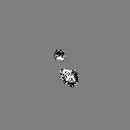
-Map
| File | Download / File: emd_52560.map.gz / Format: CCP4 / Size: 282.6 MB / Type: IMAGE STORED AS FLOATING POINT NUMBER (4 BYTES) | ||||||||||||||||||||||||||||||||||||
|---|---|---|---|---|---|---|---|---|---|---|---|---|---|---|---|---|---|---|---|---|---|---|---|---|---|---|---|---|---|---|---|---|---|---|---|---|---|
| Annotation | LocScale primary viewing map | ||||||||||||||||||||||||||||||||||||
| Projections & slices | Image control
Images are generated by Spider. | ||||||||||||||||||||||||||||||||||||
| Voxel size | X=Y=Z: 0.8336 Å | ||||||||||||||||||||||||||||||||||||
| Density |
| ||||||||||||||||||||||||||||||||||||
| Symmetry | Space group: 1 | ||||||||||||||||||||||||||||||||||||
| Details | EMDB XML:
|
 Z (Sec.)
 Y (Row.)
 X (Col.)


-Supplemental data
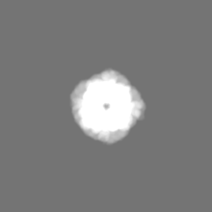
-Mask #1
| File | emd_52560_msk_1.map | ||||||||||||
|---|---|---|---|---|---|---|---|---|---|---|---|---|---|
| Projections & Slices |
| ||||||||||||
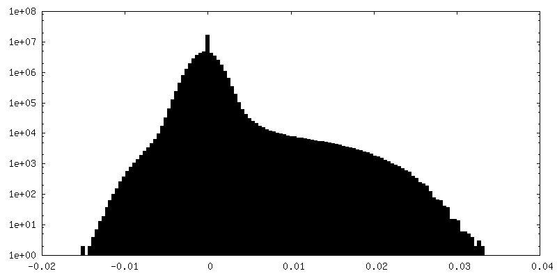
| Density Histograms |


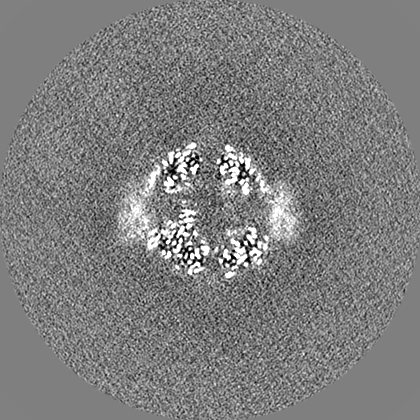
-Additional map: Original map used for refinement and validation
| File | emd_52560_additional_1.map | ||||||||||||
|---|---|---|---|---|---|---|---|---|---|---|---|---|---|
| Annotation | Original map used for refinement and validation | ||||||||||||
| Projections & Slices |
| ||||||||||||
| Density Histograms |


-Half map: #1
| File | emd_52560_half_map_1.map | ||||||||||||
|---|---|---|---|---|---|---|---|---|---|---|---|---|---|
| Projections & Slices |
| ||||||||||||
| Density Histograms |


-Half map: #2
| File | emd_52560_half_map_2.map | ||||||||||||
|---|---|---|---|---|---|---|---|---|---|---|---|---|---|
| Projections & Slices |
| ||||||||||||
| Density Histograms |


- Sample components
-Entire : Octameric assembly of inosine monophosphate dehydrogenase reactio...
| Entire | Name: Octameric assembly of inosine monophosphate dehydrogenase reaction intermediate; E-XMP* in complex with NAD+ |
|---|---|
| Components |
|
-Supramolecule #1: Octameric assembly of inosine monophosphate dehydrogenase reactio...
| Supramolecule | Name: Octameric assembly of inosine monophosphate dehydrogenase reaction intermediate; E-XMP* in complex with NAD+ type: complex / ID: 1 / Parent: 0 / Macromolecule list: #1 |
|---|---|
| Source (natural) | Organism: Mycolicibacterium smegmatis MC2 155 (bacteria) |
-Macromolecule #1: Inosine-5'-monophosphate dehydrogenase
| Macromolecule | Name: Inosine-5'-monophosphate dehydrogenase / type: protein_or_peptide / ID: 1 / Number of copies: 8 / Enantiomer: LEVO / EC number: IMP dehydrogenase |
|---|---|
| Source (natural) | Organism: Mycolicibacterium smegmatis MC2 155 (bacteria) / Strain: MC2 155 |
| Molecular weight | Theoretical: 53.388988 KDa |
| Recombinant expression | Organism: |
| Sequence | String: MSIAESSVPI AVPVPTGGDD PTKVAMLGLT FDDVLLLPAA SDVVPATADT SSQLTKRIRL RVPLVSSAMD TVTESRMAIA MARAGGMGV LHRNLPVAEQ AGQVETVKRS EAGMVTDPVT CSPDNTLAEV DAMCARFRIS GLPVVDDTGE LVGIITNRDM R FEVDQSKP ...String: MSIAESSVPI AVPVPTGGDD PTKVAMLGLT FDDVLLLPAA SDVVPATADT SSQLTKRIRL RVPLVSSAMD TVTESRMAIA MARAGGMGV LHRNLPVAEQ AGQVETVKRS EAGMVTDPVT CSPDNTLAEV DAMCARFRIS GLPVVDDTGE LVGIITNRDM R FEVDQSKP VSEVMTKAPL ITAKEGVSAE AALGLLRRHK IEKLPIVDGH GKLTGLITVK DFVKTEQFPL STKDSDGRLL VG AAVGVGD DAWTRAMTLV DAGVDVLIVD TAHAHNRGVL DMVSRLKQAV GERVDVVGGN VATRAAAAAL VEAGADAVKV GVG PGSICT TRVVAGVGAP QITAILEAVA ACKPYGVPVI ADGGLQYSGD IAKALAAGAS TAMLGSLLAG TAESPGELIF VNGK QFKSY RGMGSLGAMQ GRGAAKSYSK DRYFQDDVLS EDKLVPEGIE GRVPFRGPLG TVIHQLTGGL RAAMGYTGSA TIEQL QQAQ FVQITAAGLK ESHPHDITMT VEAPNYYTR UniProtKB: Inosine-5'-monophosphate dehydrogenase |
-Macromolecule #2: INOSINIC ACID
| Macromolecule | Name: INOSINIC ACID / type: ligand / ID: 2 / Number of copies: 8 / Formula: IMP |
|---|---|
| Molecular weight | Theoretical: 348.206 Da |
| Chemical component information | 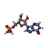 ChemComp-I: |
-Macromolecule #3: NICOTINAMIDE-ADENINE-DINUCLEOTIDE
| Macromolecule | Name: NICOTINAMIDE-ADENINE-DINUCLEOTIDE / type: ligand / ID: 3 / Number of copies: 8 / Formula: NAD |
|---|---|
| Molecular weight | Theoretical: 663.425 Da |
| Chemical component information |  ChemComp-NAD: |
-Macromolecule #4: POTASSIUM ION
| Macromolecule | Name: POTASSIUM ION / type: ligand / ID: 4 / Number of copies: 8 / Formula: K |
|---|---|
| Molecular weight | Theoretical: 39.098 Da |
-Macromolecule #5: water
| Macromolecule | Name: water / type: ligand / ID: 5 / Number of copies: 65 / Formula: HOH |
|---|---|
| Molecular weight | Theoretical: 18.015 Da |
| Chemical component information |  ChemComp-HOH: |
-Experimental details
-Structure determination
| Method | cryo EM |
|---|---|
 Processing Processing | single particle reconstruction |
| Aggregation state | particle |
-Sample preparation
| Buffer | pH: 7.5 Component:
Details: 50 mM HEPES (pH 7.5), 200 mM KCl, 5 mM DTT, 32 mM MgCl2 Ligand: 10 mM ATP + 10 mM IMP + 10 mM NAD | ||||||||||||||||||||||||
|---|---|---|---|---|---|---|---|---|---|---|---|---|---|---|---|---|---|---|---|---|---|---|---|---|---|
| Grid | Model: Quantifoil R2/1 / Material: GOLD / Mesh: 300 / Support film - Material: CARBON / Support film - topology: HOLEY ARRAY | ||||||||||||||||||||||||
| Vitrification | Cryogen name: ETHANE / Chamber humidity: 100 % / Instrument: FEI VITROBOT MARK IV |
- Electron microscopy
| Microscope | TFS KRIOS |
|---|---|
| Image recording | Film or detector model: GATAN K3 BIOQUANTUM (6k x 4k) / Number real images: 8691 / Average exposure time: 2.0 sec. / Average electron dose: 40.1 e/Å2 |
| Electron beam | Acceleration voltage: 300 kV / Electron source:  FIELD EMISSION GUN FIELD EMISSION GUN |
| Electron optics | Illumination mode: FLOOD BEAM / Imaging mode: BRIGHT FIELD / Cs: 2.7 mm / Nominal defocus max: 2.3000000000000003 µm / Nominal defocus min: 1.2 µm / Nominal magnification: 165000 |
| Sample stage | Cooling holder cryogen: NITROGEN |
| Experimental equipment |  Model: Titan Krios / Image courtesy: FEI Company |
+Image processing

-Atomic model buiding 1
| Initial model | PDB ID: Chain - Source name: PDB / Chain - Initial model type: experimental model |
|---|---|
| Details | Initial fitting was done in UCSF ChimeraX. Model refinement was done by iterative cycles of manual fitting with Coot and ISOLDE and automated fitting with phenix.real_space_refine. |
| Refinement | Protocol: RIGID BODY FIT / Overall B value: 45.82 / Target criteria: CC coefficient |
| Output model | PDB-9i0l: |