Movie
Movie Controller

[English] 日本語
 Yorodumi
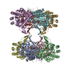
- PDB-9i0k: Mycobacterium smegmatis inosine monophosphate dehydrogenase (IMPD... -
+ Open data

- Basic information
| Entry | Database: PDB / ID: 9i0k | ||||||
|---|---|---|---|---|---|---|---|
| Title | Mycobacterium smegmatis inosine monophosphate dehydrogenase (IMPDH) E-XMP* intermediate, compressed | ||||||
 Components Components | Inosine-5'-monophosphate dehydrogenase | ||||||
 Keywords Keywords | OXIDOREDUCTASE / Octamer / reaction intermediate / Purine metabolism / IMPDH | ||||||
| Function / homology |  Function and homology information Function and homology informationIMP dehydrogenase / IMP dehydrogenase activity / GMP biosynthetic process / GTP biosynthetic process / nucleotide binding / metal ion binding Similarity search - Function | ||||||
| Biological species |  Mycolicibacterium smegmatis MC2 155 (bacteria) Mycolicibacterium smegmatis MC2 155 (bacteria) | ||||||
| Method | ELECTRON MICROSCOPY / single particle reconstruction / cryo EM / Resolution: 2.73 Å | ||||||
 Authors Authors | Bulvas, O. / Kouba, T. / Pichova, I. | ||||||
| Funding support | European Union, 1items
| ||||||
 Citation Citation | Journal: J Struct Biol / Year: 2025 Title: Conformational landscape of the mycobacterial inosine 5'-monophosphate dehydrogenase octamerization interface. Authors: Ondřej Bulvas / Zdeněk Knejzlík / Anatolij Filimoněnko / Tomáš Kouba / Iva Pichová /  Abstract: Inosine 5'-monophosphate dehydrogenase (IMPDH), a key enzyme in bacterial purine metabolism, plays an essential role in the biosynthesis of guanine nucleotides and shows promise as a target for ...Inosine 5'-monophosphate dehydrogenase (IMPDH), a key enzyme in bacterial purine metabolism, plays an essential role in the biosynthesis of guanine nucleotides and shows promise as a target for antimicrobial drug development. Despite its significance, the conformational dynamics and substrate-induced structural changes in bacterial IMPDH remain poorly understood, particularly with respect to its octameric assembly. Using cryo-EM, we present full-length structures of IMPDH from Mycobacterium smegmatis (MsmIMPDH) captured in a reaction intermediate state, revealing conformational changes upon substrate binding. The structures feature resolved flexible loops that coordinate the binding of the substrate, the cofactor, and the K ion. Our structural analysis identifies a novel octamerization interface unique to MsmIMPDH. Additionally, a previously unobserved barrel-like density suggests potential self-interactions within the C-terminal regions, hinting at a regulatory mechanism tied to assembly and function of the enzyme. These data provide insights into substrate-induced conformational dynamics and novel interaction interfaces in MsmIMPDH, potentially informing the development of IMPDH-targeted drugs. | ||||||
| History |
|
- Structure visualization
| Structure viewer | Molecule: MolmilJmol/JSmol |
|---|
- Downloads & links
-Download
| PDBx/mmCIF format | 9i0k.cif.gz | 660 KB | Display | PDBx/mmCIF format |
|---|---|---|---|---|
| PDB format | pdb9i0k.ent.gz | 434.2 KB | Display | PDB format |
| PDBx/mmJSON format | 9i0k.json.gz | Tree view | PDBx/mmJSON format | |
| Others |  Other downloads Other downloads |
-Validation report
| Arichive directory | https://data.pdbj.org/pub/pdb/validation_reports/i0/9i0kftp://data.pdbj.org/pub/pdb/validation_reports/i0/9i0k | HTTPS FTP |
|---|
-Related structure data
| Related structure data |  52559MC  9i0lC  9i0mC M: map data used to model this data C: citing same article ( |
|---|---|
| Similar structure data |


-Links
 PDBj
PDBj
- Assembly
| Deposited unit | 
|
|---|---|
| 1 |
|
-Components
| #1: Protein | Mass: 53388.988 Da / Num. of mol.: 8 Source method: isolated from a genetically manipulated source Source: (gene. exp.) Mycolicibacterium smegmatis MC2 155 (bacteria)Strain: MC2 155 / Gene: guaB, MSMEG_1602 / Plasmid: pRSFDuet-1 / Production host: #2: Chemical | ChemComp-IMP /   Mass: 348.206 Da / Num. of mol.: 8 / Source method: obtained synthetically / Formula: C10H13N4O8P Mass: 348.206 Da / Num. of mol.: 8 / Source method: obtained synthetically / Formula: C10H13N4O8P#3: Chemical | ChemComp-NAD /   Mass: 663.425 Da / Num. of mol.: 8 / Source method: obtained synthetically / Formula: C21H27N7O14P2 / Comment: NAD*YM Mass: 663.425 Da / Num. of mol.: 8 / Source method: obtained synthetically / Formula: C21H27N7O14P2 / Comment: NAD*YM#4: Chemical | ChemComp-K /   Mass: 39.098 Da / Num. of mol.: 8 / Source method: obtained synthetically / Formula: K Mass: 39.098 Da / Num. of mol.: 8 / Source method: obtained synthetically / Formula: K#5: Water | ChemComp-HOH / |  Mass: 18.015 Da / Num. of mol.: 80 / Source method: isolated from a natural source / Formula: H2O Mass: 18.015 Da / Num. of mol.: 80 / Source method: isolated from a natural source / Formula: H2OHas ligand of interest | N | Has protein modification | Y | |
|---|
-Experimental details
-Experiment
| Experiment | Method: ELECTRON MICROSCOPY |
|---|---|
| EM experiment | Aggregation state: PARTICLE / 3D reconstruction method: single particle reconstruction |
- Sample preparation
| Component | Name: Octameric assembly of inosine monophosphate dehydrogenase reaction intermediate; E-XMP* in complex with NAD+ Type: COMPLEX / Entity ID: #1 / Source: RECOMBINANT | ||||||||||||||||||||||||||||||||||||||||
|---|---|---|---|---|---|---|---|---|---|---|---|---|---|---|---|---|---|---|---|---|---|---|---|---|---|---|---|---|---|---|---|---|---|---|---|---|---|---|---|---|---|
| Molecular weight | Experimental value: NO | ||||||||||||||||||||||||||||||||||||||||
| Source (natural) | Organism: Mycolicibacterium smegmatis MC2 155 (bacteria) | ||||||||||||||||||||||||||||||||||||||||
| Source (recombinant) | Organism: | ||||||||||||||||||||||||||||||||||||||||
| Buffer solution | pH: 7.5 Details: 50 mM HEPES (pH 7.5), 200 mM KCl, 5 mM DTT, 32 mM MgCl2 Ligand: 10 mM ATP + 10 mM IMP + 10 mM NAD | ||||||||||||||||||||||||||||||||||||||||
| Buffer component |
| ||||||||||||||||||||||||||||||||||||||||
| Specimen | Embedding applied: NO / Shadowing applied: NO / Staining applied: NO / Vitrification applied: YES | ||||||||||||||||||||||||||||||||||||||||
| Specimen support | Grid material: GOLD / Grid mesh size: 300 divisions/in. / Grid type: Quantifoil R2/1 | ||||||||||||||||||||||||||||||||||||||||
| Vitrification | Instrument: FEI VITROBOT MARK IV / Cryogen name: ETHANE / Humidity: 100 % |
- Electron microscopy imaging
| Experimental equipment |  Model: Titan Krios / Image courtesy: FEI Company |
|---|---|
| Microscopy | Model: TFS KRIOS |
| Electron gun | Electron source:  FIELD EMISSION GUN / Accelerating voltage: 300 kV / Illumination mode: FLOOD BEAM FIELD EMISSION GUN / Accelerating voltage: 300 kV / Illumination mode: FLOOD BEAM |
| Electron lens | Mode: BRIGHT FIELD / Nominal magnification: 165000 X / Nominal defocus max: 3200 nm / Nominal defocus min: 1200 nm / Cs: 2.7 mm / Alignment procedure: ZEMLIN TABLEAU |
| Specimen holder | Cryogen: NITROGEN |
| Image recording | Average exposure time: 2 sec. / Electron dose: 40.1 e/Å2 / Film or detector model: GATAN K3 BIOQUANTUM (6k x 4k) / Num. of real images: 8691 |
- Processing
| EM software | Name: PHENIX / Version: 1.21rc1_5084 / Category: model refinement | ||||||||||||||||||||||||
|---|---|---|---|---|---|---|---|---|---|---|---|---|---|---|---|---|---|---|---|---|---|---|---|---|---|
| CTF correction | Type: PHASE FLIPPING ONLY | ||||||||||||||||||||||||
| Particle selection | Num. of particles selected: 4833002 | ||||||||||||||||||||||||
| 3D reconstruction | Resolution: 2.73 Å / Resolution method: FSC 0.143 CUT-OFF / Num. of particles: 124968 / Symmetry type: POINT | ||||||||||||||||||||||||
| Atomic model building | B value: 37.48 / Protocol: RIGID BODY FIT / Target criteria: CC coefficient Details: Initial fitting was done in UCSF ChimeraX. Model refinement was done by iterative cycles of manual fitting with Coot and ISOLDE and automated fitting with phenix.real_space_refine. | ||||||||||||||||||||||||
| Atomic model building | PDB-ID: 8QQV Accession code: 8QQV / Source name: PDB / Type: experimental model | ||||||||||||||||||||||||
| Refinement | Cross valid method: NONE Stereochemistry target values: GeoStd + Monomer Library + CDL v1.2 | ||||||||||||||||||||||||
| Displacement parameters | Biso mean: 38.75 Å2 | ||||||||||||||||||||||||
| Refine LS restraints |
|