 ムービー
ムービー コントローラー


+ データを開く

- 基本情報
基本情報
| 登録情報 | 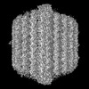 | |||||||||
|---|---|---|---|---|---|---|---|---|---|---|
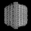
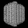
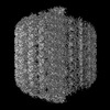
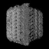
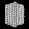
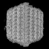
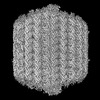
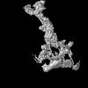
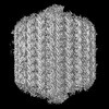
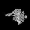
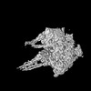
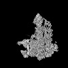
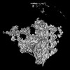
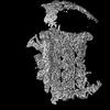
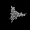
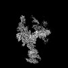
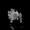
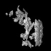
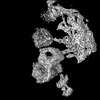
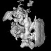
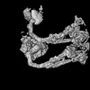
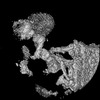
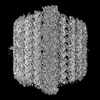
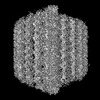
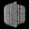
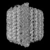
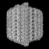
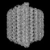
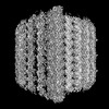
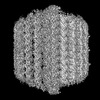
| タイトル | Focus refine map of T. brucei 48 nm repear FAP106A-KD part1-1 | |||||||||
 マップデータ マップデータ | ||||||||||
 試料 試料 |
| |||||||||
 キーワード キーワード | flagella / microtubule / MOTOR PROTEIN | |||||||||
| 生物種 |  | |||||||||
| 手法 | 単粒子再構成法 / クライオ電子顕微鏡法 / 解像度: 4.1 Å | |||||||||
 データ登録者 データ登録者 | Xia X / Shimogawa MM / Wang H / Liu S / Wijono A / Langousis G / Kassem AM / Wohlschlegel JA / Hill KL / Zhou ZH | |||||||||
| 資金援助 |  米国, 2件 米国, 2件
| |||||||||
 引用 引用 | ジャーナル: Science / 年: 2025 タイトル: Trypanosome doublet microtubule structures reveal flagellum assembly and motility mechanisms. 著者: Xian Xia / Michelle M Shimogawa / Hui Wang / Samuel Liu / Angeline Wijono / Gerasimos Langousis / Ahmad M Kassem / James A Wohlschlegel / Kent L Hill / Z Hong Zhou / 要旨: The flagellum of drives the parasite's characteristic screw-like motion and is essential for its replication, transmission, and pathogenesis. However, the molecular details of this process remain ...The flagellum of drives the parasite's characteristic screw-like motion and is essential for its replication, transmission, and pathogenesis. However, the molecular details of this process remain unclear. Here, we present high-resolution (up to 2.8 angstrom) cryo-electron microscopy structures of flagellar doublet microtubules (DMTs). Integrated modeling identified 154 different axonemal proteins inside and outside the DMT and, together with genetic and proteomic interrogation, revealed conserved and trypanosome-specific foundations of flagellum assembly and motility. We captured axonemal dynein motors in their pre-power stroke state. Comparing atomic models between pre- and post-power strokes defined how dynein structural changes drive sliding of adjacent DMTs during flagellar beating. This study illuminates structural dynamics underlying flagellar motility and identifies pathogen-specific proteins to consider for therapeutic interventions targeting neglected diseases. | |||||||||
| 履歴 |
|
- 構造の表示
構造の表示
| 添付画像 |
|---|

- ダウンロードとリンク
-EMDBアーカイブ
| マップデータ | emd_47773.map.gz | 565 MB |  EMDBマップデータ形式 EMDBマップデータ形式 | |
|---|---|---|---|---|
| ヘッダ (付随情報) | emd-47773-v30.xmlemd-47773.xml | 24.6 KB 24.6 KB | 表示 表示 | EMDBヘッダ |
| 画像 |  emd_47773.png emd_47773.png | 139.7 KB | ||
| マスクデータ | emd_47773_msk_1.map | 600.7 MB | マスクマップ | |
| Filedesc metadata | emd-47773.cif.gz | 4.7 KB | ||
| その他 | emd_47773_half_map_1.map.gzemd_47773_half_map_2.map.gz | 558.1 MB 558.1 MB | ||
| アーカイブディレクトリ |  http://ftp.pdbj.org/pub/emdb/structures/EMD-47773ftp://ftp.pdbj.org/pub/emdb/structures/EMD-47773 http://ftp.pdbj.org/pub/emdb/structures/EMD-47773ftp://ftp.pdbj.org/pub/emdb/structures/EMD-47773 | HTTPS FTP |
-関連構造データ


-リンク
| EMDBのページ | EMDB (EBI/PDBe) / EMDataResource |
|---|
-マップ
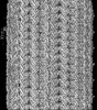
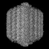
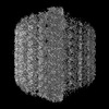
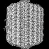
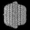
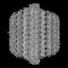
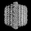
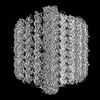
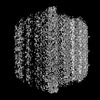
| ファイル | ダウンロード / ファイル: emd_47773.map.gz / 形式: CCP4 / 大きさ: 600.7 MB / タイプ: IMAGE STORED AS FLOATING POINT NUMBER (4 BYTES) | ||||||||||||||||||||||||||||||||||||
|---|---|---|---|---|---|---|---|---|---|---|---|---|---|---|---|---|---|---|---|---|---|---|---|---|---|---|---|---|---|---|---|---|---|---|---|---|---|
| 投影像・断面図 | 画像のコントロール
画像は Spider により作成 | ||||||||||||||||||||||||||||||||||||
| ボクセルのサイズ | X=Y=Z: 1.1 Å | ||||||||||||||||||||||||||||||||||||
| 密度 |
| ||||||||||||||||||||||||||||||||||||
| 対称性 | 空間群: 1 | ||||||||||||||||||||||||||||||||||||
| 詳細 | EMDB XML:
|
 Z (Sec.)
 Y (Row.)
 X (Col.)


-添付データ
-マスク #1
| ファイル | emd_47773_msk_1.map | ||||||||||||
|---|---|---|---|---|---|---|---|---|---|---|---|---|---|
| 投影像・断面図 |
| ||||||||||||
| 密度ヒストグラム |


-ハーフマップ: #1
| ファイル | emd_47773_half_map_1.map | ||||||||||||
|---|---|---|---|---|---|---|---|---|---|---|---|---|---|
| 投影像・断面図 |
| ||||||||||||
| 密度ヒストグラム |


-ハーフマップ: #2
| ファイル | emd_47773_half_map_2.map | ||||||||||||
|---|---|---|---|---|---|---|---|---|---|---|---|---|---|
| 投影像・断面図 |
| ||||||||||||
| 密度ヒストグラム |


- 試料の構成要素
-全体 : Trypanosoma brucei flagellum with FAP106A-KD
| 全体 | 名称: Trypanosoma brucei flagellum with FAP106A-KD |
|---|---|
| 要素 |
|
-超分子 #1: Trypanosoma brucei flagellum with FAP106A-KD
| 超分子 | 名称: Trypanosoma brucei flagellum with FAP106A-KD / タイプ: organelle_or_cellular_component / ID: 1 / 親要素: 0 含まれる分子: #60, #59, #51-#52, #56-#58, #50, #55, #49, #54, #47-#48, #53, #46, #36, #38, #34, #37, #35, #41, #43, #42, #40, #44, #39, #45, #28, #21-#22, #20, #25, #16, #27, #26, #23, #19, ...含まれる分子: #60, #59, #51-#52, #56-#58, #50, #55, #49, #54, #47-#48, #53, #46, #36, #38, #34, #37, #35, #41, #43, #42, #40, #44, #39, #45, #28, #21-#22, #20, #25, #16, #27, #26, #23, #19, #29, #32, #31, #30, #33, #8, #13, #12, #11, #10, #14, #9, #24, #18, #15, #17, #5-#7, #1-#4 詳細: Demembraned flagella were split by mild protease treatment |
|---|---|
| 由来(天然) | 生物種: |
-実験情報
-構造解析
| 手法 | クライオ電子顕微鏡法 |
|---|---|
 解析 解析 | 単粒子再構成法 |
| 試料の集合状態 | filament |
-試料調製
| 緩衝液 | pH: 7.4 |
|---|---|
| グリッド | モデル: Quantifoil R2/1 / 材質: COPPER / メッシュ: 200 / 支持フィルム - 材質: CARBON / 支持フィルム - トポロジー: HOLEY |
| 凍結 | 凍結剤: ETHANE / チャンバー内湿度: 100 % / チャンバー内温度: 277 K / 装置: FEI VITROBOT MARK IV |
- 電子顕微鏡法
| 顕微鏡 | TFS KRIOS |
|---|---|
| 特殊光学系 | エネルギーフィルター - 名称: GIF Quantum LS / エネルギーフィルター - スリット幅: 20 eV |
| ソフトウェア | 名称: SerialEM (ver. 4.1) |
| 撮影 | フィルム・検出器のモデル: GATAN K3 BIOQUANTUM (6k x 4k) 撮影したグリッド数: 2 / 実像数: 9246 / 平均露光時間: 2.0 sec. / 平均電子線量: 45.0 e/Å2 |
| 電子線 | 加速電圧: 300 kV / 電子線源:  FIELD EMISSION GUN FIELD EMISSION GUN |
| 電子光学系 | C2レンズ絞り径: 50.0 µm / 照射モード: FLOOD BEAM / 撮影モード: BRIGHT FIELD / Cs: 2.7 mm / 最大 デフォーカス(公称値): 2.4 µm / 最小 デフォーカス(公称値): 1.6 µm / 倍率(公称値): 81000 |
| 試料ステージ | 試料ホルダーモデル: FEI TITAN KRIOS AUTOGRID HOLDER ホルダー冷却材: NITROGEN |
| 実験機器 |  モデル: Titan Krios / 画像提供: FEI Company |
+画像解析

-原子モデル構築 1
| 精密化 | 空間: REAL / プロトコル: AB INITIO MODEL |
|---|
