 Movie
Movie Controller

[English] 日本語
 Yorodumi
- PDB-9e2g: Cryo-EM structure of 48 nm repeat of microtubule doublet from T. ... -
+ Open data

- Basic information
| Entry | Database: PDB / ID: 9e2g | |||||||||
|---|---|---|---|---|---|---|---|---|---|---|
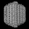
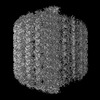
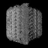
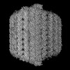
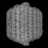
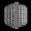
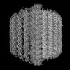
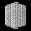
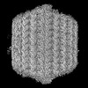
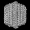
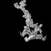
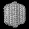
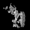
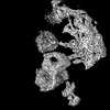
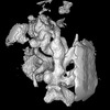
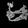
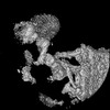
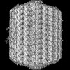
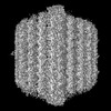
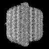
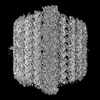
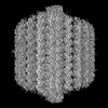
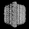
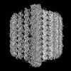
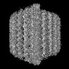
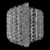
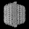
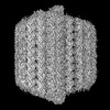
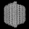
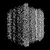
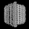
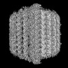
| Title | Cryo-EM structure of 48 nm repeat of microtubule doublet from T. brucei flagellum | |||||||||
 Components Components |
| |||||||||
 Keywords Keywords | MOTOR PROTEIN / flagella / microtubule | |||||||||
| Function / homology |  Function and homology information Function and homology informationparaflagellar rod / intraciliary transport particle / microtubule anchoring / cilium-dependent cell motility / regulation of cilium beat frequency involved in ciliary motility / axonemal microtubule / negative regulation of microtubule depolymerization / nucleoside-diphosphate kinase / nuclear lumen / UTP biosynthetic process ...paraflagellar rod / intraciliary transport particle / microtubule anchoring / cilium-dependent cell motility / regulation of cilium beat frequency involved in ciliary motility / axonemal microtubule / negative regulation of microtubule depolymerization / nucleoside-diphosphate kinase / nuclear lumen / UTP biosynthetic process / ciliary plasm / CTP biosynthetic process / motile cilium / nucleoside diphosphate kinase activity / positive regulation of cell motility / GTP biosynthetic process / cyclosporin A binding / post-transcriptional regulation of gene expression / phosphate ion binding / cortical cytoskeleton / mitotic cytokinesis / cilium assembly / axoneme / alpha-tubulin binding / cytoplasmic microtubule / Hsp70 protein binding / mitotic spindle organization / peptidylprolyl isomerase / meiotic cell cycle / peptidyl-prolyl cis-trans isomerase activity / Hsp90 protein binding / structural constituent of cytoskeleton / microtubule cytoskeleton organization / mitotic spindle / mitotic cell cycle / protein folding / microtubule binding / cytoskeleton / microtubule / calmodulin binding / cilium / ciliary basal body / ribosome / hydrolase activity / GTPase activity / calcium ion binding / GTP binding / mitochondrion / nucleoplasm / ATP binding / metal ion binding / nucleus / cytoplasm Similarity search - Function | |||||||||
| Biological species |  | |||||||||
| Method | ELECTRON MICROSCOPY / single particle reconstruction / cryo EM / Resolution: 2.8 Å | |||||||||
 Authors Authors | Xia, X. / Shimogawa, M.M. / Wang, H. / Liu, S. / Wijono, A. / Langousis, G. / Kassem, A.M. / Wohlschlegel, J.A. / Hill, K. / Zhou, Z.H. | |||||||||
| Funding support |  United States, 2items United States, 2items
| |||||||||
 Citation Citation | Journal: Science / Year: 2025 Title: Trypanosome doublet microtubule structures reveal flagellum assembly and motility mechanisms. Authors: Xian Xia / Michelle M Shimogawa / Hui Wang / Samuel Liu / Angeline Wijono / Gerasimos Langousis / Ahmad M Kassem / James A Wohlschlegel / Kent L Hill / Z Hong Zhou / Abstract: The flagellum of drives the parasite's characteristic screw-like motion and is essential for its replication, transmission, and pathogenesis. However, the molecular details of this process remain ...The flagellum of drives the parasite's characteristic screw-like motion and is essential for its replication, transmission, and pathogenesis. However, the molecular details of this process remain unclear. Here, we present high-resolution (up to 2.8 angstrom) cryo-electron microscopy structures of flagellar doublet microtubules (DMTs). Integrated modeling identified 154 different axonemal proteins inside and outside the DMT and, together with genetic and proteomic interrogation, revealed conserved and trypanosome-specific foundations of flagellum assembly and motility. We captured axonemal dynein motors in their pre-power stroke state. Comparing atomic models between pre- and post-power strokes defined how dynein structural changes drive sliding of adjacent DMTs during flagellar beating. This study illuminates structural dynamics underlying flagellar motility and identifies pathogen-specific proteins to consider for therapeutic interventions targeting neglected diseases. | |||||||||
| History |
|
- Structure visualization
| Structure viewer | Molecule: MolmilJmol/JSmol |
|---|
- Downloads & links
-Download
| PDBx/mmCIF format | 9e2g.cif.gz | 25.4 MB | Display | PDBx/mmCIF format |
|---|---|---|---|---|
| PDB format | pdb9e2g.ent.gz | Display | PDB format | |
| PDBx/mmJSON format | 9e2g.json.gz | Tree view | PDBx/mmJSON format | |
| Others |  Other downloads Other downloads |
-Validation report
| Arichive directory | https://data.pdbj.org/pub/pdb/validation_reports/e2/9e2gftp://data.pdbj.org/pub/pdb/validation_reports/e2/9e2g | HTTPS FTP |
|---|
-Related structure data
| Related structure data |  47451MC  9e5cC M: map data used to model this data C: citing same article ( |
|---|---|
| Similar structure data |


-Links
 PDBj
PDBj


- Assembly
| Deposited unit | 
|
|---|---|
| 1 |
|
-Components
-EF-hand domain-containing ... , 8 types, 11 molecules 0A0B0C0D0E0S0T0X0Z1B1D
| #1: Protein | Mass: 86973.781 Da / Num. of mol.: 4 / Source method: isolated from a natural source Source: (natural) References: UniProt: Q387A9 #2: Protein | | Mass: 88366.906 Da / Num. of mol.: 1 / Source method: isolated from a natural source Source: (natural) References: UniProt: Q57ZX1 #10: Protein | | Mass: 47731.914 Da / Num. of mol.: 1 / Source method: isolated from a natural source Source: (natural) References: UniProt: Q581Q7 #11: Protein | | Mass: 48071.531 Da / Num. of mol.: 1 / Source method: isolated from a natural source Source: (natural) References: UniProt: Q388A8 #15: Protein | | Mass: 30499.936 Da / Num. of mol.: 1 / Source method: isolated from a natural source Source: (natural) References: UniProt: Q57XA3 #17: Protein | | Mass: 61839.383 Da / Num. of mol.: 1 / Source method: isolated from a natural source Source: (natural) References: UniProt: Q383Y5 #19: Protein | | Mass: 34213.496 Da / Num. of mol.: 1 / Source method: isolated from a natural source Source: (natural) References: UniProt: Q38F14 #21: Protein | | Mass: 36100.395 Da / Num. of mol.: 1 / Source method: isolated from a natural source Source: (natural) References: UniProt: Q38AR7 |
|---|
+Protein , 38 types, 362 molecules 0F0G0H0I0J0K0O0P0U0V0W0Y1A1C1E1G1H1K1L1M1N1O1P4X1Q1R1S1T1U2I...
-Cilia- and flagella-associated protein ... , 4 types, 15 molecules 0M0N1V1W1X1Y1Z2A2B2C2D2O2P2Q2R
| #6: Protein | Mass: 58689.828 Da / Num. of mol.: 2 / Source method: isolated from a natural source Source: (natural) References: UniProt: Q57VE0 #32: Protein | Mass: 34593.465 Da / Num. of mol.: 6 / Source method: isolated from a natural source Source: (natural) References: UniProt: Q38C25 #33: Protein | Mass: 67770.891 Da / Num. of mol.: 3 / Source method: isolated from a natural source Source: (natural) References: UniProt: Q384Q0 #39: Protein | Mass: 58198.273 Da / Num. of mol.: 4 / Source method: isolated from a natural source Source: (natural) References: UniProt: Q57UZ3 |
|---|
-Nucleoside diphosphate kinase, ... , 2 types, 2 molecules 0Q0R
| #8: Protein | Mass: 37147.473 Da / Num. of mol.: 1 / Source method: isolated from a natural source Source: (natural) References: UniProt: Q38DQ0, nucleoside-diphosphate kinase |
|---|---|
| #9: Protein | Mass: 38993.656 Da / Num. of mol.: 1 / Source method: isolated from a natural source Source: (natural) References: UniProt: Q581Q9, nucleoside-diphosphate kinase |
-T. brucei spp.-specific ... , 2 types, 3 molecules 1F1I1J
| #23: Protein | Mass: 28844.326 Da / Num. of mol.: 1 / Source method: isolated from a natural source Source: (natural) References: UniProt: Q38EE4 |
|---|---|
| #26: Protein | Mass: 46540.539 Da / Num. of mol.: 2 / Source method: isolated from a natural source Source: (natural) References: UniProt: Q384L6 |
-Enkurin domain-containing ... , 4 types, 10 molecules 2E2F2G2H3V3W3X3Y3Z4A
| #34: Protein | Mass: 30264.260 Da / Num. of mol.: 2 / Source method: isolated from a natural source Source: (natural) References: UniProt: Q385T0 #35: Protein | Mass: 42281.645 Da / Num. of mol.: 2 / Source method: isolated from a natural source Source: (natural) References: UniProt: Q4GZB9 #47: Protein | Mass: 31393.762 Da / Num. of mol.: 3 / Source method: isolated from a natural source Source: (natural) References: UniProt: Q583F4 #48: Protein | Mass: 30823.730 Da / Num. of mol.: 3 / Source method: isolated from a natural source Source: (natural) References: UniProt: Q583F5 |
|---|
-Trichohyalin-plectin-homology domain-containing ... , 2 types, 12 molecules 2S2T2U2V2W2X2Y2Z3A3B3C3D
| #40: Protein | Mass: 61310.469 Da / Num. of mol.: 3 / Source method: isolated from a natural source Source: (natural) References: UniProt: Q386E1 #41: Protein | Mass: 41736.418 Da / Num. of mol.: 9 / Source method: isolated from a natural source Source: (natural) References: UniProt: Q383H7 |
|---|
-Non-polymers , 4 types, 456 molecules 


| #61: Chemical | ChemComp-ZN /  Mass: 65.409 Da / Num. of mol.: 24 / Source method: obtained synthetically / Formula: Zn / Feature type: SUBJECT OF INVESTIGATION Mass: 65.409 Da / Num. of mol.: 24 / Source method: obtained synthetically / Formula: Zn / Feature type: SUBJECT OF INVESTIGATION#62: Chemical | ChemComp-GTP /  Mass: 523.180 Da / Num. of mol.: 145 / Source method: obtained synthetically / Formula: C10H16N5O14P3 / Feature type: SUBJECT OF INVESTIGATION / Comment: GTP, energy-carrying molecule*YM Mass: 523.180 Da / Num. of mol.: 145 / Source method: obtained synthetically / Formula: C10H16N5O14P3 / Feature type: SUBJECT OF INVESTIGATION / Comment: GTP, energy-carrying molecule*YM#63: Chemical | ChemComp-MG /  Mass: 24.305 Da / Num. of mol.: 145 / Source method: obtained synthetically / Formula: Mg / Feature type: SUBJECT OF INVESTIGATION Mass: 24.305 Da / Num. of mol.: 145 / Source method: obtained synthetically / Formula: Mg / Feature type: SUBJECT OF INVESTIGATION#64: Chemical | ChemComp-GDP /  Type: RNA linking / Mass: 443.201 Da / Num. of mol.: 142 / Source method: obtained synthetically / Formula: C10H15N5O11P2 / Feature type: SUBJECT OF INVESTIGATION / Comment: GDP, energy-carrying molecule*YM Type: RNA linking / Mass: 443.201 Da / Num. of mol.: 142 / Source method: obtained synthetically / Formula: C10H15N5O11P2 / Feature type: SUBJECT OF INVESTIGATION / Comment: GDP, energy-carrying molecule*YM |
|---|
-Details
| Has ligand of interest | Y |
|---|---|
| Has protein modification | N |
-Experimental details
-Experiment
| Experiment | Method: ELECTRON MICROSCOPY |
|---|---|
| EM experiment | Aggregation state: FILAMENT / 3D reconstruction method: single particle reconstruction |
- Sample preparation
| Component | Name: Trypanosoma brucei flagellum / Type: ORGANELLE OR CELLULAR COMPONENT Details: Demembraned flagella were split by mild protease treatment Entity ID: #60, #59, #51-#52, #56-#58, #50, #55, #49, #54, #47-#48, #53, #46, #36, #38, #34, #37, #35, #41, #43, #42, #40, #44, #39, #45, #28, #21-#22, #20, #25, #16, #27, #26, #23, #19, #29, #32, ...Entity ID: #60, #59, #51-#52, #56-#58, #50, #55, #49, #54, #47-#48, #53, #46, #36, #38, #34, #37, #35, #41, #43, #42, #40, #44, #39, #45, #28, #21-#22, #20, #25, #16, #27, #26, #23, #19, #29, #32, #31, #30, #33, #8, #13, #12, #11, #10, #14, #9, #24, #18, #15, #17, #5-#7, #1-#4 Source: NATURAL |
|---|---|
| Molecular weight | Experimental value: NO |
| Source (natural) | Organism: |
| Buffer solution | pH: 7.4 |
| Specimen | Embedding applied: NO / Shadowing applied: NO / Staining applied: NO / Vitrification applied: YES |
| Specimen support | Grid material: COPPER / Grid mesh size: 200 divisions/in. / Grid type: Quantifoil R2/1 |
| Vitrification | Instrument: FEI VITROBOT MARK IV / Cryogen name: ETHANE / Humidity: 100 % / Chamber temperature: 277 K |
- Electron microscopy imaging
| Experimental equipment |  Model: Titan Krios / Image courtesy: FEI Company |
|---|---|
| Microscopy | Model: TFS KRIOS |
| Electron gun | Electron source:  FIELD EMISSION GUN / Accelerating voltage: 300 kV / Illumination mode: FLOOD BEAM FIELD EMISSION GUN / Accelerating voltage: 300 kV / Illumination mode: FLOOD BEAM |
| Electron lens | Mode: BRIGHT FIELD / Nominal magnification: 81000 X / Nominal defocus max: 2400 nm / Nominal defocus min: 1600 nm / Cs: 2.7 mm / C2 aperture diameter: 50 µm / Alignment procedure: COMA FREE |
| Specimen holder | Cryogen: NITROGEN / Specimen holder model: FEI TITAN KRIOS AUTOGRID HOLDER |
| Image recording | Average exposure time: 2 sec. / Electron dose: 45 e/Å2 / Film or detector model: GATAN K3 BIOQUANTUM (6k x 4k) / Num. of grids imaged: 2 / Num. of real images: 106694 |
| EM imaging optics | Energyfilter name: GIF Quantum LS / Energyfilter slit width: 20 eV |
- Processing
| EM software | Name: PHENIX / Version: 1.19.2_4158: / Category: model refinement |
|---|---|
| CTF correction | Type: PHASE FLIPPING ONLY |
| Particle selection | Num. of particles selected: 7830948 |
| Symmetry | Point symmetry: C1 (asymmetric) |
| 3D reconstruction | Resolution: 2.8 Å / Resolution method: FSC 0.143 CUT-OFF / Num. of particles: 455012 / Num. of class averages: 1 / Symmetry type: POINT |
| Atomic model building | Protocol: AB INITIO MODEL / Space: REAL |
