 Movie
Movie Controller

[English] 日本語
 Yorodumi
- EMDB-47409: Structure of 48-nm repeat DMT from Trypanosoma brucei FAP106A-KD -
+ Open data

- Basic information
| Entry |  | |||||||||
|---|---|---|---|---|---|---|---|---|---|---|
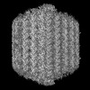
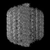
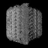
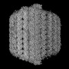
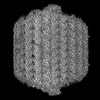
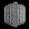
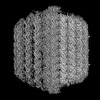
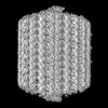
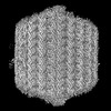
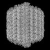
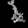
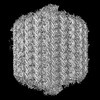
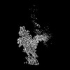
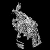
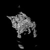
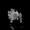
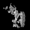
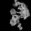
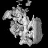
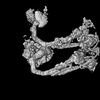
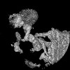
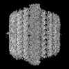
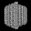
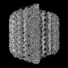
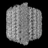
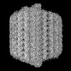
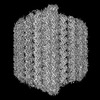
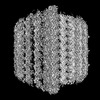
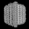
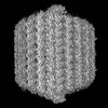
| Title | Structure of 48-nm repeat DMT from Trypanosoma brucei FAP106A-KD | |||||||||
 Map data Map data | ||||||||||
 Sample Sample |
| |||||||||
 Keywords Keywords | flagella / microtubule / MOTOR PROTEIN | |||||||||
| Biological species |  | |||||||||
| Method | single particle reconstruction / cryo EM / Resolution: 3.9 Å | |||||||||
 Authors Authors | Xia X / Shimogawa MM / Wang H / Liu S / Wijono A / Langousis G / Kassem AM / Wohlschlegel JA / Zhou ZH / Hill KL | |||||||||
| Funding support |  United States, 2 items United States, 2 items
| |||||||||
 Citation Citation | Journal: Science / Year: 2025 Title: Trypanosome doublet microtubule structures reveal flagellum assembly and motility mechanisms. Authors: Xian Xia / Michelle M Shimogawa / Hui Wang / Samuel Liu / Angeline Wijono / Gerasimos Langousis / Ahmad M Kassem / James A Wohlschlegel / Kent L Hill / Z Hong Zhou / Abstract: The flagellum of drives the parasite's characteristic screw-like motion and is essential for its replication, transmission, and pathogenesis. However, the molecular details of this process remain ...The flagellum of drives the parasite's characteristic screw-like motion and is essential for its replication, transmission, and pathogenesis. However, the molecular details of this process remain unclear. Here, we present high-resolution (up to 2.8 angstrom) cryo-electron microscopy structures of flagellar doublet microtubules (DMTs). Integrated modeling identified 154 different axonemal proteins inside and outside the DMT and, together with genetic and proteomic interrogation, revealed conserved and trypanosome-specific foundations of flagellum assembly and motility. We captured axonemal dynein motors in their pre-power stroke state. Comparing atomic models between pre- and post-power strokes defined how dynein structural changes drive sliding of adjacent DMTs during flagellar beating. This study illuminates structural dynamics underlying flagellar motility and identifies pathogen-specific proteins to consider for therapeutic interventions targeting neglected diseases. | |||||||||
| History |
|
- Structure visualization
| Supplemental images |
|---|

- Downloads & links
-EMDB archive
| Map data | emd_47409.map.gz | 146.1 MB |  EMDB map data format EMDB map data format | |
|---|---|---|---|---|
| Header (meta data) | emd-47409-v30.xmlemd-47409.xml | 51.1 KB 51.1 KB | Display Display | EMDB header |
| Images |  emd_47409.png emd_47409.png | 86.6 KB | ||
| Filedesc metadata | emd-47409.cif.gz | 5.3 KB | ||
| Others | emd_47409_additional_1.map.gzemd_47409_additional_10.map.gzemd_47409_additional_11.map.gzemd_47409_additional_12.map.gzemd_47409_additional_13.map.gzemd_47409_additional_14.map.gzemd_47409_additional_2.map.gzemd_47409_additional_3.map.gzemd_47409_additional_4.map.gzemd_47409_additional_5.map.gzemd_47409_additional_6.map.gzemd_47409_additional_7.map.gzemd_47409_additional_8.map.gzemd_47409_additional_9.map.gz | 26.5 MB 27.5 MB 28 MB 35.8 MB 30.1 MB 36.9 MB 33 MB 29.1 MB 32.9 MB 27.4 MB 28.1 MB 34.1 MB 28.8 MB 33.6 MB | ||
| Archive directory |  http://ftp.pdbj.org/pub/emdb/structures/EMD-47409ftp://ftp.pdbj.org/pub/emdb/structures/EMD-47409 http://ftp.pdbj.org/pub/emdb/structures/EMD-47409ftp://ftp.pdbj.org/pub/emdb/structures/EMD-47409 | HTTPS FTP |
-Related structure data


-Links
| EMDB pages | EMDB (EBI/PDBe) / EMDataResource |
|---|
-Map
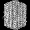
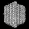
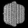
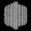
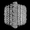
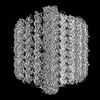
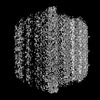
| File | Download / File: emd_47409.map.gz / Format: CCP4 / Size: 287.8 MB / Type: IMAGE STORED AS FLOATING POINT NUMBER (4 BYTES) | ||||||||||||||||||||||||||||||||||||
|---|---|---|---|---|---|---|---|---|---|---|---|---|---|---|---|---|---|---|---|---|---|---|---|---|---|---|---|---|---|---|---|---|---|---|---|---|---|
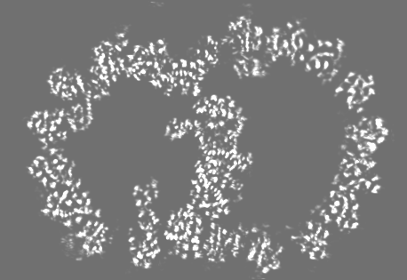
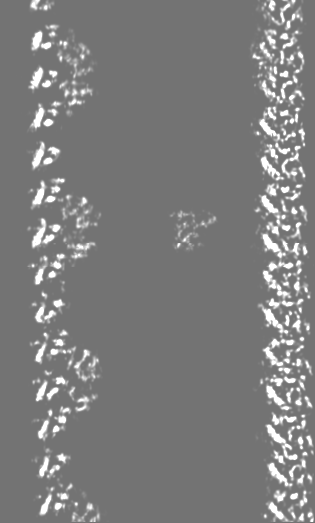
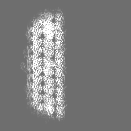
| Projections & slices | Image control
Images are generated by Spider. generated in cubic-lattice coordinate | ||||||||||||||||||||||||||||||||||||
| Voxel size | X=Y=Z: 1.1 Å | ||||||||||||||||||||||||||||||||||||
| Density |
| ||||||||||||||||||||||||||||||||||||
| Symmetry | Space group: 1 | ||||||||||||||||||||||||||||||||||||
| Details | EMDB XML:
|
 Z (Sec.)
 Y (Row.)
 X (Col.)


-Supplemental data
+Additional map: #10


+Additional map: #5


+Additional map: #12


+Additional map: #3


+Additional map: #6


+Additional map: #11


+Additional map: #9


+Additional map: #8


+Additional map: #2


+Additional map: #14


+Additional map: #4


+Additional map: #1


+Additional map: #13


+Additional map: #7


- Sample components
-Entire : Trypanosoma brucei flagellum with FAP106A-KD
| Entire | Name: Trypanosoma brucei flagellum with FAP106A-KD |
|---|---|
| Components |
|
-Supramolecule #1: Trypanosoma brucei flagellum with FAP106A-KD
| Supramolecule | Name: Trypanosoma brucei flagellum with FAP106A-KD / type: organelle_or_cellular_component / ID: 1 / Parent: 0 Macromolecule list: #60, #59, #51-#52, #56-#58, #50, #55, #49, #54, #47-#48, #53, #46, #36, #38, #34, #37, #35, #41, #43, #42, #40, #44, #39, #45, #28, #21-#22, #20, #25, #16, #27, #26, #23, #19, ...Macromolecule list: #60, #59, #51-#52, #56-#58, #50, #55, #49, #54, #47-#48, #53, #46, #36, #38, #34, #37, #35, #41, #43, #42, #40, #44, #39, #45, #28, #21-#22, #20, #25, #16, #27, #26, #23, #19, #29, #32, #31, #30, #33, #8, #13, #12, #11, #10, #14, #9, #24, #18, #15, #17, #5-#7, #1-#4 Details: Demembraned flagella were split by mild protease treatment |
|---|---|
| Source (natural) | Organism: |
-Experimental details
-Structure determination
| Method | cryo EM |
|---|---|
 Processing Processing | single particle reconstruction |
| Aggregation state | filament |
-Sample preparation
| Buffer | pH: 7.4 |
|---|---|
| Grid | Model: Quantifoil R2/1 / Material: COPPER / Mesh: 200 / Support film - Material: CARBON / Support film - topology: HOLEY / Pretreatment - Type: GLOW DISCHARGE / Pretreatment - Time: 60 sec. / Pretreatment - Atmosphere: AIR |
| Vitrification | Cryogen name: ETHANE / Chamber humidity: 100 % / Chamber temperature: 277 K / Instrument: FEI VITROBOT MARK IV |
- Electron microscopy
| Microscope | TFS KRIOS |
|---|---|
| Specialist optics | Energy filter - Name: GIF Quantum LS / Energy filter - Slit width: 20 eV |
| Software | Name: SerialEM (ver. 4.1) |
| Image recording | Film or detector model: GATAN K3 BIOQUANTUM (6k x 4k) / Number grids imaged: 2 / Number real images: 9246 / Average exposure time: 2.0 sec. / Average electron dose: 45.0 e/Å2 |
| Electron beam | Acceleration voltage: 300 kV / Electron source:  FIELD EMISSION GUN FIELD EMISSION GUN |
| Electron optics | C2 aperture diameter: 50.0 µm / Illumination mode: FLOOD BEAM / Imaging mode: BRIGHT FIELD / Cs: 2.7 mm / Nominal defocus max: 2.4 µm / Nominal defocus min: 1.6 µm / Nominal magnification: 81000 |
| Sample stage | Specimen holder model: FEI TITAN KRIOS AUTOGRID HOLDER / Cooling holder cryogen: NITROGEN |
| Experimental equipment |  Model: Titan Krios / Image courtesy: FEI Company |
+Image processing

-Atomic model buiding 1
| Refinement | Space: REAL / Protocol: AB INITIO MODEL |
|---|
