Movie
Movie Controller

[English] 日本語
 Yorodumi
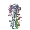
- EMDB-46477: Map of influenza hemagglutinin A/Sing/INFIMH/16 expressed in GntI... -
+ Open data

- Basic information
| Entry |  | |||||||||
|---|---|---|---|---|---|---|---|---|---|---|
| Title | Map of influenza hemagglutinin A/Sing/INFIMH/16 expressed in GntI- cells | |||||||||
 Map data Map data | map of influenza hemagglutinin A/Sing/INFIMH/16 expressed in GntI- cells | |||||||||
 Sample Sample |
| |||||||||
 Keywords Keywords | influenza / H3 / Sing16 / VIRAL PROTEIN | |||||||||
| Function / homology |  Function and homology information Function and homology informationclathrin-dependent endocytosis of virus by host cell / host cell surface receptor binding / fusion of virus membrane with host plasma membrane / fusion of virus membrane with host endosome membrane / viral envelope / virion attachment to host cell / host cell plasma membrane / virion membrane Similarity search - Function | |||||||||
| Biological species |   Influenza A virus Influenza A virus | |||||||||
| Method | single particle reconstruction / cryo EM / Resolution: 3.7 Å | |||||||||
 Authors Authors | Torrents de la Pena A / de Paiva Froes Rocha R / Ward AB | |||||||||
| Funding support |  United States, 1 items United States, 1 items
| |||||||||
 Citation Citation | Journal: Nat Commun / Year: 2025 Title: Structural and immunological characterization of the H3 influenza hemagglutinin during antigenic drift. Authors: Rebeca de Paiva Froes Rocha / Ilhan Tomris / Charles A Bowman / Emma Stevens / Jason Kantorow / Corinna M Plitt / Weiwei Peng / Svearike Oeverdieck / Thales Galdino Andrade / James A ...Authors: Rebeca de Paiva Froes Rocha / Ilhan Tomris / Charles A Bowman / Emma Stevens / Jason Kantorow / Corinna M Plitt / Weiwei Peng / Svearike Oeverdieck / Thales Galdino Andrade / James A Ferguson / Diana D Jung / Rafael Elias Marques / Sander Herfst / Joost Snijder / Srirupa Chakraborty / Alba Torrents de la Peña / Zachary T Berndsen / Robert P de Vries / Andrew B Ward /   Abstract: The quest for a universal influenza vaccine holds great promise for mitigating the global burden of influenza-related morbidity and mortality. However, challenges persist in identifying conserved ...The quest for a universal influenza vaccine holds great promise for mitigating the global burden of influenza-related morbidity and mortality. However, challenges persist in identifying conserved epitopes capable of eliciting robust and durable immune responses. In this study, we explore the influence of glycan evolution on H3 hemagglutinin from 1968 to present day and its impacts on protein structure, antigenicity and immunogenicity by using computational, biochemical and biophysical techniques. Structural characterization of HK/68 and Sing/16 by cryo-electron microscopy shows that while HK/68 is resistant to enzymatic deglycosylation, removal of glycans destabilizes the hyperglycosylated head and membrane-proximal region in Sing/16. Furthermore, the appearance of glycans in Sing/16 hemagglutinin head domain shifts the polyclonal immune response upon vaccination to target the esterase and stem. These insights expand our understanding of glycans beyond their role in protein folding and highlight the interplay among glycan integration and immune recognition to design a universal influenza vaccine. | |||||||||
| History |
|
- Structure visualization
| Supplemental images |
|---|

- Downloads & links
-EMDB archive
| Map data | emd_46477.map.gz | 168.1 MB | EMDB map data format | |
|---|---|---|---|---|
| Header (meta data) | emd-46477-v30.xmlemd-46477.xml | 22.8 KB 22.8 KB | Display Display | EMDB header |
| FSC (resolution estimation) | emd_46477_fsc.xml | 11.9 KB | Display | FSC data file |
| Images |  emd_46477.png emd_46477.png | 50.6 KB | ||
| Masks | emd_46477_msk_1.map | 178 MB | Mask map | |
| Filedesc metadata | emd-46477.cif.gz | 6.8 KB | ||
| Others | emd_46477_half_map_1.map.gzemd_46477_half_map_2.map.gz | 165.1 MB 165.1 MB | ||
| Archive directory |  http://ftp.pdbj.org/pub/emdb/structures/EMD-46477ftp://ftp.pdbj.org/pub/emdb/structures/EMD-46477 http://ftp.pdbj.org/pub/emdb/structures/EMD-46477ftp://ftp.pdbj.org/pub/emdb/structures/EMD-46477 | HTTPS FTP |
-Related structure data
| Related structure data |  9d1uMC  9cxtC  9cxuC  9d0yC  9d2mC M: atomic model generated by this map C: citing same article ( |
|---|---|
| Similar structure data |


-Links
| EMDB pages | EMDB (EBI/PDBe) / EMDataResource |
|---|---|
| Related items in Molecule of the Month |


-Map
| File | Download / File: emd_46477.map.gz / Format: CCP4 / Size: 178 MB / Type: IMAGE STORED AS FLOATING POINT NUMBER (4 BYTES) | ||||||||||||||||||||||||||||||||||||
|---|---|---|---|---|---|---|---|---|---|---|---|---|---|---|---|---|---|---|---|---|---|---|---|---|---|---|---|---|---|---|---|---|---|---|---|---|---|
| Annotation | map of influenza hemagglutinin A/Sing/INFIMH/16 expressed in GntI- cells | ||||||||||||||||||||||||||||||||||||
| Projections & slices | Image control
Images are generated by Spider. | ||||||||||||||||||||||||||||||||||||
| Voxel size | X=Y=Z: 1.15 Å | ||||||||||||||||||||||||||||||||||||
| Density |
| ||||||||||||||||||||||||||||||||||||
| Symmetry | Space group: 1 | ||||||||||||||||||||||||||||||||||||
| Details | EMDB XML:
|
 Z (Sec.)
 Y (Row.)
 X (Col.)


-Supplemental data
-Mask #1
| File | emd_46477_msk_1.map | ||||||||||||
|---|---|---|---|---|---|---|---|---|---|---|---|---|---|
| Projections & Slices |
| ||||||||||||
| Density Histograms |


-Half map: half map of influenza hemagglutinin A/Sing/INFIMH/16 expressed in...
| File | emd_46477_half_map_1.map | ||||||||||||
|---|---|---|---|---|---|---|---|---|---|---|---|---|---|
| Annotation | half map of influenza hemagglutinin A/Sing/INFIMH/16 expressed in GntI- cells | ||||||||||||
| Projections & Slices |
| ||||||||||||
| Density Histograms |


-Half map: half map of influenza hemagglutinin A/Sing/INFIMH/16 expressed in...
| File | emd_46477_half_map_2.map | ||||||||||||
|---|---|---|---|---|---|---|---|---|---|---|---|---|---|
| Annotation | half map of influenza hemagglutinin A/Sing/INFIMH/16 expressed in GntI- cells | ||||||||||||
| Projections & Slices |
| ||||||||||||
| Density Histograms |


- Sample components
-Entire : influenza hemagglutinin A/Sing/INFIMH/16 expressed in GntI- cells...
| Entire | Name: influenza hemagglutinin A/Sing/INFIMH/16 expressed in GntI- cells trimer |
|---|---|
| Components |
|
-Supramolecule #1: influenza hemagglutinin A/Sing/INFIMH/16 expressed in GntI- cells...
| Supramolecule | Name: influenza hemagglutinin A/Sing/INFIMH/16 expressed in GntI- cells trimer type: complex / ID: 1 / Parent: 0 / Macromolecule list: all |
|---|---|
| Source (natural) | Organism: Influenza A virus |
-Macromolecule #1: Hemagglutinin
| Macromolecule | Name: Hemagglutinin / type: protein_or_peptide / ID: 1 / Number of copies: 3 / Enantiomer: LEVO |
|---|---|
| Source (natural) | Organism: Influenza A virus |
| Molecular weight | Theoretical: 92.743625 KDa |
| Recombinant expression | Organism:  Homo sapiens (human) Homo sapiens (human) |
| Sequence | String: MPMGSLQPLA TLYLLGMLVA SVLAATLCLG HHAVPNGTIV KTITNDRIEV TNATELVQNS SIGEICDSPH QILDGENCTL IDALLGDPQ CDGFQNKKWD LFVERSKAYS NCYPYDVPDY ASLRSLVASS GTLEFKNESF NWTGVTQNGT SSACIRGSSS S FFSRLNWL ...String: MPMGSLQPLA TLYLLGMLVA SVLAATLCLG HHAVPNGTIV KTITNDRIEV TNATELVQNS SIGEICDSPH QILDGENCTL IDALLGDPQ CDGFQNKKWD LFVERSKAYS NCYPYDVPDY ASLRSLVASS GTLEFKNESF NWTGVTQNGT SSACIRGSSS S FFSRLNWL THLNYTYPAL NVTMPNKEQF DKLYIWGVHH PGTDKDQIFL YAQSSGRITV STKRSQQAVI PNIGSRPRIR DI PSRISIY WTIVKPGDIL LINSTGNLIA PRGYFKIRSG KSSIMRSDAP IGKCKSECIT PNGSIPNDKP FQNVNRITYG ACP RYVKHS TLKLATGMRN VPEKQTRGIF GAIAGFIENG WEGMVDGWYG FRHQNSEGRG QAADLKSTQA AIDQINGKLN RLIG KTNEK FHQIEKEFSE VEGRVQDLEK YVEDTKIDLW SYNAELLVAL ENQHTIDLTD SEMNKLFEKT KKQLRENAED MGNGC FKIY HKCDNACIES IRNETYDHNV YRDEALNNRF QIKRMKQIED KIEEIESKQK KIENEIARIK KIKLVPRGSV DENLYF QAM SKGEELFTGV VPILVELDGD VNGHKFSVRG EGEGDATNGK LTLKFICTTG KLPVPWPTLV TTLTYGVQCF SRYPDHM KR HDFFKSAMPE GYVQERTISF KDDGTYKTRA EVKFEGDTLV NRIELKGIDF KEDGNILGHK LEYNFNSHNV YITADKQK N GIKANFKIRH NVEDGSVQLA DHYQQNTPIG DGPVLLPDNH YLSTQSVLSK DPNEKRDHMV LLEFVTAAGI THGMSSAWS HPQFEKGGGS GGGSGGSAWS HPQFEK UniProtKB: Hemagglutinin |
-Experimental details
-Structure determination
| Method | cryo EM |
|---|---|
 Processing Processing | single particle reconstruction |
| Aggregation state | particle |
-Sample preparation
| Concentration | 1 mg/mL |
|---|---|
| Buffer | pH: 7.4 / Details: TBS |
| Grid | Model: EMS Lacey Carbon / Material: COPPER / Mesh: 300 / Pretreatment - Type: PLASMA CLEANING / Pretreatment - Time: 20 sec. |
| Vitrification | Cryogen name: ETHANE / Chamber humidity: 100 % / Chamber temperature: 277 K / Instrument: FEI VITROBOT MARK IV |
- Electron microscopy
| Microscope | FEI TALOS ARCTICA |
|---|---|
| Image recording | Film or detector model: GATAN K2 SUMMIT (4k x 4k) / Average electron dose: 50.0 e/Å2 |
| Electron beam | Acceleration voltage: 200 kV / Electron source:  FIELD EMISSION GUN FIELD EMISSION GUN |
| Electron optics | C2 aperture diameter: 100.0 µm / Illumination mode: FLOOD BEAM / Imaging mode: DIFFRACTION / Cs: 2.7 mm / Nominal defocus max: 1.5 µm / Nominal defocus min: 0.8 µm |
| Experimental equipment |  Model: Talos Arctica / Image courtesy: FEI Company |