 Movie
Movie Controller

[English] 日本語
 Yorodumi
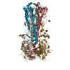
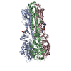
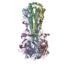
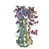
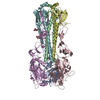
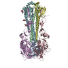
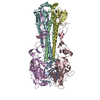
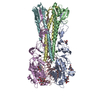
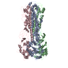
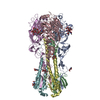
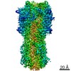
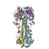
- PDB-4m4y: Crystal structure of a 2009 H1N1 influenza virus hemagglutinin wi... -
+ Open data

- Basic information
| Entry | Database: PDB / ID: 4m4y | |||||||||
|---|---|---|---|---|---|---|---|---|---|---|
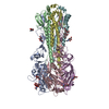
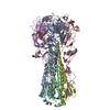
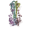
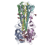
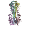
| Title | Crystal structure of a 2009 H1N1 influenza virus hemagglutinin with a stabilization mutation HA2 E47G | |||||||||
 Components Components | (Hemagglutinin ...) x 2 | |||||||||
 Keywords Keywords | VIRAL PROTEIN / viral envelope protein / viral fusion protein / stabilization mutant | |||||||||
| Function / homology |  Function and homology information Function and homology informationviral budding from plasma membrane / clathrin-dependent endocytosis of virus by host cell / host cell surface receptor binding / fusion of virus membrane with host plasma membrane / fusion of virus membrane with host endosome membrane / viral envelope / virion attachment to host cell / host cell plasma membrane / virion membrane / membrane / metal ion binding Similarity search - Function | |||||||||
| Biological species |   Influenza A virus Influenza A virus | |||||||||
| Method |  X-RAY DIFFRACTION / SYNCHROTRON / MOLECULAR REPLACEMENT / Resolution: 2.2 Å X-RAY DIFFRACTION / SYNCHROTRON / MOLECULAR REPLACEMENT / Resolution: 2.2 Å | |||||||||
 Authors Authors | Zhu, X. / Wilson, I.A. | |||||||||
 Citation Citation | Journal: J Virol / Year: 2013 Title: Antibody recognition of the pandemic H1N1 Influenza virus hemagglutinin receptor binding site. Authors: Minsun Hong / Peter S Lee / Ryan M B Hoffman / Xueyong Zhu / Jens C Krause / Nick S Laursen / Sung-Il Yoon / Langzhou Song / Lynda Tussey / James E Crowe / Andrew B Ward / Ian A Wilson Abstract: Influenza virus is a global health concern due to its unpredictable pandemic potential. This potential threat was realized in 2009 when an H1N1 virus emerged that resembled the 1918 virus in ...Influenza virus is a global health concern due to its unpredictable pandemic potential. This potential threat was realized in 2009 when an H1N1 virus emerged that resembled the 1918 virus in antigenicity but fortunately was not nearly as deadly. 5J8 is a human antibody that potently neutralizes a broad spectrum of H1N1 viruses, including the 1918 and 2009 pandemic viruses. Here, we present the crystal structure of 5J8 Fab in complex with a bacterially expressed and refolded globular head domain from the hemagglutinin (HA) of the A/California/07/2009 (H1N1) pandemic virus. 5J8 recognizes a conserved epitope in and around the receptor binding site (RBS), and its HCDR3 closely mimics interactions of the sialic acid receptor. Electron microscopy (EM) reconstructions of 5J8 Fab in complex with an HA trimer from a 1986 H1 strain and with an engineered stabilized HA trimer from the 2009 H1 pandemic virus showed a similar mode of binding. As for other characterized RBS-targeted antibodies, 5J8 uses avidity to extend its breadth and affinity against divergent H1 strains. 5J8 selectively interacts with HA insertion residue 133a, which is conserved in pandemic H1 strains and has precluded binding of other RBS-targeted antibodies. Thus, the RBS of divergent HAs is targeted by 5J8 and adds to the growing arsenal of common recognition motifs for design of therapeutics and vaccines. Moreover, consistent with previous studies, the bacterially expressed H1 HA properly refolds, retaining its antigenic structure, and presents a low-cost and rapid alternative for engineering and manufacturing candidate flu vaccines. | |||||||||
| History |
|
- Structure visualization
| Structure viewer | Molecule: MolmilJmol/JSmol |
|---|
- Downloads & links
-Download
| PDBx/mmCIF format | 4m4y.cif.gz | 326.3 KB | Display | PDBx/mmCIF format |
|---|---|---|---|---|
| PDB format | pdb4m4y.ent.gz | 262.4 KB | Display | PDB format |
| PDBx/mmJSON format | 4m4y.json.gz | Tree view | PDBx/mmJSON format | |
| Others |  Other downloads Other downloads |
-Validation report
| Arichive directory | https://data.pdbj.org/pub/pdb/validation_reports/m4/4m4yftp://data.pdbj.org/pub/pdb/validation_reports/m4/4m4y | HTTPS FTP |
|---|
-Related structure data
| Related structure data |  5731C  5733C  4m5yC  4m5zC 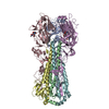 3lzgS S: Starting model for refinement C: citing same article ( |
|---|---|
| Similar structure data |


-Links
 PDBj
PDBj


- Assembly
| Deposited unit | 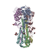
| ||||||||
|---|---|---|---|---|---|---|---|---|---|
| 1 |
| ||||||||
| Unit cell |
|
-Components
-Hemagglutinin ... , 2 types, 6 molecules ACEBDF
| #1: Protein | Mass: 36729.402 Da / Num. of mol.: 3 / Fragment: ectodomain (residues 18-344) Source method: isolated from a genetically manipulated source Source: (gene. exp.) Influenza A virus / Strain: A/California/04/2009 / Gene: HA, hemagglutinin / Plasmid: pFASTBAC-HT / Production host:  Trichoplusia ni (cabbage looper) / Strain (production host): Hi5 / References: UniProt: C3W5S1 Trichoplusia ni (cabbage looper) / Strain (production host): Hi5 / References: UniProt: C3W5S1#2: Protein | Mass: 20206.320 Da / Num. of mol.: 3 / Fragment: ectodomain (residues 345-518) / Mutation: E47G Source method: isolated from a genetically manipulated source Source: (gene. exp.) Influenza A virus / Strain: A/California/04/2009 / Gene: HA, hemagglutinin / Plasmid: pFASTBAC-HT / Production host: Trichoplusia ni (cabbage looper) / Strain (production host): Hi5 / References: UniProt: C3W5S1 |
|---|
-Sugars , 3 types, 10 molecules 
| #3: Polysaccharide | alpha-D-mannopyranose-(1-3)-beta-D-mannopyranose-(1-4)-2-acetamido-2-deoxy-beta-D-glucopyranose-(1- ...alpha-D-mannopyranose-(1-3)-beta-D-mannopyranose-(1-4)-2-acetamido-2-deoxy-beta-D-glucopyranose-(1-4)-2-acetamido-2-deoxy-beta-D-glucopyranose Source method: isolated from a genetically manipulated source |
|---|---|
| #4: Polysaccharide | beta-L-fucopyranose-(1-3)-[2-acetamido-2-deoxy-beta-D-glucopyranose-(1-4)]2-acetamido-2-deoxy-beta- ...beta-L-fucopyranose-(1-3)-[2-acetamido-2-deoxy-beta-D-glucopyranose-(1-4)]2-acetamido-2-deoxy-beta-D-glucopyranose Source method: isolated from a genetically manipulated source |
| #5: Sugar | ChemComp-NAG /  Type: D-saccharide, beta linking / Mass: 221.208 Da / Num. of mol.: 8 Type: D-saccharide, beta linking / Mass: 221.208 Da / Num. of mol.: 8Source method: isolated from a genetically manipulated source Formula: C8H15NO6 |
-Non-polymers , 1 types, 940 molecules 
| #6: Water | ChemComp-HOH / Mass: 18.015 Da / Num. of mol.: 940 / Source method: isolated from a natural source / Formula: H2O |
|---|
-Details
| Has protein modification | Y |
|---|
-Experimental details
-Experiment
| Experiment | Method: X-RAY DIFFRACTION / Number of used crystals: 1 |
|---|
- Sample preparation
| Crystal | Density Matthews: 2.79 Å3/Da / Density % sol: 55.85 % |
|---|---|
| Crystal grow | Temperature: 295 K / Method: vapor diffusion, sitting drop / pH: 8.8 Details: 0.1 M Tris, pH 8.8, 25% w/v MPEG2000, VAPOR DIFFUSION, SITTING DROP, temperature 295K |
-Data collection
| Diffraction | Mean temperature: 100 K |
|---|---|
| Diffraction source | Source: SYNCHROTRON / Site: SSRL  / Beamline: BL12-2 / Wavelength: 0.9795 Å / Beamline: BL12-2 / Wavelength: 0.9795 Å |
| Detector | Type: DECTRIS PILATUS 6M / Detector: PIXEL / Date: May 22, 2012 |
| Radiation | Monochromator: Liquid nitrogen-cooled double crystal Si(111) Protocol: SINGLE WAVELENGTH / Monochromatic (M) / Laue (L): M / Scattering type: x-ray |
| Radiation wavelength | Wavelength: 0.9795 Å / Relative weight: 1 |
| Reflection | Resolution: 2.089→110.57 Å / Num. all: 112741 / Num. obs: 95740 / % possible obs: 96.3 % / Observed criterion σ(I): -3 |
| Reflection shell | Resolution: 2.2→2.24 Å / Redundancy: 3.2 % / Mean I/σ(I) obs: 1.5 / Rsym value: 0.71 / % possible all: 98.9 |
- Processing
| Software |
| |||||||||||||||||||||||||||||||||||||||||||||||||||||||||||||||||||||||||||||||||||||||||||||||||||||||||||||||||||||||||||||||||||||||||||||||||||||||||||||||||||||||||||||||||||||||||||||||||||||||||||||||||||||||||
|---|---|---|---|---|---|---|---|---|---|---|---|---|---|---|---|---|---|---|---|---|---|---|---|---|---|---|---|---|---|---|---|---|---|---|---|---|---|---|---|---|---|---|---|---|---|---|---|---|---|---|---|---|---|---|---|---|---|---|---|---|---|---|---|---|---|---|---|---|---|---|---|---|---|---|---|---|---|---|---|---|---|---|---|---|---|---|---|---|---|---|---|---|---|---|---|---|---|---|---|---|---|---|---|---|---|---|---|---|---|---|---|---|---|---|---|---|---|---|---|---|---|---|---|---|---|---|---|---|---|---|---|---|---|---|---|---|---|---|---|---|---|---|---|---|---|---|---|---|---|---|---|---|---|---|---|---|---|---|---|---|---|---|---|---|---|---|---|---|---|---|---|---|---|---|---|---|---|---|---|---|---|---|---|---|---|---|---|---|---|---|---|---|---|---|---|---|---|---|---|---|---|---|---|---|---|---|---|---|---|---|---|---|---|---|---|---|---|---|
| Refinement | Method to determine structure: MOLECULAR REPLACEMENT Starting model: PDB ENTRY 3LZG Resolution: 2.2→48.49 Å / SU ML: 0.25 / Cross valid method: THROUGHOUT / σ(F): 1.34 / Phase error: 23.68 / Stereochemistry target values: ML
| |||||||||||||||||||||||||||||||||||||||||||||||||||||||||||||||||||||||||||||||||||||||||||||||||||||||||||||||||||||||||||||||||||||||||||||||||||||||||||||||||||||||||||||||||||||||||||||||||||||||||||||||||||||||||
| Solvent computation | Shrinkage radii: 0.9 Å / VDW probe radii: 1.11 Å / Solvent model: FLAT BULK SOLVENT MODEL | |||||||||||||||||||||||||||||||||||||||||||||||||||||||||||||||||||||||||||||||||||||||||||||||||||||||||||||||||||||||||||||||||||||||||||||||||||||||||||||||||||||||||||||||||||||||||||||||||||||||||||||||||||||||||
| Refinement step | Cycle: LAST / Resolution: 2.2→48.49 Å
| |||||||||||||||||||||||||||||||||||||||||||||||||||||||||||||||||||||||||||||||||||||||||||||||||||||||||||||||||||||||||||||||||||||||||||||||||||||||||||||||||||||||||||||||||||||||||||||||||||||||||||||||||||||||||
| Refine LS restraints |
| |||||||||||||||||||||||||||||||||||||||||||||||||||||||||||||||||||||||||||||||||||||||||||||||||||||||||||||||||||||||||||||||||||||||||||||||||||||||||||||||||||||||||||||||||||||||||||||||||||||||||||||||||||||||||
| LS refinement shell |
|
