 Movie
Movie Controller


+ Open data

- Basic information
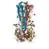
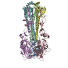
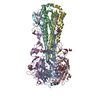
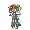
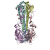
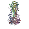
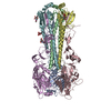
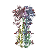
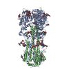
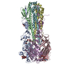
| Entry | Database: PDB / ID: 3lzg | |||||||||
|---|---|---|---|---|---|---|---|---|---|---|
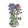
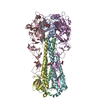
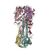
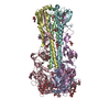
| Title | Crystal structure of a 2009 H1N1 influenza virus hemagglutinin | |||||||||
 Components Components | (Hemagglutinin, ...) x 2 | |||||||||
 Keywords Keywords | VIRAL PROTEIN/IMMUNE SYSTEM / viral envelope protein / hemagglutinin / viral fusion protein / VIRAL PROTEIN-IMMUNE SYSTEM complex | |||||||||
| Function / homology |  Function and homology information Function and homology informationviral budding from plasma membrane / clathrin-dependent endocytosis of virus by host cell / host cell surface receptor binding / fusion of virus membrane with host plasma membrane / fusion of virus membrane with host endosome membrane / viral envelope / virion attachment to host cell / host cell plasma membrane / virion membrane / membrane / metal ion binding Similarity search - Function | |||||||||
| Biological species |   Influenza A virus Influenza A virus | |||||||||
| Method |  X-RAY DIFFRACTION / SYNCHROTRON / MOLECULAR REPLACEMENT / Resolution: 2.6 Å X-RAY DIFFRACTION / SYNCHROTRON / MOLECULAR REPLACEMENT / Resolution: 2.6 Å | |||||||||
 Authors Authors | Xu, R. / Wilson, I.A. | |||||||||
 Citation Citation | Journal: Science / Year: 2010 Title: Structural basis of preexisting immunity to the 2009 H1N1 pandemic influenza virus. Authors: Xu, R. / Ekiert, D.C. / Krause, J.C. / Hai, R. / Crowe, J.E. / Wilson, I.A. | |||||||||
| History |
|
- Structure visualization
| Structure viewer | Molecule: MolmilJmol/JSmol |
|---|
- Downloads & links
-Download
| PDBx/mmCIF format | 3lzg.cif.gz | 1.2 MB | Display | PDBx/mmCIF format |
|---|---|---|---|---|
| PDB format | pdb3lzg.ent.gz | 994.9 KB | Display | PDB format |
| PDBx/mmJSON format | 3lzg.json.gz | Tree view | PDBx/mmJSON format | |
| Others |  Other downloads Other downloads |
-Validation report
| Arichive directory | https://data.pdbj.org/pub/pdb/validation_reports/lz/3lzgftp://data.pdbj.org/pub/pdb/validation_reports/lz/3lzg | HTTPS FTP |
|---|
-Related structure data
| Related structure data |  3lzfC 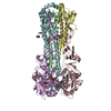 1rd8S C: citing same article ( S: Starting model for refinement |
|---|---|
| Similar structure data |


-Links
 PDBj
PDBj


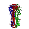
- Assembly
| Deposited unit | 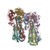
| ||||||||
|---|---|---|---|---|---|---|---|---|---|
| 1 | 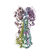
| ||||||||
| 2 | 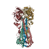
| ||||||||
| Unit cell |
|
-Components
-Hemagglutinin, ... , 2 types, 12 molecules ACEGIKBDFHJL
| #1: Protein | Mass: 36543.238 Da / Num. of mol.: 6 / Fragment: Ectodomain HA1, residues 18-344 Source method: isolated from a genetically manipulated source Source: (gene. exp.) Influenza A virus / Strain: A/California/04/2009 / Gene: HA, hemagglutinin / Plasmid: pFASTbac-HT / Production host:  Trichoplusia ni (cabbage looper) / Strain (production host): Hi5 / References: UniProt: C3W5S1 Trichoplusia ni (cabbage looper) / Strain (production host): Hi5 / References: UniProt: C3W5S1#2: Protein | Mass: 20278.383 Da / Num. of mol.: 6 / Fragment: Ectodomain HA2, residues 345-520 Source method: isolated from a genetically manipulated source Source: (gene. exp.) Influenza A virus / Strain: A/California/04/2009 / Gene: HA, hemagglutinin / Plasmid: pFastbac-HT / Production host: Trichoplusia ni (cabbage looper) / Strain (production host): Hi5 / References: UniProt: C3W5S1 |
|---|
-Sugars , 4 types, 6 molecules 
| #3: Polysaccharide | beta-D-mannopyranose-(1-4)-2-acetamido-2-deoxy-beta-D-glucopyranose-(1-4)-2-acetamido-2-deoxy-beta- ...beta-D-mannopyranose-(1-4)-2-acetamido-2-deoxy-beta-D-glucopyranose-(1-4)-2-acetamido-2-deoxy-beta-D-glucopyranose Source method: isolated from a genetically manipulated source | ||||
|---|---|---|---|---|---|
| #4: Polysaccharide | Source method: isolated from a genetically manipulated source #5: Polysaccharide | alpha-D-mannopyranose-(1-3)-[alpha-D-mannopyranose-(1-6)]beta-D-mannopyranose-(1-4)-2-acetamido-2- ...alpha-D-mannopyranose-(1-3)-[alpha-D-mannopyranose-(1-6)]beta-D-mannopyranose-(1-4)-2-acetamido-2-deoxy-beta-D-glucopyranose-(1-4)-2-acetamido-2-deoxy-beta-D-glucopyranose | Source method: isolated from a genetically manipulated source #6: Sugar |  Type: D-saccharide, beta linking / Mass: 221.208 Da / Num. of mol.: 2 Type: D-saccharide, beta linking / Mass: 221.208 Da / Num. of mol.: 2Source method: isolated from a genetically manipulated source Formula: C8H15NO6 |
-Non-polymers , 1 types, 463 molecules 
| #7: Water | ChemComp-HOH / Mass: 18.015 Da / Num. of mol.: 463 / Source method: isolated from a natural source / Formula: H2O |
|---|
-Details
| Has protein modification | Y |
|---|
-Experimental details
-Experiment
| Experiment | Method: X-RAY DIFFRACTION / Number of used crystals: 1 |
|---|
- Sample preparation
| Crystal | Density Matthews: 2.43 Å3/Da / Density % sol: 49.4 % |
|---|---|
| Crystal grow | Temperature: 295.5 K / Method: vapor diffusion, sitting drop / pH: 8.8 Details: 27% PEG-MME 2000, 0.1M Tris pH 8.8, VAPOR DIFFUSION, SITTING DROP, temperature 295.5K |
-Data collection
| Diffraction | Mean temperature: 110 K |
|---|---|
| Diffraction source | Source: SYNCHROTRON / Site: SSRL  / Beamline: BL9-2 / Wavelength: 1.03317 Å / Beamline: BL9-2 / Wavelength: 1.03317 Å |
| Detector | Type: MARMOSAIC 325 mm CCD / Detector: CCD / Date: Jun 26, 2009 Details: Flat collimating mirror, double crystal monochromator, toroid focusing mirror |
| Radiation | Monochromator: Double crystal monochromator / Protocol: SINGLE WAVELENGTH / Monochromatic (M) / Laue (L): M / Scattering type: x-ray |
| Radiation wavelength | Wavelength: 1.03317 Å / Relative weight: 1 |
| Reflection | Resolution: 2.6→45 Å / Num. all: 102429 / Num. obs: 93210 / % possible obs: 91 % / Observed criterion σ(I): 1 / Redundancy: 1.8 % / Biso Wilson estimate: 61.66 Å2 / Rmerge(I) obs: 0.078 / Net I/σ(I): 9.5 |
| Reflection shell | Resolution: 2.6→2.69 Å / Redundancy: 1.6 % / Mean I/σ(I) obs: 1.8 / Num. unique all: 7120 / Rsym value: 0.375 / % possible all: 69.7 |
- Processing
| Software |
| |||||||||||||||||||||||||
|---|---|---|---|---|---|---|---|---|---|---|---|---|---|---|---|---|---|---|---|---|---|---|---|---|---|---|
| Refinement | Method to determine structure: MOLECULAR REPLACEMENT Starting model: PDB entry 1RD8 Resolution: 2.6→45 Å / Cross valid method: THROUGHOUT / σ(F): 0 / Stereochemistry target values: Engh & Huber
| |||||||||||||||||||||||||
| Displacement parameters | Biso mean: 65.71 Å2
| |||||||||||||||||||||||||
| Refine analyze | Luzzati coordinate error obs: 0.36 Å | |||||||||||||||||||||||||
| Refinement step | Cycle: LAST / Resolution: 2.6→45 Å
| |||||||||||||||||||||||||
| Refine LS restraints |
| |||||||||||||||||||||||||
| LS refinement shell | Resolution: 2.6→2.67 Å / Total num. of bins used: 20
|
