 Movie
Movie Controller

[English] 日本語
 Yorodumi
- PDB-6aoq: Crystal structure of the A/Brisbane/10/2007 (H3N2) influenza viru... -
+ Open data

- Basic information
| Entry | Database: PDB / ID: 6aoq | ||||||||||||
|---|---|---|---|---|---|---|---|---|---|---|---|---|---|
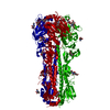
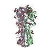
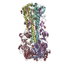
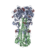
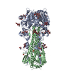
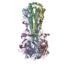
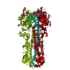
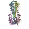
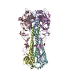
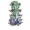
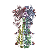
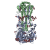
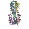
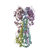
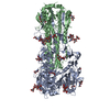
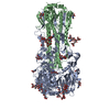
| Title | Crystal structure of the A/Brisbane/10/2007 (H3N2) influenza virus hemagglutinin apo form | ||||||||||||
 Components Components | (Hemagglutinin ...) x 2 | ||||||||||||
 Keywords Keywords | VIRAL PROTEIN / Influenza A virus / hemagglutinin / mutant / receptor binding / antigenicity | ||||||||||||
| Function / homology |  Function and homology information Function and homology informationviral budding from plasma membrane / clathrin-dependent endocytosis of virus by host cell / host cell surface receptor binding / fusion of virus membrane with host plasma membrane / fusion of virus membrane with host endosome membrane / viral envelope / virion attachment to host cell / host cell plasma membrane / virion membrane / membrane Similarity search - Function | ||||||||||||
| Biological species |   Influenza A virus Influenza A virus | ||||||||||||
| Method |  X-RAY DIFFRACTION / SYNCHROTRON / MOLECULAR REPLACEMENT / Resolution: 2.35 Å X-RAY DIFFRACTION / SYNCHROTRON / MOLECULAR REPLACEMENT / Resolution: 2.35 Å | ||||||||||||
 Authors Authors | Wu, N.C. / Wilson, I.A. | ||||||||||||
| Funding support |  United States, 3items United States, 3items
| ||||||||||||
 Citation Citation | Journal: PLoS Pathog. / Year: 2017 Title: A structural explanation for the low effectiveness of the seasonal influenza H3N2 vaccine. Authors: Wu, N.C. / Zost, S.J. / Thompson, A.J. / Oyen, D. / Nycholat, C.M. / McBride, R. / Paulson, J.C. / Hensley, S.E. / Wilson, I.A. | ||||||||||||
| History |
|
- Structure visualization
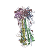
| Structure viewer | Molecule: MolmilJmol/JSmol |
|---|
- Downloads & links
-Download
| PDBx/mmCIF format | 6aoq.cif.gz | 220 KB | Display | PDBx/mmCIF format |
|---|---|---|---|---|
| PDB format | pdb6aoq.ent.gz | 176.1 KB | Display | PDB format |
| PDBx/mmJSON format | 6aoq.json.gz | Tree view | PDBx/mmJSON format | |
| Others |  Other downloads Other downloads |
-Validation report
| Arichive directory | https://data.pdbj.org/pub/pdb/validation_reports/ao/6aoqftp://data.pdbj.org/pub/pdb/validation_reports/ao/6aoq | HTTPS FTP |
|---|
-Related structure data
| Related structure data |  6aopC 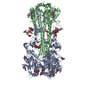 6aorC  6aosC  6aotC  6aouC 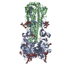 6aovC  4o5nS S: Starting model for refinement C: citing same article ( |
|---|---|
| Similar structure data |

-Links
 PDBj
PDBj


- Assembly
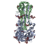
| Deposited unit | 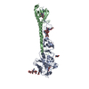
| ||||||||
|---|---|---|---|---|---|---|---|---|---|
| 1 | 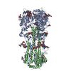
| ||||||||
| Unit cell |
|
-Components
-Hemagglutinin ... , 2 types, 2 molecules AB
| #1: Protein | Mass: 35922.480 Da / Num. of mol.: 1 Source method: isolated from a genetically manipulated source Source: (gene. exp.) Influenza A virus / Strain: A/Brisbane/10/2007(H3N2) / Gene: HA / Production host:  Trichoplusia ni (cabbage looper) / References: UniProt: A8W891, UniProt: B2VNK2*PLUS Trichoplusia ni (cabbage looper) / References: UniProt: A8W891, UniProt: B2VNK2*PLUS |
|---|---|
| #2: Protein | Mass: 19953.174 Da / Num. of mol.: 1 Source method: isolated from a genetically manipulated source Source: (gene. exp.) Influenza A virus (A/Brisbane/10/2007(H3N2))Strain: A/Brisbane/10/2007(H3N2) / Gene: HA / Production host: Trichoplusia ni (cabbage looper) / References: UniProt: A8W891, UniProt: C3PR70*PLUS |
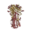
-Sugars , 4 types, 8 molecules 
| #3: Polysaccharide | alpha-D-mannopyranose-(1-3)-[alpha-D-mannopyranose-(1-6)]beta-D-mannopyranose-(1-4)-2-acetamido-2- ...alpha-D-mannopyranose-(1-3)-[alpha-D-mannopyranose-(1-6)]beta-D-mannopyranose-(1-4)-2-acetamido-2-deoxy-beta-D-glucopyranose-(1-4)-2-acetamido-2-deoxy-beta-D-glucopyranose Source method: isolated from a genetically manipulated source |
|---|---|
| #4: Polysaccharide | beta-D-mannopyranose-(1-4)-2-acetamido-2-deoxy-beta-D-glucopyranose-(1-4)-2-acetamido-2-deoxy-beta- ...beta-D-mannopyranose-(1-4)-2-acetamido-2-deoxy-beta-D-glucopyranose-(1-4)-2-acetamido-2-deoxy-beta-D-glucopyranose Source method: isolated from a genetically manipulated source |
| #5: Polysaccharide | 2-acetamido-2-deoxy-beta-D-glucopyranose-(1-4)-2-acetamido-2-deoxy-beta-D-glucopyranose Source method: isolated from a genetically manipulated source |
| #6: Sugar | ChemComp-NAG /  Type: D-saccharide, beta linking / Mass: 221.208 Da / Num. of mol.: 5 Type: D-saccharide, beta linking / Mass: 221.208 Da / Num. of mol.: 5Source method: isolated from a genetically manipulated source Formula: C8H15NO6 |
-Non-polymers , 1 types, 127 molecules 
| #7: Water | ChemComp-HOH / Mass: 18.015 Da / Num. of mol.: 127 / Source method: isolated from a natural source / Formula: H2O |
|---|
-Details
| Has protein modification | Y |
|---|
-Experimental details
-Experiment
| Experiment | Method: X-RAY DIFFRACTION / Number of used crystals: 1 |
|---|
- Sample preparation
| Crystal | Density Matthews: 3.34 Å3/Da / Density % sol: 63.12 % |
|---|---|
| Crystal grow | Temperature: 293 K / Method: vapor diffusion, sitting drop / pH: 10.5 / Details: 0.1 M CAPS pH 10.5 and 29% PEG 400 |
-Data collection
| Diffraction | Mean temperature: 100 K |
|---|---|
| Diffraction source | Source: SYNCHROTRON / Site: SSRL / Beamline: BL12-2 / Wavelength: 0.9795 Å |
| Detector | Type: DECTRIS PILATUS 6M / Detector: PIXEL / Date: Feb 22, 2017 |
| Radiation | Protocol: SINGLE WAVELENGTH / Monochromatic (M) / Laue (L): M / Scattering type: x-ray |
| Radiation wavelength | Wavelength: 0.9795 Å / Relative weight: 1 |
| Reflection | Resolution: 2.35→50 Å / Num. obs: 31953 / % possible obs: 99.9 % / Redundancy: 15.5 % / Biso Wilson estimate: 43 Å2 / CC1/2: 1 / Rpim(I) all: 0.03 / Rsym value: 0.11 / Net I/σ(I): 34.1 |
| Reflection shell | Resolution: 2.35→2.43 Å / Redundancy: 15.2 % / Mean I/σ(I) obs: 2.8 / Num. unique obs: 3148 / CC1/2: 0.93 / Rpim(I) all: 0.22 / Rsym value: 0.84 / % possible all: 100 |
- Processing
| Software |
| ||||||||||||||||||||||||||||||||||||||||||||||||||||||||||||||||||||||||||||||||||||||||||||||||||||||||||||||||||||||||||||||||||||||||||||||||||||||||||||||||||||||||||||||||||||||
|---|---|---|---|---|---|---|---|---|---|---|---|---|---|---|---|---|---|---|---|---|---|---|---|---|---|---|---|---|---|---|---|---|---|---|---|---|---|---|---|---|---|---|---|---|---|---|---|---|---|---|---|---|---|---|---|---|---|---|---|---|---|---|---|---|---|---|---|---|---|---|---|---|---|---|---|---|---|---|---|---|---|---|---|---|---|---|---|---|---|---|---|---|---|---|---|---|---|---|---|---|---|---|---|---|---|---|---|---|---|---|---|---|---|---|---|---|---|---|---|---|---|---|---|---|---|---|---|---|---|---|---|---|---|---|---|---|---|---|---|---|---|---|---|---|---|---|---|---|---|---|---|---|---|---|---|---|---|---|---|---|---|---|---|---|---|---|---|---|---|---|---|---|---|---|---|---|---|---|---|---|---|---|---|
| Refinement | Method to determine structure: MOLECULAR REPLACEMENT Starting model: 4O5N Resolution: 2.35→50 Å / Cor.coef. Fo:Fc: 0.945 / Cor.coef. Fo:Fc free: 0.878 / SU B: 18.637 / SU ML: 0.215 / Cross valid method: THROUGHOUT / ESU R: 0.289 / ESU R Free: 0.233 / Details: HYDROGENS HAVE BEEN ADDED IN THE RIDING POSITIONS
| ||||||||||||||||||||||||||||||||||||||||||||||||||||||||||||||||||||||||||||||||||||||||||||||||||||||||||||||||||||||||||||||||||||||||||||||||||||||||||||||||||||||||||||||||||||||
| Solvent computation | Ion probe radii: 0.8 Å / Shrinkage radii: 0.8 Å / VDW probe radii: 1.2 Å | ||||||||||||||||||||||||||||||||||||||||||||||||||||||||||||||||||||||||||||||||||||||||||||||||||||||||||||||||||||||||||||||||||||||||||||||||||||||||||||||||||||||||||||||||||||||
| Displacement parameters | Biso mean: 66.439 Å2
| ||||||||||||||||||||||||||||||||||||||||||||||||||||||||||||||||||||||||||||||||||||||||||||||||||||||||||||||||||||||||||||||||||||||||||||||||||||||||||||||||||||||||||||||||||||||
| Refinement step | Cycle: 1 / Resolution: 2.35→50 Å
| ||||||||||||||||||||||||||||||||||||||||||||||||||||||||||||||||||||||||||||||||||||||||||||||||||||||||||||||||||||||||||||||||||||||||||||||||||||||||||||||||||||||||||||||||||||||
| Refine LS restraints |
|