Movie
Movie Controller


+ Open data

- Basic information
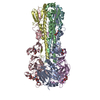
| Entry | Database: PDB / ID: 9cxu | |||||||||||||||||||||||||||
|---|---|---|---|---|---|---|---|---|---|---|---|---|---|---|---|---|---|---|---|---|---|---|---|---|---|---|---|---|
| Title | Endo H-treated hemagglutinin A/Hong Kong/1/68 | |||||||||||||||||||||||||||
 Components Components |
| |||||||||||||||||||||||||||
 Keywords Keywords | VIRAL PROTEIN / hemagglutinin / glycoprotein / H3 | |||||||||||||||||||||||||||
| Function / homology |  Function and homology information Function and homology informationviral budding from plasma membrane / bioluminescence / generation of precursor metabolites and energy / clathrin-dependent endocytosis of virus by host cell / host cell surface receptor binding / fusion of virus membrane with host plasma membrane / fusion of virus membrane with host endosome membrane / viral envelope / virion attachment to host cell / host cell plasma membrane ...viral budding from plasma membrane / bioluminescence / generation of precursor metabolites and energy / clathrin-dependent endocytosis of virus by host cell / host cell surface receptor binding / fusion of virus membrane with host plasma membrane / fusion of virus membrane with host endosome membrane / viral envelope / virion attachment to host cell / host cell plasma membrane / virion membrane / membrane Similarity search - Function | |||||||||||||||||||||||||||
| Biological species |   Influenza A virus Influenza A virus  Aequorea victoria (jellyfish) Aequorea victoria (jellyfish) | |||||||||||||||||||||||||||
| Method | ELECTRON MICROSCOPY / single particle reconstruction / cryo EM / Resolution: 2.3 Å | |||||||||||||||||||||||||||
 Authors Authors | Torrents de la Pena, A. / de Paiva Froes Rocha, R. / Ward, A.B. | |||||||||||||||||||||||||||
| Funding support |  United States, 1items United States, 1items
| |||||||||||||||||||||||||||
 Citation Citation | Journal: Nat Commun / Year: 2025 Title: Structural and immunological characterization of the H3 influenza hemagglutinin during antigenic drift. Authors: Rebeca de Paiva Froes Rocha / Ilhan Tomris / Charles A Bowman / Emma Stevens / Jason Kantorow / Corinna M Plitt / Weiwei Peng / Svearike Oeverdieck / Thales Galdino Andrade / James A ...Authors: Rebeca de Paiva Froes Rocha / Ilhan Tomris / Charles A Bowman / Emma Stevens / Jason Kantorow / Corinna M Plitt / Weiwei Peng / Svearike Oeverdieck / Thales Galdino Andrade / James A Ferguson / Diana D Jung / Rafael Elias Marques / Sander Herfst / Joost Snijder / Srirupa Chakraborty / Alba Torrents de la Peña / Zachary T Berndsen / Robert P de Vries / Andrew B Ward /   Abstract: The quest for a universal influenza vaccine holds great promise for mitigating the global burden of influenza-related morbidity and mortality. However, challenges persist in identifying conserved ...The quest for a universal influenza vaccine holds great promise for mitigating the global burden of influenza-related morbidity and mortality. However, challenges persist in identifying conserved epitopes capable of eliciting robust and durable immune responses. In this study, we explore the influence of glycan evolution on H3 hemagglutinin from 1968 to present day and its impacts on protein structure, antigenicity and immunogenicity by using computational, biochemical and biophysical techniques. Structural characterization of HK/68 and Sing/16 by cryo-electron microscopy shows that while HK/68 is resistant to enzymatic deglycosylation, removal of glycans destabilizes the hyperglycosylated head and membrane-proximal region in Sing/16. Furthermore, the appearance of glycans in Sing/16 hemagglutinin head domain shifts the polyclonal immune response upon vaccination to target the esterase and stem. These insights expand our understanding of glycans beyond their role in protein folding and highlight the interplay among glycan integration and immune recognition to design a universal influenza vaccine. | |||||||||||||||||||||||||||
| History |
|
- Structure visualization
| Structure viewer | Molecule: MolmilJmol/JSmol |
|---|
- Downloads & links
-Download
| PDBx/mmCIF format | 9cxu.cif.gz | 309.1 KB | Display | PDBx/mmCIF format |
|---|---|---|---|---|
| PDB format | pdb9cxu.ent.gz | 237.6 KB | Display | PDB format |
| PDBx/mmJSON format | 9cxu.json.gz | Tree view | PDBx/mmJSON format | |
| Others |  Other downloads Other downloads |
-Validation report
| Arichive directory | https://data.pdbj.org/pub/pdb/validation_reports/cx/9cxuftp://data.pdbj.org/pub/pdb/validation_reports/cx/9cxu | HTTPS FTP |
|---|
-Related structure data
| Related structure data |  45998MC  9cxtC  9d0yC  9d1uC  9d2mC M: map data used to model this data C: citing same article ( |
|---|---|
| Similar structure data |


-Links
 PDBj
PDBj


- Assembly
| Deposited unit | 
|
|---|---|
| 1 |
|
-Components
| #1: Protein | Mass: 38523.496 Da / Num. of mol.: 3 Source method: isolated from a genetically manipulated source Source: (gene. exp.) Influenza A virus (strain A/Hong Kong/1/1968 H3N2)Strain: A/Hong Kong/1/1968 H3N2 / Gene: HA / Production host:  Homo sapiens (human) / References: UniProt: Q91MA7 Homo sapiens (human) / References: UniProt: Q91MA7#2: Protein | Mass: 55973.602 Da / Num. of mol.: 3 Source method: isolated from a genetically manipulated source Source: (gene. exp.) Influenza A virus (strain A/Hong Kong/1/1968 H3N2), (gene. exp.) Aequorea victoria (jellyfish)Strain: A/Hong Kong/1/1968 H3N2 / Gene: HA, GFP / Production host: Homo sapiens (human) / References: UniProt: Q91MA7, UniProt: P42212#3: Sugar | ChemComp-NAG /   Type: D-saccharide, beta linking / Mass: 221.208 Da / Num. of mol.: 12 / Source method: obtained synthetically / Formula: C8H15NO6 Type: D-saccharide, beta linking / Mass: 221.208 Da / Num. of mol.: 12 / Source method: obtained synthetically / Formula: C8H15NO6Has ligand of interest | N | Has protein modification | Y | |
|---|
-Experimental details
-Experiment
| Experiment | Method: ELECTRON MICROSCOPY |
|---|---|
| EM experiment | Aggregation state: PARTICLE / 3D reconstruction method: single particle reconstruction |
- Sample preparation
| Component | Name: hemagglutinin A/Hong Kong/1/68 trimer / Type: COMPLEX / Entity ID: #1 / Source: MULTIPLE SOURCES |
|---|---|
| Source (natural) | Organism: Influenza A virus / Strain: A/Hong Kong/1/1968 H3N2 |
| Source (recombinant) | Organism: Homo sapiens (human) |
| Buffer solution | pH: 7.4 / Details: TBS |
| Specimen | Conc.: 1 mg/ml / Embedding applied: NO / Shadowing applied: NO / Staining applied: NO / Vitrification applied: YES |
| Vitrification | Instrument: FEI VITROBOT MARK IV / Cryogen name: ETHANE / Humidity: 100 % / Chamber temperature: 277 K |
- Electron microscopy imaging
| Experimental equipment |  Model: Talos Arctica / Image courtesy: FEI Company |
|---|---|
| Microscopy | Model: FEI TECNAI ARCTICA |
| Electron gun | Electron source:  FIELD EMISSION GUN / Accelerating voltage: 200 kV / Illumination mode: FLOOD BEAM FIELD EMISSION GUN / Accelerating voltage: 200 kV / Illumination mode: FLOOD BEAM |
| Electron lens | Mode: BRIGHT FIELD / Nominal defocus max: 1500 nm / Nominal defocus min: 800 nm |
| Image recording | Electron dose: 50 e/Å2 / Detector mode: COUNTING / Film or detector model: GATAN K2 SUMMIT (4k x 4k) |
- Processing
| EM software |
| ||||||||||||||||||||||||
|---|---|---|---|---|---|---|---|---|---|---|---|---|---|---|---|---|---|---|---|---|---|---|---|---|---|
| CTF correction | Type: PHASE FLIPPING AND AMPLITUDE CORRECTION | ||||||||||||||||||||||||
| Symmetry | Point symmetry: C3 (3 fold cyclic) | ||||||||||||||||||||||||
| 3D reconstruction | Resolution: 2.3 Å / Resolution method: FSC 0.143 CUT-OFF / Num. of particles: 332294 / Symmetry type: POINT | ||||||||||||||||||||||||
| Atomic model building | PDB-ID: 4ZCJ Accession code: 4ZCJ / Source name: PDB / Type: experimental model | ||||||||||||||||||||||||
| Refine LS restraints |
|