 Movie
Movie Controller

[English] 日本語
 Yorodumi
- EMDB-45726: Cryo-EM model derived from localized reconstruction of human aden... -
+ Open data

- Basic information
| Entry | 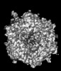 | |||||||||
|---|---|---|---|---|---|---|---|---|---|---|
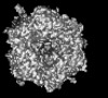
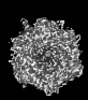
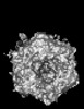
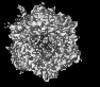
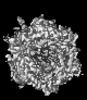
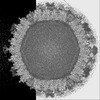
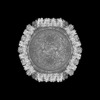
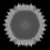
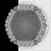
| Title | Cryo-EM model derived from localized reconstruction of human adenovirus 5 (Ad5)-hexon-FII complex at 3.9A resolution | |||||||||
 Map data Map data | ||||||||||
 Sample Sample |
| |||||||||
 Keywords Keywords | Adenovirus / Hexon / Coagulation factor X / Coagulation factor II / Prothrombin / Complex / Interactions / VIRUS | |||||||||
| Function / homology |  Function and homology information Function and homology informationT=25 icosahedral viral capsid / microtubule-dependent intracellular transport of viral material towards nucleus / : / thrombospondin receptor activity / thrombin / thrombin-activated receptor signaling pathway / Defective factor XII causes hereditary angioedema / negative regulation of astrocyte differentiation / regulation of blood coagulation / neutrophil-mediated killing of gram-negative bacterium ...T=25 icosahedral viral capsid / microtubule-dependent intracellular transport of viral material towards nucleus / : / thrombospondin receptor activity / thrombin / thrombin-activated receptor signaling pathway / Defective factor XII causes hereditary angioedema / negative regulation of astrocyte differentiation / regulation of blood coagulation / neutrophil-mediated killing of gram-negative bacterium / positive regulation of phospholipase C-activating G protein-coupled receptor signaling pathway / Defective F8 cleavage by thrombin / ligand-gated ion channel signaling pathway / Platelet Aggregation (Plug Formation) / positive regulation of collagen biosynthetic process / negative regulation of platelet activation / negative regulation of blood coagulation / negative regulation of fibrinolysis / blood coagulation, fibrin clot formation / positive regulation of blood coagulation / Transport of gamma-carboxylated protein precursors from the endoplasmic reticulum to the Golgi apparatus / : / Gamma-carboxylation of protein precursors / Removal of aminoterminal propeptides from gamma-carboxylated proteins / regulation of cytosolic calcium ion concentration / fibrinolysis / : / negative regulation of proteolysis / negative regulation of cytokine production involved in inflammatory response / Regulation of Complement cascade / acute-phase response / Cell surface interactions at the vascular wall / positive regulation of release of sequestered calcium ion into cytosol / Peptide ligand-binding receptors / growth factor activity / positive regulation of receptor signaling pathway via JAK-STAT / lipopolysaccharide binding / platelet activation / response to wounding / positive regulation of protein localization to nucleus / Golgi lumen / Regulation of Insulin-like Growth Factor (IGF) transport and uptake by Insulin-like Growth Factor Binding Proteins (IGFBPs) / positive regulation of reactive oxygen species metabolic process / blood coagulation / positive regulation of insulin secretion / viral capsid / regulation of cell shape / antimicrobial humoral immune response mediated by antimicrobial peptide / host cell / heparin binding / Thrombin signalling through proteinase activated receptors (PARs) / positive regulation of cell growth / blood microparticle / G alpha (q) signalling events / cell surface receptor signaling pathway / positive regulation of phosphatidylinositol 3-kinase/protein kinase B signal transduction / endoplasmic reticulum lumen / receptor ligand activity / signaling receptor binding / serine-type endopeptidase activity / calcium ion binding / positive regulation of cell population proliferation / symbiont entry into host cell / host cell nucleus / structural molecule activity / proteolysis / : / extracellular exosome / extracellular region / plasma membrane Similarity search - Function | |||||||||
| Biological species |   Human adenovirus 5 / Human adenovirus 5 /  Homo sapiens (human) Homo sapiens (human) | |||||||||
| Method | single particle reconstruction / cryo EM / Resolution: 4.13 Å | |||||||||
 Authors Authors | Reddy VS / Ma OX | |||||||||
| Funding support |  United States, 1 items United States, 1 items
| |||||||||
 Citation Citation | Journal: Nat Commun / Year: 2024 Title: Structure-derived insights from blood factors binding to the surfaces of different adenoviruses. Authors: Haley E Mudrick / Shao-Chia Lu / Janarjan Bhandari / Mary E Barry / Jack R Hemsath / Felix G M Andres / Olivia X Ma / Michael A Barry / Vijay S Reddy / Abstract: The tropism of adenoviruses (Ads) is significantly influenced by the binding of various blood factors. To investigate differences in their binding, we conducted cryo-EM analysis on complexes of ...The tropism of adenoviruses (Ads) is significantly influenced by the binding of various blood factors. To investigate differences in their binding, we conducted cryo-EM analysis on complexes of several human adenoviruses with human platelet factor-4 (PF4), coagulation factors FII (Prothrombin), and FX. While we observed EM densities for FII and FX bound to all the species-C adenoviruses examined, no densities were seen for PF4, even though PF4 can co-pellet with various Ads. Similar to FX, the γ-carboxyglutamic acid (Gla) domain of FII binds within the surface cavity of hexon trimers. While FII binds equally to species-C Ads: Ad5, Ad6, and Ad657, FX exhibits significantly better binding to Ad5 and Ad657 compared to Ad6. Although only the FX-Gla domain is observed at high-resolution (3.7 Å), the entire FX is visible at low-resolution bound to Ad5 in three equivalent binding modes consistent with the 3-fold symmetric hexon. Only the Gla and kringle-1 domains of FII are visible on all the species-C adenoviruses, where the rigid FII binds in an upright fashion, in contrast to the flexible and bent FX. These data suggest that differential binding of FII and FX may shield certain species-C adenoviruses differently against immune molecules, thereby modulating their tropism. | |||||||||
| History |
|
- Structure visualization
| Supplemental images |
|---|

- Downloads & links
-EMDB archive
| Map data | emd_45726.map.gz | 767.1 KB | EMDB map data format | |
|---|---|---|---|---|
| Header (meta data) | emd-45726-v30.xmlemd-45726.xml | 19.8 KB 19.8 KB | Display Display | EMDB header |
| FSC (resolution estimation) | emd_45726_fsc.xml | 7.2 KB | Display | FSC data file |
| Images |  emd_45726.png emd_45726.png | 44.5 KB | ||
| Filedesc metadata | emd-45726.cif.gz | 7 KB | ||
| Others | emd_45726_half_map_1.map.gzemd_45726_half_map_2.map.gz | 23 MB 23 MB | ||
| Archive directory |  http://ftp.pdbj.org/pub/emdb/structures/EMD-45726ftp://ftp.pdbj.org/pub/emdb/structures/EMD-45726 http://ftp.pdbj.org/pub/emdb/structures/EMD-45726ftp://ftp.pdbj.org/pub/emdb/structures/EMD-45726 | HTTPS FTP |
-Related structure data
| Related structure data | 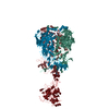 9clnMC 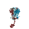 9cliC 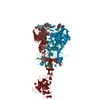 9clsC 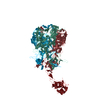 9cm2C 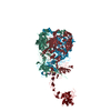 9cm9C 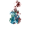 9cmoC M: atomic model generated by this map C: citing same article ( |
|---|---|
| Similar structure data |
-Links
| EMDB pages | EMDB (EBI/PDBe) / EMDataResource |
|---|---|
| Related items in Molecule of the Month |


-Map
| File | Download / File: emd_45726.map.gz / Format: CCP4 / Size: 4.2 MB / Type: IMAGE STORED AS FLOATING POINT NUMBER (4 BYTES) | ||||||||||||||||||||||||||||||||||||
|---|---|---|---|---|---|---|---|---|---|---|---|---|---|---|---|---|---|---|---|---|---|---|---|---|---|---|---|---|---|---|---|---|---|---|---|---|---|
| Projections & slices | Image control
Images are generated by Spider. generated in cubic-lattice coordinate | ||||||||||||||||||||||||||||||||||||
| Voxel size | X=Y=Z: 1.408 Å | ||||||||||||||||||||||||||||||||||||
| Density |
| ||||||||||||||||||||||||||||||||||||
| Symmetry | Space group: 1 | ||||||||||||||||||||||||||||||||||||
| Details | EMDB XML:
|
 Y (Sec.)
 X (Row.)
 Z (Col.)


-Supplemental data
-Half map: #2
| File | emd_45726_half_map_1.map | ||||||||||||
|---|---|---|---|---|---|---|---|---|---|---|---|---|---|
| Projections & Slices |
| ||||||||||||
| Density Histograms |


-Half map: #1
| File | emd_45726_half_map_2.map | ||||||||||||
|---|---|---|---|---|---|---|---|---|---|---|---|---|---|
| Projections & Slices |
| ||||||||||||
| Density Histograms |


- Sample components
-Entire : Human adenovirus 5 (Ad5) in complex with Coagulation factor FII
| Entire | Name: Human adenovirus 5 (Ad5) in complex with Coagulation factor FII |
|---|---|
| Components |
|
-Supramolecule #1: Human adenovirus 5 (Ad5) in complex with Coagulation factor FII
| Supramolecule | Name: Human adenovirus 5 (Ad5) in complex with Coagulation factor FII type: complex / ID: 1 / Parent: 0 / Macromolecule list: #1-#2 Details: Cryo-EM model derived from localized reconstruction |
|---|---|
| Source (natural) | Organism: Human adenovirus 5 |
| Molecular weight | Theoretical: 400 KDa |
-Supramolecule #2: Hexon protein
| Supramolecule | Name: Hexon protein / type: organelle_or_cellular_component / ID: 2 / Parent: 1 / Macromolecule list: #1 |
|---|---|
| Source (natural) | Organism: Human adenovirus 5 |
-Supramolecule #3: Human Coagulation Factor II
| Supramolecule | Name: Human Coagulation Factor II / type: organelle_or_cellular_component / ID: 3 / Parent: 2 / Macromolecule list: #2 |
|---|---|
| Source (natural) | Organism: Homo sapiens (human) |
-Macromolecule #1: Hexon protein
| Macromolecule | Name: Hexon protein / type: protein_or_peptide / ID: 1 / Number of copies: 3 / Enantiomer: LEVO |
|---|---|
| Source (natural) | Organism: Human adenovirus 5 |
| Molecular weight | Theoretical: 108.107617 KDa |
| Sequence | String: MATPSMMPQW SYMHISGQDA SEYLSPGLVQ FARATETYFS LNNKFRNPTV APTHDVTTDR SQRLTLRFIP VDREDTAYSY KARFTLAVG DNRVLDMAST YFDIRGVLDR GPTFKPYSGT AYNALAPKGA PNPCEWDEAA TALEINLEEE DDDNEDEVDE Q AEQQKTHV ...String: MATPSMMPQW SYMHISGQDA SEYLSPGLVQ FARATETYFS LNNKFRNPTV APTHDVTTDR SQRLTLRFIP VDREDTAYSY KARFTLAVG DNRVLDMAST YFDIRGVLDR GPTFKPYSGT AYNALAPKGA PNPCEWDEAA TALEINLEEE DDDNEDEVDE Q AEQQKTHV FGQAPYSGIN ITKEGIQIGV EGQTPKYADK TFQPEPQIGE SQWYETEINH AAGRVLKKTT PMKPCYGSYA KP TNENGGQ GILVKQQNGK LESQVEMQFF STTEATAGNG DNLTPKVVLY SEDVDIETPD THISYMPTIK EGNSRELMGQ QSM PNRPNY IAFRDNFIGL MYYNSTGNMG VLAGQASQLN AVVDLQDRNT ELSYQLLLDS IGDRTRYFSM WNQAVDSYDP DVRI IENHG TEDELPNYCF PLGGVINTET LTKVKPKTGQ ENGWEKDATE FSDKNEIRVG NNFAMEINLN ANLWRNFLYS NIALY LPDK LKYSPSNVKI SDNPNTYDYM NKRVVAPGLV DCYINLGARW SLDYMDNVNP FNHHRNAGLR YRSMLLGNGR YVPFHI QVP QKFFAIKNLL LLPGSYTYEW NFRKDVNMVL QSSLGNDLRV DGASIKFDSI CLYATFFPMA HNTASTLEAM LRNDTND QS FNDYLSAANM LYPIPANATN VPISIPSRNW AAFRGWAFTR LKTKETPSLG SGYDPYYTYS GSIPYLDGTF YLNHTFKK V AITFDSSVSW PGNDRLLTPN EFEIKRSVDG EGYNVAQCNM TKDWFLVQML ANYNIGYQGF YIPESYKDRM YSFFRNFQP MSRQVVDDTK YKDYQQVGIL HQHNNSGFVG YLAPTMREGQ AYPANFPYPL IGKTAVDSIT QKKFLCDRTL WRIPFSSNFM SMGALTDLG QNLLYANSAH ALDMTFEVDP MDEPTLLYVL FEVFDVVRVH RPHRGVIETV YLRTPFSAGN ATT UniProtKB: Hexon protein |
-Macromolecule #2: Prothrombin
| Macromolecule | Name: Prothrombin / type: protein_or_peptide / ID: 2 / Number of copies: 1 / Enantiomer: LEVO / EC number: thrombin |
|---|---|
| Source (natural) | Organism: Homo sapiens (human) |
| Molecular weight | Theoretical: 70.56293 KDa |
| Sequence | String: MAHVRGLQLP GCLALAALCS LVHSQHVFLA PQQARSLLQR VRRANTFL(CGU)(CGU) VRKGNL(CGU)R(CGU)C V (CGU)(CGU)TCSY(CGU)(CGU) AF(CGU)AL(CGU)SSTA TDVFWAKYTA CETARTPRDK LAACLEGNCA EGLGTNYR G HVNITRSGIE ...String: MAHVRGLQLP GCLALAALCS LVHSQHVFLA PQQARSLLQR VRRANTFL(CGU)(CGU) VRKGNL(CGU)R(CGU)C V (CGU)(CGU)TCSY(CGU)(CGU) AF(CGU)AL(CGU)SSTA TDVFWAKYTA CETARTPRDK LAACLEGNCA EGLGTNYR G HVNITRSGIE CQLWRSRYPH KPEINSTTHP GADLQENFCR NPDSSTTGPW CYTTDPTVRR QECSIPVCGQ DQVTVAMTP RSEGSSVNLS PPLEQCVPDR GQQYQGRLAV TTHGLPCLAW ASAQAKALSK HQDFNSAVQL VENFCRNPDG DEEGVWCYVA GKPGDFGYC DLNYCEEAVE EETGDGLDED SDRAIEGRTA TSEYQTFFNP RTFGSGEADC GLRPLFEKKS LEDKTERELL E SYIDGRIV EGSDAEIGMS PWQVMLFRKS PQELLCGASL ISDRWVLTAA HCLLYPPWDK NFTENDLLVR IGKHSRTRYE RN IEKISML EKIYIHPRYN WRENLDRDIA LMKLKKPVAF SDYIHPVCLP DRETAASLLQ AGYKGRVTGW GNLKETWTAN VGK GQPSVL QVVNLPIVER PVCKDSTRIR ITDNMFCAGY KPDEGKRGDA CEGDSGGPFV MKSPFNNRWY QMGIVSWGEG CDRD GKYGF YTHVFRLKKW IQKVIDQFGE UniProtKB: Prothrombin |
-Macromolecule #3: CALCIUM ION
| Macromolecule | Name: CALCIUM ION / type: ligand / ID: 3 / Number of copies: 7 / Formula: CA |
|---|---|
| Molecular weight | Theoretical: 40.078 Da |
-Experimental details
-Structure determination
| Method | cryo EM |
|---|---|
 Processing Processing | single particle reconstruction |
| Aggregation state | particle |
-Sample preparation
| Concentration | 2 mg/mL |
|---|---|
| Buffer | pH: 7.2 / Component - Concentration: 20.0 mM / Component - Name: Hepes |
| Vitrification | Cryogen name: ETHANE |
- Electron microscopy
| Microscope | FEI TITAN KRIOS |
|---|---|
| Image recording | Film or detector model: GATAN K3 BIOCONTINUUM (6k x 4k) / Number grids imaged: 3 / Number real images: 10000 / Average electron dose: 81.0 e/Å2 |
| Electron beam | Acceleration voltage: 300 kV / Electron source:  FIELD EMISSION GUN FIELD EMISSION GUN |
| Electron optics | C2 aperture diameter: 100.0 µm / Calibrated defocus max: 5.0 µm / Calibrated defocus min: 1.0 µm / Calibrated magnification: 81000 / Illumination mode: FLOOD BEAM / Imaging mode: BRIGHT FIELD / Cs: 2.7 mm / Nominal defocus max: 4.0 µm / Nominal defocus min: 2.5 µm / Nominal magnification: 81000 |
| Sample stage | Specimen holder model: FEI TITAN KRIOS AUTOGRID HOLDER / Cooling holder cryogen: NITROGEN |
| Experimental equipment |  Model: Titan Krios / Image courtesy: FEI Company |


