 Movie
Movie Controller

[English] 日本語
 Yorodumi
- EMDB-4364: Prespliceosome structure provides insight into spliceosome assemb... -
+ Open data

- Basic information
| Entry | Database: EMDB / ID: EMD-4364 | |||||||||
|---|---|---|---|---|---|---|---|---|---|---|
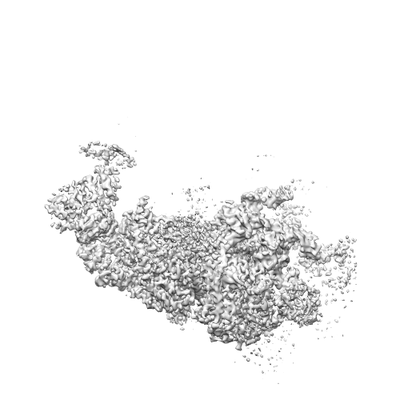
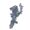
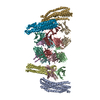
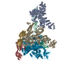
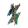
| Title | Prespliceosome structure provides insight into spliceosome assembly and regulation (map A2) | |||||||||
 Map data Map data | Sharpened A2 map | |||||||||
 Sample Sample |
| |||||||||
 Keywords Keywords | Spliceosome / A complex / RNA processing / RNA-protein complex / SPLICING | |||||||||
| Function / homology |  Function and homology information Function and homology informationmRNA splice site recognition / U4/U6 snRNP / 7-methylguanosine cap hypermethylation / pICln-Sm protein complex / positive regulation of mRNA splicing, via spliceosome / small nuclear ribonucleoprotein complex / SMN-Sm protein complex / spliceosomal tri-snRNP complex / splicing factor binding / snRNP binding ...mRNA splice site recognition / U4/U6 snRNP / 7-methylguanosine cap hypermethylation / pICln-Sm protein complex / positive regulation of mRNA splicing, via spliceosome / small nuclear ribonucleoprotein complex / SMN-Sm protein complex / spliceosomal tri-snRNP complex / splicing factor binding / snRNP binding / commitment complex / mRNA cis splicing, via spliceosome / U2-type prespliceosome assembly / U2-type spliceosomal complex / U1 snRNP / U2 snRNP / U4 snRNP / pre-mRNA 5'-splice site binding / U2-type prespliceosome / poly(U) RNA binding / precatalytic spliceosome / mRNA 5'-splice site recognition / spliceosomal complex assembly / Prp19 complex / U5 snRNP / spliceosomal snRNP assembly / U2 snRNA binding / U1 snRNA binding / U4/U6 x U5 tri-snRNP complex / catalytic step 2 spliceosome / spliceosomal complex / mRNA splicing, via spliceosome / response to xenobiotic stimulus / mRNA binding / RNA binding / zinc ion binding / nucleus / cytoplasm / cytosol Similarity search - Function | |||||||||
| Biological species |  | |||||||||
| Method | single particle reconstruction / cryo EM / Resolution: 4.0 Å | |||||||||
 Authors Authors | Plaschka C / Lin P-C | |||||||||
| Funding support |  United Kingdom, 2 items United Kingdom, 2 items
| |||||||||
 Citation Citation | Journal: Nature / Year: 2018 Title: Prespliceosome structure provides insights into spliceosome assembly and regulation. Authors: Clemens Plaschka / Pei-Chun Lin / Clément Charenton / Kiyoshi Nagai /  Abstract: The spliceosome catalyses the excision of introns from pre-mRNA in two steps, branching and exon ligation, and is assembled from five small nuclear ribonucleoprotein particles (snRNPs; U1, U2, U4, ...The spliceosome catalyses the excision of introns from pre-mRNA in two steps, branching and exon ligation, and is assembled from five small nuclear ribonucleoprotein particles (snRNPs; U1, U2, U4, U5, U6) and numerous non-snRNP factors. For branching, the intron 5' splice site and the branch point sequence are selected and brought by the U1 and U2 snRNPs into the prespliceosome, which is a focal point for regulation by alternative splicing factors. The U4/U6.U5 tri-snRNP subsequently joins the prespliceosome to form the complete pre-catalytic spliceosome. Recent studies have revealed the structural basis of the branching and exon-ligation reactions, however, the structural basis of the early events in spliceosome assembly remains poorly understood. Here we report the cryo-electron microscopy structure of the yeast Saccharomyces cerevisiae prespliceosome at near-atomic resolution. The structure reveals an induced stabilization of the 5' splice site in the U1 snRNP, and provides structural insights into the functions of the human alternative splicing factors LUC7-like (yeast Luc7) and TIA-1 (yeast Nam8), both of which have been linked to human disease. In the prespliceosome, the U1 snRNP associates with the U2 snRNP through a stable contact with the U2 3' domain and a transient yeast-specific contact with the U2 SF3b-containing 5' region, leaving its tri-snRNP-binding interface fully exposed. The results suggest mechanisms for 5' splice site transfer to the U6 ACAGAGA region within the assembled spliceosome and for its subsequent conversion to the activation-competent B-complex spliceosome. Taken together, the data provide a working model to investigate the early steps of spliceosome assembly. | |||||||||
| History |
|
- Structure visualization
| Movie |
Movie viewer |
|---|---|
| Structure viewer | EM map: SurfViewMolmilJmol/JSmol |
| Supplemental images |
- Downloads & links
-EMDB archive
| Map data | emd_4364.map.gz | 627.1 MB | EMDB map data format | |
|---|---|---|---|---|
| Header (meta data) | emd-4364-v30.xmlemd-4364.xml | 51.4 KB 51.4 KB | Display Display | EMDB header |
| Images |  emd_4364.png emd_4364.png | 34.8 KB | ||
| Filedesc metadata | emd-4364.cif.gz | 13.8 KB | ||
| Others | emd_4364_additional.map.gz | 613.8 MB | ||
| Archive directory |  http://ftp.pdbj.org/pub/emdb/structures/EMD-4364ftp://ftp.pdbj.org/pub/emdb/structures/EMD-4364 http://ftp.pdbj.org/pub/emdb/structures/EMD-4364ftp://ftp.pdbj.org/pub/emdb/structures/EMD-4364 | HTTPS FTP |
-Related structure data
| Related structure data |  6g90MC  4363C  4365C M: atomic model generated by this map C: citing same article ( |
|---|---|
| Similar structure data |


-Links
| EMDB pages | EMDB (EBI/PDBe) / EMDataResource |
|---|---|
| Related items in Molecule of the Month |


-Map
| File | Download / File: emd_4364.map.gz / Format: CCP4 / Size: 669.9 MB / Type: IMAGE STORED AS FLOATING POINT NUMBER (4 BYTES) | ||||||||||||||||||||||||||||||||||||||||||||||||||||||||||||
|---|---|---|---|---|---|---|---|---|---|---|---|---|---|---|---|---|---|---|---|---|---|---|---|---|---|---|---|---|---|---|---|---|---|---|---|---|---|---|---|---|---|---|---|---|---|---|---|---|---|---|---|---|---|---|---|---|---|---|---|---|---|
| Annotation | Sharpened A2 map | ||||||||||||||||||||||||||||||||||||||||||||||||||||||||||||
| Projections & slices | Image control
Images are generated by Spider. | ||||||||||||||||||||||||||||||||||||||||||||||||||||||||||||
| Voxel size | X=Y=Z: 1.13 Å | ||||||||||||||||||||||||||||||||||||||||||||||||||||||||||||
| Density |
| ||||||||||||||||||||||||||||||||||||||||||||||||||||||||||||
| Symmetry | Space group: 1 | ||||||||||||||||||||||||||||||||||||||||||||||||||||||||||||
| Details | EMDB XML:
CCP4 map header:
| ||||||||||||||||||||||||||||||||||||||||||||||||||||||||||||
 Z (Sec.)
 Y (Row.)
 X (Col.)


-Supplemental data
-Additional map: Unsharpened A2 map
| File | emd_4364_additional.map | ||||||||||||
|---|---|---|---|---|---|---|---|---|---|---|---|---|---|
| Annotation | Unsharpened A2 map | ||||||||||||
| Projections & Slices |
| ||||||||||||
| Density Histograms |


- Sample components
+Entire : Prespliceosome A complex
+Supramolecule #1: Prespliceosome A complex
+Macromolecule #1: U1 snRNA,U1 snRNA,U1 snRNA,U1 snRNA,U1 snRNA
+Macromolecule #2: U2 snRNA
+Macromolecule #11: Yeast UBC4 pre-mRNA (mutant)
+Macromolecule #3: U1 small nuclear ribonucleoprotein A,U1 small nuclear ribonucleop...
+Macromolecule #4: U1 small nuclear ribonucleoprotein 70 kDa homolog,U1 small nuclea...
+Macromolecule #5: U1 small nuclear ribonucleoprotein C
+Macromolecule #6: Pre-mRNA-processing factor 39
+Macromolecule #7: U1 small nuclear ribonucleoprotein component PRP42
+Macromolecule #8: Protein NAM8
+Macromolecule #9: 56 kDa U1 small nuclear ribonucleoprotein component
+Macromolecule #10: Protein LUC7,Protein LUC7,Protein LUC7
+Macromolecule #12: U1 small nuclear ribonucleoprotein component SNU71
+Macromolecule #13: U2 snRNP component HSH155
+Macromolecule #14: Pre-mRNA-splicing factor RSE1
+Macromolecule #15: Cold sensitive U2 snRNA suppressor 1
+Macromolecule #16: Protein HSH49
+Macromolecule #17: Pre-mRNA-splicing factor RDS3
+Macromolecule #18: Pre-mRNA-splicing factor PRP9
+Macromolecule #19: Pre-mRNA-splicing factor PRP11,Pre-mRNA-splicing factor PRP11,Pre...
+Macromolecule #20: Pre-mRNA-splicing factor PRP21
+Macromolecule #21: U2 small nuclear ribonucleoprotein A'
+Macromolecule #22: Unknown
+Macromolecule #23: U2 small nuclear ribonucleoprotein B''
+Macromolecule #24: RDS3 complex subunit 10
+Macromolecule #25: Small nuclear ribonucleoprotein-associated protein B
+Macromolecule #26: Small nuclear ribonucleoprotein Sm D3
+Macromolecule #27: Small nuclear ribonucleoprotein E
+Macromolecule #28: Small nuclear ribonucleoprotein F
+Macromolecule #29: Small nuclear ribonucleoprotein G
+Macromolecule #30: Small nuclear ribonucleoprotein Sm D1
+Macromolecule #31: Small nuclear ribonucleoprotein Sm D2
+Macromolecule #32: ZINC ION
-Experimental details
-Structure determination
| Method | cryo EM |
|---|---|
 Processing Processing | single particle reconstruction |
| Aggregation state | particle |
-Sample preparation
| Concentration | 0.4 mg/mL | |||||||||||||||||||||
|---|---|---|---|---|---|---|---|---|---|---|---|---|---|---|---|---|---|---|---|---|---|---|
| Buffer | pH: 7.9 Component:
| |||||||||||||||||||||
| Grid | Model: Quantifoil R2/2 / Material: COPPER / Mesh: 400 / Support film - Material: CARBON / Support film - topology: HOLEY / Pretreatment - Type: GLOW DISCHARGE / Pretreatment - Time: 20 sec. | |||||||||||||||||||||
| Vitrification | Cryogen name: ETHANE / Chamber humidity: 100 % / Instrument: FEI VITROBOT MARK III Details: Grids were blotted for 2-3.5 s and vitrified by plunging into liquid ethane with a FEI Vitrobot Mark III operated at 4 degree Celsius and 100% humidity.. |
- Electron microscopy
| Microscope | FEI TITAN KRIOS |
|---|---|
| Image recording | Film or detector model: GATAN K2 SUMMIT (4k x 4k) / Detector mode: COUNTING / Number real images: 9407 / Average electron dose: 43.0 e/Å2 |
| Electron beam | Acceleration voltage: 300 kV / Electron source:  FIELD EMISSION GUN FIELD EMISSION GUN |
| Electron optics | Illumination mode: FLOOD BEAM / Imaging mode: BRIGHT FIELD / Cs: 2.7 mm / Nominal magnification: 105000 |
| Sample stage | Specimen holder model: FEI TITAN KRIOS AUTOGRID HOLDER / Cooling holder cryogen: NITROGEN |
| Experimental equipment |  Model: Titan Krios / Image courtesy: FEI Company |
-Image processing
| Startup model | Type of model: OTHER Details: The initial model was generated from 22319 particles using cryoSPARC. |
|---|---|
| Final reconstruction | Applied symmetry - Point group: C1 (asymmetric) / Resolution.type: BY AUTHOR / Resolution: 4.0 Å / Resolution method: FSC 0.143 CUT-OFF / Software - Name: RELION (ver. 2.1) / Number images used: 153556 |
| Initial angle assignment | Type: PROJECTION MATCHING |
| Final angle assignment | Type: PROJECTION MATCHING / Software - Name: RELION (ver. 2.1) |
-Atomic model buiding 1
| Refinement | Space: REAL |
|---|---|
| Output model | PDB-6g90: |
