 Movie
Movie Controller


+ Open data

- Basic information
| Entry |  | |||||||||
|---|---|---|---|---|---|---|---|---|---|---|
| Title | Cryo-EM structure of fascin crosslinked F-actin | |||||||||
 Map data Map data | Composite map used for model building and refinement | |||||||||
 Sample Sample |
| |||||||||
 Keywords Keywords | Cytoskeleton / F-actin crosslinker / F-actin bundle / STRUCTURAL PROTEIN | |||||||||
| Function / homology |  Function and homology information Function and homology informationmicrospike / parallel actin filament bundle assembly / regulation of microvillus assembly / microspike assembly / establishment of apical/basal cell polarity / positive regulation of extracellular matrix disassembly / positive regulation of podosome assembly / cell projection membrane / cell-cell junction assembly / podosome ...microspike / parallel actin filament bundle assembly / regulation of microvillus assembly / microspike assembly / establishment of apical/basal cell polarity / positive regulation of extracellular matrix disassembly / positive regulation of podosome assembly / cell projection membrane / cell-cell junction assembly / podosome / positive regulation of filopodium assembly / microvillus / establishment or maintenance of cell polarity / striated muscle thin filament / actin filament bundle assembly / skeletal muscle thin filament assembly / positive regulation of lamellipodium assembly / skeletal muscle fiber development / ruffle / stress fiber / regulation of actin cytoskeleton organization / cell motility / actin filament / filopodium / Hydrolases; Acting on acid anhydrides; Acting on acid anhydrides to facilitate cellular and subcellular movement / structural constituent of cytoskeleton / cell-cell junction / actin filament binding / actin cytoskeleton / cell migration / lamellipodium / growth cone / actin cytoskeleton organization / actin binding / Interleukin-4 and Interleukin-13 signaling / cell cortex / cytoskeleton / protein-macromolecule adaptor activity / cadherin binding / hydrolase activity / RNA binding / extracellular exosome / ATP binding / cytosol / cytoplasm Similarity search - Function | |||||||||
| Biological species |  Homo sapiens (human) / Homo sapiens (human) /  | |||||||||
| Method | single particle reconstruction / cryo EM / Resolution: 3.1 Å | |||||||||
 Authors Authors | Gong R / Reynolds MJ / Alushin GM | |||||||||
| Funding support |  United States, 2 items United States, 2 items
| |||||||||
 Citation Citation | Journal: Nat Struct Mol Biol / Year: 2025 Title: Fascin structural plasticity mediates flexible actin bundle construction. Authors: Rui Gong / Matthew J Reynolds / Keith R Carney / Keith Hamilton / Tamara C Bidone / Gregory M Alushin / Abstract: Fascin cross-links actin filaments (F-actin) into bundles that support tubular membrane protrusions including filopodia and stereocilia. Fascin dysregulation drives aberrant cell migration during ...Fascin cross-links actin filaments (F-actin) into bundles that support tubular membrane protrusions including filopodia and stereocilia. Fascin dysregulation drives aberrant cell migration during metastasis, and fascin inhibitors are under development as cancer therapeutics. Here, we use cryo-EM, cryo-electron tomography coupled with custom denoising and computational modeling to probe human fascin-1's F-actin cross-linking mechanisms across spatial scales. Our fascin cross-bridge structure reveals an asymmetric F-actin binding conformation that is allosterically blocked by the inhibitor G2. Reconstructions of seven-filament hexagonal bundle elements, variability analysis and simulations show how structural plasticity enables fascin to bridge varied interfilament orientations, accommodating mismatches between F-actin's helical symmetry and bundle hexagonal packing. Tomography of many-filament bundles and modeling uncover geometric rules underlying emergent fascin binding patterns, as well as the accumulation of unfavorable cross-links that limit bundle size. Collectively, this work shows how fascin harnesses fine-tuned nanoscale structural dynamics to build and regulate micron-scale F-actin bundles. | |||||||||
| History |
|
- Structure visualization
| Supplemental images |
|---|

- Downloads & links
-EMDB archive
| Map data | emd_43366.map.gz | 2.1 MB | EMDB map data format | |
|---|---|---|---|---|
| Header (meta data) | emd-43366-v30.xmlemd-43366.xml | 14.6 KB 14.6 KB | Display Display | EMDB header |
| Images |  emd_43366.png emd_43366.png | 110.6 KB | ||
| Filedesc metadata | emd-43366.cif.gz | 6.5 KB | ||
| Archive directory |  http://ftp.pdbj.org/pub/emdb/structures/EMD-43366ftp://ftp.pdbj.org/pub/emdb/structures/EMD-43366 http://ftp.pdbj.org/pub/emdb/structures/EMD-43366ftp://ftp.pdbj.org/pub/emdb/structures/EMD-43366 | HTTPS FTP |
-Related structure data
| Related structure data | 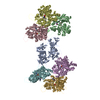 8vo7MC  8vo5C  8vo6C  8vo8C  8vo9C  8voaC C: citing same article ( M: atomic model generated by this map |
|---|---|
| Similar structure data |


-Links
| EMDB pages | EMDB (EBI/PDBe) / EMDataResource |
|---|---|
| Related items in Molecule of the Month |


-Map
| File | Download / File: emd_43366.map.gz / Format: CCP4 / Size: 343 MB / Type: IMAGE STORED AS FLOATING POINT NUMBER (4 BYTES) | ||||||||||||||||||||||||||||||||||||
|---|---|---|---|---|---|---|---|---|---|---|---|---|---|---|---|---|---|---|---|---|---|---|---|---|---|---|---|---|---|---|---|---|---|---|---|---|---|
| Annotation | Composite map used for model building and refinement | ||||||||||||||||||||||||||||||||||||
| Projections & slices | Image control
Images are generated by Spider. | ||||||||||||||||||||||||||||||||||||
| Voxel size | X=Y=Z: 1.03 Å | ||||||||||||||||||||||||||||||||||||
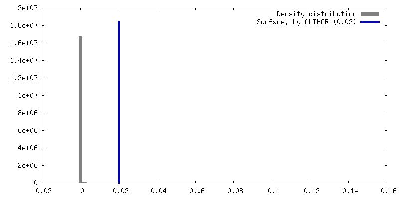
| Density |
| ||||||||||||||||||||||||||||||||||||
| Symmetry | Space group: 1 | ||||||||||||||||||||||||||||||||||||
| Details | EMDB XML:
|
 Z (Sec.)
 Y (Row.)
 X (Col.)


-Supplemental data
- Sample components
-Entire : Fascin crosslinked F-actin
| Entire | Name: Fascin crosslinked F-actin |
|---|---|
| Components |
|
-Supramolecule #1: Fascin crosslinked F-actin
| Supramolecule | Name: Fascin crosslinked F-actin / type: complex / ID: 1 / Parent: 0 / Macromolecule list: #1-#2 |
|---|---|
| Source (natural) | Organism: Homo sapiens (human) |
| Molecular weight | Theoretical: 5.4 kDa/nm |
-Macromolecule #1: Fascin
| Macromolecule | Name: Fascin / type: protein_or_peptide / ID: 1 / Number of copies: 1 / Enantiomer: LEVO |
|---|---|
| Source (natural) | Organism: Homo sapiens (human) |
| Molecular weight | Theoretical: 55.013328 KDa |
| Recombinant expression | Organism:  |
| Sequence | String: GPLGSMTANG TAEAVQIQFG LINCGNKYLT AEAFGFKVNA SASSLKKKQI WTLEQPPDEA GSAAVCLRSH LGRYLAADKD GNVTCEREV PGPDCRFLIV AHDDGRWSLQ SEAHRRYFGG TEDRLSCFAQ TVSPAEKWSV HIAMHPQVNI YSVTRKRYAH L SARPADEI ...String: GPLGSMTANG TAEAVQIQFG LINCGNKYLT AEAFGFKVNA SASSLKKKQI WTLEQPPDEA GSAAVCLRSH LGRYLAADKD GNVTCEREV PGPDCRFLIV AHDDGRWSLQ SEAHRRYFGG TEDRLSCFAQ TVSPAEKWSV HIAMHPQVNI YSVTRKRYAH L SARPADEI AVDRDVPWGV DSLITLAFQD QRYSVQTADH RFLRHDGRLV ARPEPATGYT LEFRSGKVAF RDCEGRYLAP SG PSGTLKA GKATKVGKDE LFALEQSCAQ VVLQAANERN VSTRQGMDLS ANQDEETDQE TFQLEIDRDT KKCAFRTHTG KYW TLTATG GVQSTASSKN ASCYFDIEWR DRRITLRASN GKFVTSKKNG QLAASVETAG DSELFLMKLI NRPIIVFRGE HGFI GCRKV TGTLDANRSS YDVFQLEFND GAYNIKDSTG KYWTVGSDSA VTSSGDTPVD FFFEFCDYNK VAIKVGGRYL KGDHA GVLK ASAETVDPAS LWEY UniProtKB: Fascin |
-Macromolecule #2: Actin, alpha skeletal muscle
| Macromolecule | Name: Actin, alpha skeletal muscle / type: protein_or_peptide / ID: 2 / Number of copies: 6 / Enantiomer: LEVO |
|---|---|
| Source (natural) | Organism: |
| Molecular weight | Theoretical: 41.63143 KDa |
| Sequence | String: DETTALVCDN GSGLVKAGFA GDDAPRAVFP SIVGRPRHQG VMVGMGQKDS YVGDEAQSKR GILTLKYPIE (HIC)GIITN WDD MEKIWHHTFY NELRVAPEEH PTLLTEAPLN PKANREKMTQ IMFETFNVPA MYVAIQAVLS LYASGRTTGI VLDSGDG VT HNVPIYEGYA ...String: DETTALVCDN GSGLVKAGFA GDDAPRAVFP SIVGRPRHQG VMVGMGQKDS YVGDEAQSKR GILTLKYPIE (HIC)GIITN WDD MEKIWHHTFY NELRVAPEEH PTLLTEAPLN PKANREKMTQ IMFETFNVPA MYVAIQAVLS LYASGRTTGI VLDSGDG VT HNVPIYEGYA LPHAIMRLDL AGRDLTDYLM KILTERGYSF VTTAEREIVR DIKEKLCYVA LDFENEMATA ASSSSLEK S YELPDGQVIT IGNERFRCPE TLFQPSFIGM ESAGIHETTY NSIMKCDIDI RKDLYANNVM SGGTTMYPGI ADRMQKEIT ALAPSTMKIK IIAPPERKYS VWIGGSILAS LSTFQQMWIT KQEYDEAGPS IVHRKCF UniProtKB: Actin, alpha skeletal muscle |
-Macromolecule #3: ADENOSINE-5'-DIPHOSPHATE
| Macromolecule | Name: ADENOSINE-5'-DIPHOSPHATE / type: ligand / ID: 3 / Number of copies: 6 / Formula: ADP |
|---|---|
| Molecular weight | Theoretical: 427.201 Da |
| Chemical component information |  ChemComp-ADP: |
-Macromolecule #4: MAGNESIUM ION
| Macromolecule | Name: MAGNESIUM ION / type: ligand / ID: 4 / Number of copies: 6 / Formula: MG |
|---|---|
| Molecular weight | Theoretical: 24.305 Da |
-Experimental details
-Structure determination
| Method | cryo EM |
|---|---|
 Processing Processing | single particle reconstruction |
| Aggregation state | filament |
-Sample preparation
| Buffer | pH: 8 |
|---|---|
| Vitrification | Cryogen name: ETHANE |
- Electron microscopy
| Microscope | FEI TITAN KRIOS |
|---|---|
| Image recording | Film or detector model: GATAN K2 SUMMIT (4k x 4k) / Average electron dose: 61.26 e/Å2 |
| Electron beam | Acceleration voltage: 300 kV / Electron source:  FIELD EMISSION GUN FIELD EMISSION GUN |
| Electron optics | Illumination mode: FLOOD BEAM / Imaging mode: BRIGHT FIELD / Nominal defocus max: 2.8000000000000003 µm / Nominal defocus min: 0.8 µm |
| Experimental equipment |  Model: Titan Krios / Image courtesy: FEI Company |

