 Movie
Movie Controller


+ Open data

- Basic information
| Entry |  | |||||||||
|---|---|---|---|---|---|---|---|---|---|---|
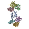
| Title | Subtomogram averaging structure of fascin bound to F-actin | |||||||||
 Map data Map data | Local resolution filtered main map | |||||||||
 Sample Sample |
| |||||||||
 Keywords Keywords | Cytoskeleton / F-actin crosslinker / F-actin bundle / STRUCTURAL PROTEIN | |||||||||
| Biological species |  Homo sapiens (human) / Homo sapiens (human) /  | |||||||||
| Method | subtomogram averaging / cryo EM / Resolution: 6.7 Å | |||||||||
 Authors Authors | Gong R / Reynolds MJ / Alushin GM | |||||||||
| Funding support |  United States, 2 items United States, 2 items
| |||||||||
 Citation Citation | Journal: Nat Struct Mol Biol / Year: 2025 Title: Fascin structural plasticity mediates flexible actin bundle construction. Authors: Rui Gong / Matthew J Reynolds / Keith R Carney / Keith Hamilton / Tamara C Bidone / Gregory M Alushin / Abstract: Fascin cross-links actin filaments (F-actin) into bundles that support tubular membrane protrusions including filopodia and stereocilia. Fascin dysregulation drives aberrant cell migration during ...Fascin cross-links actin filaments (F-actin) into bundles that support tubular membrane protrusions including filopodia and stereocilia. Fascin dysregulation drives aberrant cell migration during metastasis, and fascin inhibitors are under development as cancer therapeutics. Here, we use cryo-EM, cryo-electron tomography coupled with custom denoising and computational modeling to probe human fascin-1's F-actin cross-linking mechanisms across spatial scales. Our fascin cross-bridge structure reveals an asymmetric F-actin binding conformation that is allosterically blocked by the inhibitor G2. Reconstructions of seven-filament hexagonal bundle elements, variability analysis and simulations show how structural plasticity enables fascin to bridge varied interfilament orientations, accommodating mismatches between F-actin's helical symmetry and bundle hexagonal packing. Tomography of many-filament bundles and modeling uncover geometric rules underlying emergent fascin binding patterns, as well as the accumulation of unfavorable cross-links that limit bundle size. Collectively, this work shows how fascin harnesses fine-tuned nanoscale structural dynamics to build and regulate micron-scale F-actin bundles. | |||||||||
| History |
|
- Structure visualization
| Supplemental images |
|---|

- Downloads & links
-EMDB archive
| Map data | emd_43372.map.gz | 15.7 MB |  EMDB map data format EMDB map data format | |
|---|---|---|---|---|
| Header (meta data) | emd-43372-v30.xmlemd-43372.xml | 21.4 KB 21.4 KB | Display Display | EMDB header |
| FSC (resolution estimation) | emd_43372_fsc.xml | 6.9 KB | Display | FSC data file |
| Images |  emd_43372.png emd_43372.png | 91.8 KB | ||
| Masks | emd_43372_msk_1.map | 27 MB | Mask map | |
| Filedesc metadata | emd-43372.cif.gz | 5.8 KB | ||
| Others | emd_43372_additional_1.map.gzemd_43372_additional_2.map.gzemd_43372_additional_3.map.gzemd_43372_half_map_1.map.gzemd_43372_half_map_2.map.gz | 25.4 MB 1.7 MB 1.7 MB 13.3 MB 13.3 MB | ||
| Archive directory |  http://ftp.pdbj.org/pub/emdb/structures/EMD-43372ftp://ftp.pdbj.org/pub/emdb/structures/EMD-43372 http://ftp.pdbj.org/pub/emdb/structures/EMD-43372ftp://ftp.pdbj.org/pub/emdb/structures/EMD-43372 | HTTPS FTP |
-Related structure data


-Links
| EMDB pages | EMDB (EBI/PDBe) / EMDataResource |
|---|
-Map
| File | Download / File: emd_43372.map.gz / Format: CCP4 / Size: 27 MB / Type: IMAGE STORED AS FLOATING POINT NUMBER (4 BYTES) | ||||||||||||||||||||||||||||||||||||
|---|---|---|---|---|---|---|---|---|---|---|---|---|---|---|---|---|---|---|---|---|---|---|---|---|---|---|---|---|---|---|---|---|---|---|---|---|---|
| Annotation | Local resolution filtered main map | ||||||||||||||||||||||||||||||||||||
| Projections & slices | Image control
Images are generated by Spider. | ||||||||||||||||||||||||||||||||||||
| Voxel size | X=Y=Z: 2.6 Å | ||||||||||||||||||||||||||||||||||||
| Density |
| ||||||||||||||||||||||||||||||||||||
| Symmetry | Space group: 1 | ||||||||||||||||||||||||||||||||||||
| Details | EMDB XML:
|
 Z (Sec.)
 Y (Row.)
 X (Col.)


-Supplemental data
-Mask #1
| File | emd_43372_msk_1.map | ||||||||||||
|---|---|---|---|---|---|---|---|---|---|---|---|---|---|
| Projections & Slices |
| ||||||||||||
| Density Histograms |


-Additional map: Sharpened, postprocessed map
| File | emd_43372_additional_1.map | ||||||||||||
|---|---|---|---|---|---|---|---|---|---|---|---|---|---|
| Annotation | Sharpened, postprocessed map | ||||||||||||
| Projections & Slices |
| ||||||||||||
| Density Histograms |


-Additional map: Multibody refinement body 2. Postprocessed, masked, sharpened map
| File | emd_43372_additional_2.map | ||||||||||||
|---|---|---|---|---|---|---|---|---|---|---|---|---|---|
| Annotation | Multibody refinement body 2. Postprocessed, masked, sharpened map | ||||||||||||
| Projections & Slices |
| ||||||||||||
| Density Histograms |


-Additional map: Multibody refinement body 1. Postprocessed, masked, sharpened map
| File | emd_43372_additional_3.map | ||||||||||||
|---|---|---|---|---|---|---|---|---|---|---|---|---|---|
| Annotation | Multibody refinement body 1. Postprocessed, masked, sharpened map | ||||||||||||
| Projections & Slices |
| ||||||||||||
| Density Histograms |


-Half map: Half map 2
| File | emd_43372_half_map_1.map | ||||||||||||
|---|---|---|---|---|---|---|---|---|---|---|---|---|---|
| Annotation | Half map 2 | ||||||||||||
| Projections & Slices |
| ||||||||||||
| Density Histograms |


-Half map: Half map 1
| File | emd_43372_half_map_2.map | ||||||||||||
|---|---|---|---|---|---|---|---|---|---|---|---|---|---|
| Annotation | Half map 1 | ||||||||||||
| Projections & Slices |
| ||||||||||||
| Density Histograms |


- Sample components
-Entire : Fascin crosslinked F-actin
| Entire | Name: Fascin crosslinked F-actin |
|---|---|
| Components |
|
-Supramolecule #1: Fascin crosslinked F-actin
| Supramolecule | Name: Fascin crosslinked F-actin / type: complex / ID: 1 / Parent: 0 / Macromolecule list: all |
|---|---|
| Source (natural) | Organism: Homo sapiens (human) |
| Molecular weight | Theoretical: 5.4 kDa/nm |
-Macromolecule #1: Fascin 1
| Macromolecule | Name: Fascin 1 / type: protein_or_peptide / ID: 1 / Enantiomer: LEVO |
|---|---|
| Source (natural) | Organism: Homo sapiens (human) |
| Recombinant expression | Organism:  |
| Sequence | String: GPLGSMTANG TAEAVQIQFG LINCGNKYLT AEAFGFKVNA SASSLKKKQI WTLEQPPDEA GSAAVCLRSH LGRYLAADKD GNVTCEREVP GPDCRFLIVA HDDGRWSLQS EAHRRYFGGT EDRLSCFAQT VSPAEKWSVH IAMHPQVNIY SVTRKRYAHL SARPADEIAV ...String: GPLGSMTANG TAEAVQIQFG LINCGNKYLT AEAFGFKVNA SASSLKKKQI WTLEQPPDEA GSAAVCLRSH LGRYLAADKD GNVTCEREVP GPDCRFLIVA HDDGRWSLQS EAHRRYFGGT EDRLSCFAQT VSPAEKWSVH IAMHPQVNIY SVTRKRYAHL SARPADEIAV DRDVPWGVDS LITLAFQDQR YSVQTADHRF LRHDGRLVAR PEPATGYTLE FRSGKVAFRD CEGRYLAPSG PSGTLKAGKA TKVGKDELFA LEQSCAQVVL QAANERNVST RQGMDLSANQ DEETDQETFQ LEIDRDTKKC AFRTHTGKYW TLTATGGVQS TASSKNASCY FDIEWRDRRI TLRASNGKFV TSKKNGQLAA SVETAGDSEL FLMKLINRPI IVFRGEHGFI GCRKVTGTLD ANRSSYDVFQ LEFNDGAYNI KDSTGKYWTV GSDSAVTSSG DTPVDFFFEF CDYNKVAIKV GGRYLKGDHA GVLKASAETV DPASLWEY |
-Macromolecule #2: Alpha actin
| Macromolecule | Name: Alpha actin / type: protein_or_peptide / ID: 2 / Enantiomer: LEVO |
|---|---|
| Source (natural) | Organism: |
| Sequence | String: DETTALVCDN GSGLVKAGFA GDDAPRAVFP SIVGRPRHQG VMVGMGQKDS YVGDEAQSKR GILTLKYPIE (HIC)GIITNWDDM EKIWHHTFYN ELRVAPEEHP TLLTEAPLNP KANREKMTQI MFETFNVPAM YVAIQAVLSL YASGRTTGIV LDSGDGVTHN VPIYEGYALP ...String: DETTALVCDN GSGLVKAGFA GDDAPRAVFP SIVGRPRHQG VMVGMGQKDS YVGDEAQSKR GILTLKYPIE (HIC)GIITNWDDM EKIWHHTFYN ELRVAPEEHP TLLTEAPLNP KANREKMTQI MFETFNVPAM YVAIQAVLSL YASGRTTGIV LDSGDGVTHN VPIYEGYALP HAIMRLDLAG RDLTDYLMKI LTERGYSFVT TAEREIVRDI KEKLCYVALD FENEMATAAS SSSLEKSYEL PDGQVITIGN ERFRCPETLF QPSFIGMESA GIHETTYNSI MKCDIDIRKD LYANNVMSGG TTMYPGIADR MQKEITALAP STMKIKIIAP PERKYSVWIG GSILASLSTF QQMWITKQEY DEAGPSIVHR KCF |
-Experimental details
-Structure determination
| Method | cryo EM |
|---|---|
 Processing Processing | subtomogram averaging |
| Aggregation state | filament |
-Sample preparation
| Buffer | pH: 8 |
|---|---|
| Vitrification | Cryogen name: ETHANE |
- Electron microscopy
| Microscope | FEI TITAN KRIOS |
|---|---|
| Image recording | Film or detector model: GATAN K3 BIOQUANTUM (6k x 4k) / Average electron dose: 2.64 e/Å2 |
| Electron beam | Acceleration voltage: 300 kV / Electron source:  FIELD EMISSION GUN FIELD EMISSION GUN |
| Electron optics | Illumination mode: FLOOD BEAM / Imaging mode: BRIGHT FIELD / Nominal defocus max: 4.0 µm / Nominal defocus min: 4.0 µm / Nominal magnification: 26000 |
| Experimental equipment |  Model: Titan Krios / Image courtesy: FEI Company |

