 Movie
Movie Controller


+ Open data

- Basic information
| Entry | Database: PDB / ID: 8vo8 | |||||||||
|---|---|---|---|---|---|---|---|---|---|---|
| Title | Cryo-EM structure of fascin crosslinked F-actin (Eigen_left) | |||||||||
 Components Components |
| |||||||||
 Keywords Keywords | STRUCTURAL PROTEIN / Cytoskeleton / F-actin crosslinker / F-actin bundle | |||||||||
| Function / homology |  Function and homology information Function and homology informationmicrospike / parallel actin filament bundle assembly / regulation of microvillus assembly / microspike assembly / establishment of apical/basal cell polarity / positive regulation of extracellular matrix disassembly / positive regulation of podosome assembly / cell projection membrane / cell-cell junction assembly / podosome ...microspike / parallel actin filament bundle assembly / regulation of microvillus assembly / microspike assembly / establishment of apical/basal cell polarity / positive regulation of extracellular matrix disassembly / positive regulation of podosome assembly / cell projection membrane / cell-cell junction assembly / podosome / positive regulation of filopodium assembly / microvillus / establishment or maintenance of cell polarity / striated muscle thin filament / actin filament bundle assembly / skeletal muscle thin filament assembly / positive regulation of lamellipodium assembly / skeletal muscle fiber development / ruffle / stress fiber / regulation of actin cytoskeleton organization / cell motility / actin filament / filopodium / Hydrolases; Acting on acid anhydrides; Acting on acid anhydrides to facilitate cellular and subcellular movement / structural constituent of cytoskeleton / cell-cell junction / actin filament binding / actin cytoskeleton / cell migration / lamellipodium / growth cone / actin cytoskeleton organization / actin binding / Interleukin-4 and Interleukin-13 signaling / cell cortex / cytoskeleton / protein-macromolecule adaptor activity / cadherin binding / hydrolase activity / RNA binding / extracellular exosome / ATP binding / cytosol / cytoplasm Similarity search - Function | |||||||||
| Biological species |  Homo sapiens (human) Homo sapiens (human) | |||||||||
| Method | ELECTRON MICROSCOPY / single particle reconstruction / cryo EM / Resolution: 3.9 Å | |||||||||
 Authors Authors | Gong, R. / Reynolds, M.J. / Alushin, G.M. | |||||||||
| Funding support |  United States, 2items United States, 2items
| |||||||||
 Citation Citation | Journal: Nat Struct Mol Biol / Year: 2025 Title: Fascin structural plasticity mediates flexible actin bundle construction. Authors: Rui Gong / Matthew J Reynolds / Keith R Carney / Keith Hamilton / Tamara C Bidone / Gregory M Alushin / Abstract: Fascin cross-links actin filaments (F-actin) into bundles that support tubular membrane protrusions including filopodia and stereocilia. Fascin dysregulation drives aberrant cell migration during ...Fascin cross-links actin filaments (F-actin) into bundles that support tubular membrane protrusions including filopodia and stereocilia. Fascin dysregulation drives aberrant cell migration during metastasis, and fascin inhibitors are under development as cancer therapeutics. Here, we use cryo-EM, cryo-electron tomography coupled with custom denoising and computational modeling to probe human fascin-1's F-actin cross-linking mechanisms across spatial scales. Our fascin cross-bridge structure reveals an asymmetric F-actin binding conformation that is allosterically blocked by the inhibitor G2. Reconstructions of seven-filament hexagonal bundle elements, variability analysis and simulations show how structural plasticity enables fascin to bridge varied interfilament orientations, accommodating mismatches between F-actin's helical symmetry and bundle hexagonal packing. Tomography of many-filament bundles and modeling uncover geometric rules underlying emergent fascin binding patterns, as well as the accumulation of unfavorable cross-links that limit bundle size. Collectively, this work shows how fascin harnesses fine-tuned nanoscale structural dynamics to build and regulate micron-scale F-actin bundles. | |||||||||
| History |
|
- Structure visualization
| Structure viewer | Molecule: MolmilJmol/JSmol |
|---|
- Downloads & links
-Download
| PDBx/mmCIF format | 8vo8.cif.gz | 470.3 KB | Display | PDBx/mmCIF format |
|---|---|---|---|---|
| PDB format | pdb8vo8.ent.gz | 388.6 KB | Display | PDB format |
| PDBx/mmJSON format | 8vo8.json.gz | Tree view | PDBx/mmJSON format | |
| Others |  Other downloads Other downloads |
-Validation report
| Arichive directory | https://data.pdbj.org/pub/pdb/validation_reports/vo/8vo8ftp://data.pdbj.org/pub/pdb/validation_reports/vo/8vo8 | HTTPS FTP |
|---|
-Related structure data
| Related structure data |  43367MC  8vo5C  8vo6C 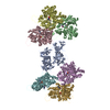 8vo7C  8vo9C  8voaC M: map data used to model this data C: citing same article ( |
|---|---|
| Similar structure data |


-Links
 PDBj
PDBj


- Assembly
| Deposited unit | 
|
|---|---|
| 1 |
|
-Components
| #1: Protein | Mass: 55013.328 Da / Num. of mol.: 1 Source method: isolated from a genetically manipulated source Source: (gene. exp.) Homo sapiens (human) / Gene: FSCN1, FAN1, HSN, SNL / Production host:  | ||||||||
|---|---|---|---|---|---|---|---|---|---|
| #2: Protein | Mass: 41631.430 Da / Num. of mol.: 6 / Source method: isolated from a natural source / Source: (natural) #3: Chemical | ChemComp-ADP /   Mass: 427.201 Da / Num. of mol.: 6 / Source method: obtained synthetically / Formula: C10H15N5O10P2 / Feature type: SUBJECT OF INVESTIGATION / Comment: ADP, energy-carrying molecule*YM Mass: 427.201 Da / Num. of mol.: 6 / Source method: obtained synthetically / Formula: C10H15N5O10P2 / Feature type: SUBJECT OF INVESTIGATION / Comment: ADP, energy-carrying molecule*YM#4: Chemical | ChemComp-MG /   Mass: 24.305 Da / Num. of mol.: 6 / Source method: obtained synthetically / Formula: Mg / Feature type: SUBJECT OF INVESTIGATION Mass: 24.305 Da / Num. of mol.: 6 / Source method: obtained synthetically / Formula: Mg / Feature type: SUBJECT OF INVESTIGATIONHas ligand of interest | Y | Has protein modification | Y | |
-Experimental details
-Experiment
| Experiment | Method: ELECTRON MICROSCOPY |
|---|---|
| EM experiment | Aggregation state: FILAMENT / 3D reconstruction method: single particle reconstruction |
- Sample preparation
| Component | Name: Fascin crosslinked F-actin / Type: COMPLEX / Entity ID: #1-#2 / Source: RECOMBINANT |
|---|---|
| Molecular weight | Value: 5.4 kDa/nm / Experimental value: YES |
| Source (natural) | Organism: Homo sapiens (human) |
| Source (recombinant) | Organism: |
| Buffer solution | pH: 8 |
| Specimen | Embedding applied: NO / Shadowing applied: NO / Staining applied: NO / Vitrification applied: YES |
| Vitrification | Cryogen name: ETHANE |
- Electron microscopy imaging
| Experimental equipment |  Model: Titan Krios / Image courtesy: FEI Company |
|---|---|
| Microscopy | Model: FEI TITAN KRIOS |
| Electron gun | Electron source:  FIELD EMISSION GUN / Accelerating voltage: 300 kV / Illumination mode: FLOOD BEAM FIELD EMISSION GUN / Accelerating voltage: 300 kV / Illumination mode: FLOOD BEAM |
| Electron lens | Mode: BRIGHT FIELD / Nominal defocus max: 2800 nm / Nominal defocus min: 800 nm |
| Image recording | Electron dose: 61.26 e/Å2 / Film or detector model: GATAN K2 SUMMIT (4k x 4k) |
- Processing
| CTF correction | Type: PHASE FLIPPING AND AMPLITUDE CORRECTION |
|---|---|
| 3D reconstruction | Resolution: 3.9 Å / Resolution method: FSC 0.143 CUT-OFF / Num. of particles: 17271 / Symmetry type: POINT |
