Movie
Movie Controller


+ Open data

- Basic information
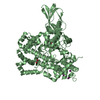
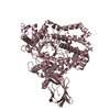
| Entry | Database: EMDB / ID: EMD-3959 | |||||||||
|---|---|---|---|---|---|---|---|---|---|---|
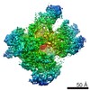
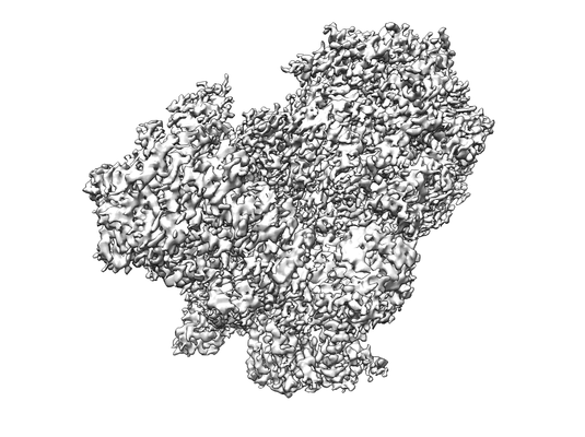
| Title | Apo RNA Polymerase III | |||||||||
 Map data Map data | Apo RNA Polymerase III | |||||||||
 Sample Sample |
| |||||||||
| Biological species |  | |||||||||
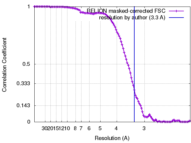
| Method | single particle reconstruction / cryo EM / Resolution: 3.3 Å | |||||||||
 Authors Authors | Abascal-Palacios G / Ramsay EP / Beuron F / Morris E / Vannini A | |||||||||
 Citation Citation | Journal: Nature / Year: 2018 Title: Structural basis of RNA polymerase III transcription initiation. Authors: Guillermo Abascal-Palacios / Ewan Phillip Ramsay / Fabienne Beuron / Edward Morris / Alessandro Vannini /  Abstract: RNA polymerase (Pol) III transcribes essential non-coding RNAs, including the entire pool of transfer RNAs, the 5S ribosomal RNA and the U6 spliceosomal RNA, and is often deregulated in cancer cells. ...RNA polymerase (Pol) III transcribes essential non-coding RNAs, including the entire pool of transfer RNAs, the 5S ribosomal RNA and the U6 spliceosomal RNA, and is often deregulated in cancer cells. The initiation of gene transcription by Pol III requires the activity of the transcription factor TFIIIB to form a transcriptionally active Pol III preinitiation complex (PIC). Here we present electron microscopy reconstructions of Pol III PICs at 3.4-4.0 Å and a reconstruction of unbound apo-Pol III at 3.1 Å. TFIIIB fully encircles the DNA and restructures Pol III. In particular, binding of the TFIIIB subunit Bdp1 rearranges the Pol III-specific subunits C37 and C34, thereby promoting DNA opening. The unwound DNA directly contacts both sides of the Pol III cleft. Topologically, the Pol III PIC resembles the Pol II PIC, whereas the Pol I PIC is more divergent. The structures presented unravel the molecular mechanisms underlying the first steps of Pol III transcription and also the general conserved mechanisms of gene transcription initiation. | |||||||||
| History |
|
- Structure visualization
| Movie |
 Movie viewer Movie viewer |
|---|---|
| Structure viewer | EM map: SurfViewMolmilJmol/JSmol |
| Supplemental images |
- Downloads & links
-EMDB archive
| Map data | emd_3959.map.gz | 10.1 MB | EMDB map data format | |
|---|---|---|---|---|
| Header (meta data) | emd-3959-v30.xmlemd-3959.xml | 11.4 KB 11.4 KB | Display Display | EMDB header |
| FSC (resolution estimation) | emd_3959_fsc.xml | 10.8 KB | Display | FSC data file |
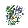
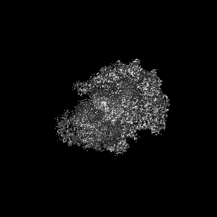
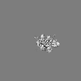
| Images |  emd_3959.png emd_3959.png | 77 KB | ||
| Archive directory |  http://ftp.pdbj.org/pub/emdb/structures/EMD-3959ftp://ftp.pdbj.org/pub/emdb/structures/EMD-3959 http://ftp.pdbj.org/pub/emdb/structures/EMD-3959ftp://ftp.pdbj.org/pub/emdb/structures/EMD-3959 | HTTPS FTP |
-Related structure data
| Related structure data | 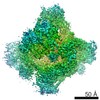 3955C 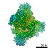 3956C 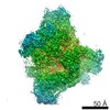 3957C 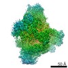 3958C 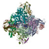 6eu0C 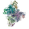 6eu1C  6eu2C 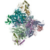 6eu3C C: citing same article ( |
|---|---|
| Similar structure data |


-Links
| EMDB pages | EMDB (EBI/PDBe) / EMDataResource |
|---|
-Map
| File | Download / File: emd_3959.map.gz / Format: CCP4 / Size: 111.5 MB / Type: IMAGE STORED AS FLOATING POINT NUMBER (4 BYTES) | ||||||||||||||||||||||||||||||||||||||||||||||||||||||||||||
|---|---|---|---|---|---|---|---|---|---|---|---|---|---|---|---|---|---|---|---|---|---|---|---|---|---|---|---|---|---|---|---|---|---|---|---|---|---|---|---|---|---|---|---|---|---|---|---|---|---|---|---|---|---|---|---|---|---|---|---|---|---|
| Annotation | Apo RNA Polymerase III | ||||||||||||||||||||||||||||||||||||||||||||||||||||||||||||
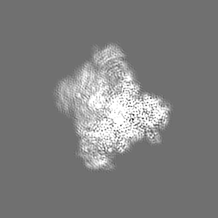
| Projections & slices | Image control
Images are generated by Spider. | ||||||||||||||||||||||||||||||||||||||||||||||||||||||||||||
| Voxel size |
| ||||||||||||||||||||||||||||||||||||||||||||||||||||||||||||
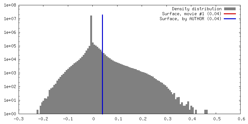
| Density |
| ||||||||||||||||||||||||||||||||||||||||||||||||||||||||||||
| Symmetry | Space group: 1 | ||||||||||||||||||||||||||||||||||||||||||||||||||||||||||||
| Details | EMDB XML:
CCP4 map header:
| ||||||||||||||||||||||||||||||||||||||||||||||||||||||||||||
 Z (Sec.)
 Y (Row.)
 X (Col.)


-Supplemental data
- Sample components
-Entire : Apo RNA Polymerase III
| Entire | Name: Apo RNA Polymerase III |
|---|---|
| Components |
|
-Supramolecule #1: Apo RNA Polymerase III
| Supramolecule | Name: Apo RNA Polymerase III / type: complex / ID: 1 / Parent: 0 / Macromolecule list: #1-#17 |
|---|---|
| Source (natural) | Organism: |
-Experimental details
-Structure determination
| Method | cryo EM |
|---|---|
 Processing Processing | single particle reconstruction |
| Aggregation state | particle |
-Sample preparation
| Concentration | 0.1 mg/mL |
|---|---|
| Buffer | pH: 8 |
| Grid | Model: Quantifoil R2/2 / Material: MOLYBDENUM / Pretreatment - Type: GLOW DISCHARGE |
| Vitrification | Cryogen name: ETHANE / Chamber humidity: 100 % |
- Electron microscopy
| Microscope | FEI TITAN KRIOS |
|---|---|
| Image recording | Film or detector model: GATAN K2 SUMMIT (4k x 4k) / Detector mode: SUPER-RESOLUTION / Average electron dose: 40.0 e/Å2 |
| Electron beam | Acceleration voltage: 300 kV / Electron source:  FIELD EMISSION GUN FIELD EMISSION GUN |
| Electron optics | Illumination mode: FLOOD BEAM / Imaging mode: BRIGHT FIELD |
| Experimental equipment |  Model: Titan Krios / Image courtesy: FEI Company |