 Movie
Movie Controller

[English] 日本語
 Yorodumi
- PDB-2hek: Crystal structure of O67745, a hypothetical protein from Aquifex ... -
+ Open data

- Basic information
| Entry | Database: PDB / ID: 2hek | ||||||
|---|---|---|---|---|---|---|---|
| Title | Crystal structure of O67745, a hypothetical protein from Aquifex aeolicus at 2.0 A resolution. | ||||||
 Components Components | Hypothetical protein | ||||||
 Keywords Keywords | Structural Genomics / Unknown Function / Predominantly alpha helical protein with GDP binding site and active site being far from each other / PSI / Protein Structure Initiative / Berkeley Structural Genomics Center / BSGC | ||||||
| Function / homology |  Function and homology information Function and homology information | ||||||
| Biological species |   Aquifex aeolicus (bacteria) Aquifex aeolicus (bacteria) | ||||||
| Method |  X-RAY DIFFRACTION / SYNCHROTRON / SAD / Resolution: 1.997 Å X-RAY DIFFRACTION / SYNCHROTRON / SAD / Resolution: 1.997 Å | ||||||
 Authors Authors | Oganesyan, V. / Jancarik, J. / Adams, P.D. / Kim, R. / Kim, S.H. / Berkeley Structural Genomics Center (BSGC) | ||||||
 Citation Citation | Journal: Acta Crystallogr.,Sect.F / Year: 2007 Title: Structure of O67745_AQUAE, a hypothetical protein from Aquifex aeolicus. Authors: Oganesyan, V. / Adams, P.D. / Jancarik, J. / Kim, R. / Kim, S.H. | ||||||
| History |
|
- Structure visualization
| Structure viewer | Molecule: MolmilJmol/JSmol |
|---|
- Downloads & links
-Download
| PDBx/mmCIF format | 2hek.cif.gz | 335.8 KB | Display | PDBx/mmCIF format |
|---|---|---|---|---|
| PDB format | pdb2hek.ent.gz | 273.2 KB | Display | PDB format |
| PDBx/mmJSON format | 2hek.json.gz | Tree view | PDBx/mmJSON format | |
| Others |  Other downloads Other downloads |
-Validation report
| Arichive directory | https://data.pdbj.org/pub/pdb/validation_reports/he/2hekftp://data.pdbj.org/pub/pdb/validation_reports/he/2hek | HTTPS FTP |
|---|
-Related structure data
| Similar structure data | |
|---|---|
| Other databases |
-Links
 PDBj
PDBj


- Assembly
| Deposited unit | 
| ||||||||
|---|---|---|---|---|---|---|---|---|---|
| 1 | 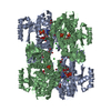
| ||||||||
| Unit cell |
| ||||||||
| Details | Asymmetric unit contains 2 polypeptides and each of two GDP molecules have contacts with both polypeptides. This information is used to call the asymmetric unit contents a biological assembly. Although it should be mentioned here that two such homodimers have also very intensive contact area. That symmetry operation is: -x,y,-z+1/2. |
-Components
-Protein , 1 types, 2 molecules AB
| #1: Protein | Mass: 44186.695 Da / Num. of mol.: 2 Source method: isolated from a genetically manipulated source Source: (gene. exp.) Aquifex aeolicus (bacteria) / Plasmid: pB2.1275B / Species (production host): Escherichia coli / Production host: |
|---|
-Non-polymers , 7 types, 377 molecules 


| #2: Chemical | ChemComp-PO4 /  Mass: 94.971 Da / Num. of mol.: 4 / Source method: obtained synthetically / Formula: PO4 Mass: 94.971 Da / Num. of mol.: 4 / Source method: obtained synthetically / Formula: PO4#3: Chemical |  Mass: 65.409 Da / Num. of mol.: 2 / Source method: obtained synthetically / Formula: Zn Mass: 65.409 Da / Num. of mol.: 2 / Source method: obtained synthetically / Formula: Zn#4: Chemical | ChemComp-CL /  Mass: 35.453 Da / Num. of mol.: 5 / Source method: obtained synthetically / Formula: Cl Mass: 35.453 Da / Num. of mol.: 5 / Source method: obtained synthetically / Formula: Cl#5: Chemical |  Type: RNA linking / Mass: 443.201 Da / Num. of mol.: 2 / Source method: obtained synthetically / Formula: C10H15N5O11P2 / Comment: GDP, energy-carrying molecule*YM Type: RNA linking / Mass: 443.201 Da / Num. of mol.: 2 / Source method: obtained synthetically / Formula: C10H15N5O11P2 / Comment: GDP, energy-carrying molecule*YM#6: Chemical | ChemComp-GOL /  Mass: 92.094 Da / Num. of mol.: 8 / Source method: obtained synthetically / Formula: C3H8O3 Mass: 92.094 Da / Num. of mol.: 8 / Source method: obtained synthetically / Formula: C3H8O3#7: Chemical | ChemComp-BR / |  Mass: 79.904 Da / Num. of mol.: 1 / Source method: obtained synthetically / Formula: Br Mass: 79.904 Da / Num. of mol.: 1 / Source method: obtained synthetically / Formula: Br#8: Water | ChemComp-HOH / | Mass: 18.015 Da / Num. of mol.: 355 / Source method: isolated from a natural source / Formula: H2O |
|---|
-Experimental details
-Experiment
| Experiment | Method: X-RAY DIFFRACTION / Number of used crystals: 1 |
|---|
- Sample preparation
| Crystal | Density Matthews: 3.18 Å3/Da / Density % sol: 61.34 % |
|---|---|
| Crystal grow | Temperature: 293 K / Method: vapor diffusion, hanging drop / pH: 4.2 Details: 10 mg/ml protein was mixed with 1:1 ratio with reservoir consisted of: 200 mM NaCl, 100 mM Phosphate-Citrate buffer, 10% PEG 3000, 100 mM NaBr, 5% Glycerol, pH 4.2, VAPOR DIFFUSION, HANGING ...Details: 10 mg/ml protein was mixed with 1:1 ratio with reservoir consisted of: 200 mM NaCl, 100 mM Phosphate-Citrate buffer, 10% PEG 3000, 100 mM NaBr, 5% Glycerol, pH 4.2, VAPOR DIFFUSION, HANGING DROP, temperature 293K |
-Data collection
| Diffraction | Mean temperature: 100 K |
|---|---|
| Diffraction source | Source: SYNCHROTRON / Site: ALS  / Beamline: 8.2.1 / Wavelength: 1 Å / Beamline: 8.2.1 / Wavelength: 1 Å |
| Detector | Type: ADSC QUANTUM 210 / Detector: CCD / Date: Jun 26, 2004 Details: Divergence: 3.0(h)x0.35(v) mrad Spot size: 0.140(h)x0.150(v) mm |
| Radiation | Monochromator: Double crystal, Si(111) / Protocol: SINGLE WAVELENGTH / Monochromatic (M) / Laue (L): M / Scattering type: x-ray |
| Radiation wavelength | Wavelength: 1 Å / Relative weight: 1 |
| Reflection | Resolution: 1.997→43 Å / Num. all: 80115 / Num. obs: 76300 / % possible obs: 95.7 % / Observed criterion σ(F): 1 / Observed criterion σ(I): 1 / Redundancy: 4.7 % / Biso Wilson estimate: 44 Å2 / Rsym value: 0.044 / Net I/σ(I): 21.4 |
| Reflection shell | Resolution: 1.997→2.07 Å / Redundancy: 3.8 % / Rmerge(I) obs: 0.52 / Mean I/σ(I) obs: 2.5 / Num. unique all: 8061 / Rsym value: 0.518 / % possible all: 95.7 |
- Processing
| Software |
| ||||||||||||||||||||||||||||||||||||||||||||||||||||||||||||||||||||||||||||||||||||||||||||||||||||||||||||||
|---|---|---|---|---|---|---|---|---|---|---|---|---|---|---|---|---|---|---|---|---|---|---|---|---|---|---|---|---|---|---|---|---|---|---|---|---|---|---|---|---|---|---|---|---|---|---|---|---|---|---|---|---|---|---|---|---|---|---|---|---|---|---|---|---|---|---|---|---|---|---|---|---|---|---|---|---|---|---|---|---|---|---|---|---|---|---|---|---|---|---|---|---|---|---|---|---|---|---|---|---|---|---|---|---|---|---|---|---|---|---|---|
| Refinement | Method to determine structure: SAD / Resolution: 1.997→12 Å / Cor.coef. Fo:Fc: 0.962 / Cor.coef. Fo:Fc free: 0.945 / SU B: 6.99 / SU ML: 0.09 / Cross valid method: THROUGHOUT / σ(F): 0 / σ(I): 0 / ESU R: 0.273 / ESU R Free: 0.137 / Stereochemistry target values: MAXIMUM LIKELIHOOD
| ||||||||||||||||||||||||||||||||||||||||||||||||||||||||||||||||||||||||||||||||||||||||||||||||||||||||||||||
| Solvent computation | Ion probe radii: 0.8 Å / Shrinkage radii: 0.8 Å / VDW probe radii: 1.4 Å / Solvent model: BABINET MODEL WITH MASK | ||||||||||||||||||||||||||||||||||||||||||||||||||||||||||||||||||||||||||||||||||||||||||||||||||||||||||||||
| Displacement parameters | Biso mean: 46.142 Å2
| ||||||||||||||||||||||||||||||||||||||||||||||||||||||||||||||||||||||||||||||||||||||||||||||||||||||||||||||
| Refine analyze | Luzzati coordinate error obs: 0.22 Å | ||||||||||||||||||||||||||||||||||||||||||||||||||||||||||||||||||||||||||||||||||||||||||||||||||||||||||||||
| Refinement step | Cycle: LAST / Resolution: 1.997→12 Å
| ||||||||||||||||||||||||||||||||||||||||||||||||||||||||||||||||||||||||||||||||||||||||||||||||||||||||||||||
| Refine LS restraints |
| ||||||||||||||||||||||||||||||||||||||||||||||||||||||||||||||||||||||||||||||||||||||||||||||||||||||||||||||
| LS refinement shell | Resolution: 1.997→2.048 Å / Total num. of bins used: 20
|
