 Movie
Movie Controller


+ Open data

- Basic information
| Entry |  | |||||||||||||||||||||
|---|---|---|---|---|---|---|---|---|---|---|---|---|---|---|---|---|---|---|---|---|---|---|
| Title | Cryo EM structure of human phosphate channel XPR1 at apo state | |||||||||||||||||||||
 Map data Map data | ||||||||||||||||||||||
 Sample Sample |
| |||||||||||||||||||||
 Keywords Keywords | phosphate channel / membrane protein / phosphate homeostasis / TRANSPORT PROTEIN | |||||||||||||||||||||
| Function / homology |  Function and homology information Function and homology informationphosphate transmembrane transporter activity / phosphate ion transport / cellular response to phosphate starvation / intracellular phosphate ion homeostasis / phosphate ion transmembrane transport / inositol hexakisphosphate binding / efflux transmembrane transporter activity / response to virus / virus receptor activity / plasma membrane Similarity search - Function | |||||||||||||||||||||
| Biological species |  Homo sapiens (human) Homo sapiens (human) | |||||||||||||||||||||
| Method | single particle reconstruction / cryo EM / Resolution: 3.68 Å | |||||||||||||||||||||
 Authors Authors | Lu Y / Yue C / Zhang L / Yao D / Yu Y / Cao Y | |||||||||||||||||||||
| Funding support |  China, 6 items China, 6 items
| |||||||||||||||||||||
 Citation Citation | Journal: Science / Year: 2024 Title: Structural basis for inositol pyrophosphate gating of the phosphate channel XPR1. Authors: Yi Lu / Chen-Xi Yue / Li Zhang / Deqiang Yao / Ying Xia / Qing Zhang / Xinchen Zhang / Shaobai Li / Yafeng Shen / Mi Cao / Chang-Run Guo / An Qin / Jie Zhao / Lu Zhou / Ye Yu / Yu Cao /  Abstract: Precise regulation of intracellular phosphate (Pi) is critical for cellular function, with xenotropic and polytropic retrovirus receptor 1 (XPR1) serving as the sole Pi exporter in humans. The ...Precise regulation of intracellular phosphate (Pi) is critical for cellular function, with xenotropic and polytropic retrovirus receptor 1 (XPR1) serving as the sole Pi exporter in humans. The mechanism of Pi efflux, activated by inositol pyrophosphates (PP-IPs), has remained unclear. This study presents cryo-electron microscopy structures of XPR1 in multiple conformations, revealing a transmembrane pathway for Pi export and a dual-binding activation pattern for PP-IPs. A canonical binding site is located at the dimeric interface of Syg1/Pho81/XPR1 (SPX) domains, and a second site, biased toward PP-IPs, is found between the transmembrane and SPX domains. By integrating structural studies with electrophysiological analyses, we characterized XPR1 as an inositol phosphates (IPs)/PP-IPs-activated phosphate channel. The interplay among its transmembrane domains, SPX domains, and IPs/PP-IPs orchestrates the conformational transition between its closed and open states. | |||||||||||||||||||||
| History |
|
- Structure visualization
| Supplemental images |
|---|

- Downloads & links
-EMDB archive
| Map data | emd_39204.map.gz | 59.6 MB | EMDB map data format | |
|---|---|---|---|---|
| Header (meta data) | emd-39204-v30.xmlemd-39204.xml | 15.3 KB 15.3 KB | Display Display | EMDB header |
| FSC (resolution estimation) | emd_39204_fsc.xml | 8.5 KB | Display | FSC data file |
| Images |  emd_39204.png emd_39204.png | 111.4 KB | ||
| Filedesc metadata | emd-39204.cif.gz | 5.5 KB | ||
| Others | emd_39204_half_map_1.map.gzemd_39204_half_map_2.map.gz | 59.4 MB 59.4 MB | ||
| Archive directory |  http://ftp.pdbj.org/pub/emdb/structures/EMD-39204ftp://ftp.pdbj.org/pub/emdb/structures/EMD-39204 http://ftp.pdbj.org/pub/emdb/structures/EMD-39204ftp://ftp.pdbj.org/pub/emdb/structures/EMD-39204 | HTTPS FTP |
-Related structure data
| Related structure data |  8yexMC  8yetC 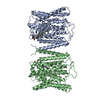 8yf4C 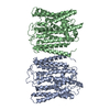 8yfdC  8yfuC  8yfwC  8yfxC  9iwsC M: atomic model generated by this map C: citing same article ( |
|---|---|
| Similar structure data |


-Links
| EMDB pages | EMDB (EBI/PDBe) / EMDataResource |
|---|
-Map
| File | Download / File: emd_39204.map.gz / Format: CCP4 / Size: 64 MB / Type: IMAGE STORED AS FLOATING POINT NUMBER (4 BYTES) | ||||||||||||||||||||||||||||||||||||
|---|---|---|---|---|---|---|---|---|---|---|---|---|---|---|---|---|---|---|---|---|---|---|---|---|---|---|---|---|---|---|---|---|---|---|---|---|---|
| Projections & slices | Image control
Images are generated by Spider. | ||||||||||||||||||||||||||||||||||||
| Voxel size | X=Y=Z: 1.1 Å | ||||||||||||||||||||||||||||||||||||
| Density |
| ||||||||||||||||||||||||||||||||||||
| Symmetry | Space group: 1 | ||||||||||||||||||||||||||||||||||||
| Details | EMDB XML:
|
 Z (Sec.)
 Y (Row.)
 X (Col.)


-Supplemental data
-Half map: #1
| File | emd_39204_half_map_1.map | ||||||||||||
|---|---|---|---|---|---|---|---|---|---|---|---|---|---|
| Projections & Slices |
| ||||||||||||
| Density Histograms |


-Half map: #2
| File | emd_39204_half_map_2.map | ||||||||||||
|---|---|---|---|---|---|---|---|---|---|---|---|---|---|
| Projections & Slices |
| ||||||||||||
| Density Histograms |


- Sample components
-Entire : Cryo EM structure of human phosphate channel XPR1 at apo state
| Entire | Name: Cryo EM structure of human phosphate channel XPR1 at apo state |
|---|---|
| Components |
|
-Supramolecule #1: Cryo EM structure of human phosphate channel XPR1 at apo state
| Supramolecule | Name: Cryo EM structure of human phosphate channel XPR1 at apo state type: complex / ID: 1 / Parent: 0 / Macromolecule list: all |
|---|---|
| Source (natural) | Organism: Homo sapiens (human) |
-Macromolecule #1: Solute carrier family 53 member 1
| Macromolecule | Name: Solute carrier family 53 member 1 / type: protein_or_peptide / ID: 1 / Number of copies: 2 / Enantiomer: LEVO |
|---|---|
| Source (natural) | Organism: Homo sapiens (human) |
| Molecular weight | Theoretical: 46.051754 KDa |
| Recombinant expression | Organism: Homo sapiens (human) |
| Sequence | String: PAPAWTTFRV GLFCGIFIVL NITLVLAAVF KLETDRSIWP LIRIYRGGFL LIEFLFLLGI NTYGWRQAGV NHVLIFELNP RSNLSHQHL FEIAGFLGIL WCLSLLACFF APISVIPTYV YPLALYGFMV FFLINPTKTF YYKSRFWLLK LLFRVFTAPF H KVGFADFW ...String: PAPAWTTFRV GLFCGIFIVL NITLVLAAVF KLETDRSIWP LIRIYRGGFL LIEFLFLLGI NTYGWRQAGV NHVLIFELNP RSNLSHQHL FEIAGFLGIL WCLSLLACFF APISVIPTYV YPLALYGFMV FFLINPTKTF YYKSRFWLLK LLFRVFTAPF H KVGFADFW LADQLNSLSV ILMDLEYMIC FYSLELKWDE SKGLLPNNSE ESGICHKYTY GVRAIVQCIP AWLRFIQCLR RY RDTKRAF PHLVNAGKYS TTFFMVTFAA LYSTHKERGH SDTMVFFYLW IVFYIISSCY TLIWDLKMDW GLFDKNAGEN TFL REEIVY PQKAYYYCAI IEDVILRFAW TIQISITSTT LLPHSGDIIA TVFAPLEVFR RFVWNFFRLE NEHLNN UniProtKB: Solute carrier family 53 member 1 |
-Experimental details
-Structure determination
| Method | cryo EM |
|---|---|
 Processing Processing | single particle reconstruction |
| Aggregation state | particle |
-Sample preparation
| Buffer | pH: 7.5 |
|---|---|
| Vitrification | Cryogen name: ETHANE |
- Electron microscopy
| Microscope | FEI TITAN KRIOS |
|---|---|
| Image recording | Film or detector model: GATAN K3 (6k x 4k) / Average electron dose: 50.0 e/Å2 |
| Electron beam | Acceleration voltage: 300 kV / Electron source:  FIELD EMISSION GUN FIELD EMISSION GUN |
| Electron optics | Illumination mode: SPOT SCAN / Imaging mode: BRIGHT FIELD / Nominal defocus max: 2.6 µm / Nominal defocus min: 1.0 µm |
| Experimental equipment |  Model: Titan Krios / Image courtesy: FEI Company |

