ムービー
ムービー コントローラー

[日本語] English
 万見
万見- EMDB-38539: Cryo-EM structure of the tethered agonist-bound human PAR1-Gi complex -
+ データを開く

- 基本情報
基本情報
| 登録情報 |  | |||||||||
|---|---|---|---|---|---|---|---|---|---|---|
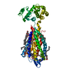
| タイトル | Cryo-EM structure of the tethered agonist-bound human PAR1-Gi complex | |||||||||
 マップデータ マップデータ | ||||||||||
 試料 試料 |
| |||||||||
 キーワード キーワード | GPCR / MEMBRANE PROTEIN / protease-activated receptor | |||||||||
| 機能・相同性 |  機能・相同性情報 機能・相同性情報negative regulation of renin secretion into blood stream / dendritic cell homeostasis / trans-synaptic signaling by endocannabinoid, modulating synaptic transmission / platelet dense tubular network / establishment of synaptic specificity at neuromuscular junction / thrombin-activated receptor activity / regulation of interleukin-1 beta production / platelet dense granule organization / connective tissue replacement involved in inflammatory response wound healing / cell-cell junction maintenance ...negative regulation of renin secretion into blood stream / dendritic cell homeostasis / trans-synaptic signaling by endocannabinoid, modulating synaptic transmission / platelet dense tubular network / establishment of synaptic specificity at neuromuscular junction / thrombin-activated receptor activity / regulation of interleukin-1 beta production / platelet dense granule organization / connective tissue replacement involved in inflammatory response wound healing / cell-cell junction maintenance / positive regulation of smooth muscle contraction / positive regulation of calcium ion transport / G-protein activation / Activation of the phototransduction cascade / Glucagon-type ligand receptors / Thromboxane signalling through TP receptor / Sensory perception of sweet, bitter, and umami (glutamate) taste / G beta:gamma signalling through PI3Kgamma / G beta:gamma signalling through CDC42 / Cooperation of PDCL (PhLP1) and TRiC/CCT in G-protein beta folding / Activation of G protein gated Potassium channels / Inhibition of voltage gated Ca2+ channels via Gbeta/gamma subunits / Ca2+ pathway / G alpha (z) signalling events / High laminar flow shear stress activates signaling by PIEZO1 and PECAM1:CDH5:KDR in endothelial cells / Glucagon-like Peptide-1 (GLP1) regulates insulin secretion / Vasopressin regulates renal water homeostasis via Aquaporins / Adrenaline,noradrenaline inhibits insulin secretion / ADP signalling through P2Y purinoceptor 12 / G alpha (q) signalling events / G alpha (i) signalling events / Thrombin signalling through proteinase activated receptors (PARs) / Activation of G protein gated Potassium channels / G-protein activation / G beta:gamma signalling through PI3Kgamma / Prostacyclin signalling through prostacyclin receptor / G beta:gamma signalling through PLC beta / ADP signalling through P2Y purinoceptor 1 / Thromboxane signalling through TP receptor / Presynaptic function of Kainate receptors / G beta:gamma signalling through CDC42 / Inhibition of voltage gated Ca2+ channels via Gbeta/gamma subunits / G alpha (12/13) signalling events / Glucagon-type ligand receptors / G beta:gamma signalling through BTK / ADP signalling through P2Y purinoceptor 12 / Adrenaline,noradrenaline inhibits insulin secretion / Cooperation of PDCL (PhLP1) and TRiC/CCT in G-protein beta folding / Ca2+ pathway / Thrombin signalling through proteinase activated receptors (PARs) / G alpha (z) signalling events / Extra-nuclear estrogen signaling / photoreceptor outer segment membrane / G alpha (s) signalling events / G alpha (q) signalling events / negative regulation of glomerular filtration / spectrin binding / G alpha (i) signalling events / thrombin-activated receptor signaling pathway / Glucagon-like Peptide-1 (GLP1) regulates insulin secretion / positive regulation of Rho protein signal transduction / High laminar flow shear stress activates signaling by PIEZO1 and PECAM1:CDH5:KDR in endothelial cells / Vasopressin regulates renal water homeostasis via Aquaporins / positive regulation of vasoconstriction / regulation of blood coagulation / alkylglycerophosphoethanolamine phosphodiesterase activity / positive regulation of collagen biosynthetic process / anatomical structure morphogenesis / G-protein alpha-subunit binding / positive regulation of blood coagulation / photoreceptor outer segment / Common Pathway of Fibrin Clot Formation / adenylate cyclase inhibitor activity / positive regulation of protein localization to cell cortex / Adenylate cyclase inhibitory pathway / T cell migration / D2 dopamine receptor binding / response to prostaglandin E / adenylate cyclase regulator activity / G protein-coupled serotonin receptor binding / adenylate cyclase-inhibiting serotonin receptor signaling pathway / release of sequestered calcium ion into cytosol / cardiac muscle cell apoptotic process / photoreceptor inner segment / cellular response to forskolin / homeostasis of number of cells within a tissue / regulation of mitotic spindle organization / Peptide ligand-binding receptors / positive regulation of release of sequestered calcium ion into cytosol / positive regulation of interleukin-8 production / positive regulation of receptor signaling pathway via JAK-STAT / Regulation of insulin secretion / neuromuscular junction / positive regulation of cholesterol biosynthetic process / negative regulation of insulin secretion / G protein-coupled receptor binding / G protein-coupled receptor activity / response to peptide hormone / adenylate cyclase-inhibiting G protein-coupled receptor signaling pathway / regulation of synaptic plasticity 類似検索 - 分子機能 | |||||||||
| 生物種 |  Homo sapiens (ヒト) / Homo sapiens (ヒト) /  | |||||||||
| 手法 | 単粒子再構成法 / クライオ電子顕微鏡法 / 解像度: 3.2 Å | |||||||||
 データ登録者 データ登録者 | Guo J / Zhang Y | |||||||||
| 資金援助 |  中国, 1件 中国, 1件
| |||||||||
 引用 引用 | ジャーナル: Cell Res / 年: 2024 タイトル: Structural basis of tethered agonism and G protein coupling of protease-activated receptors. 著者: Jia Guo / Yun-Li Zhou / Yixin Yang / Shimeng Guo / Erli You / Xin Xie / Yi Jiang / Chunyou Mao / H Eric Xu / Yan Zhang / 要旨: Protease-activated receptors (PARs) are a unique group within the G protein-coupled receptor superfamily, orchestrating cellular responses to extracellular proteases via enzymatic cleavage, which ...Protease-activated receptors (PARs) are a unique group within the G protein-coupled receptor superfamily, orchestrating cellular responses to extracellular proteases via enzymatic cleavage, which triggers intracellular signaling pathways. Protease-activated receptor 1 (PAR1) is a key member of this family and is recognized as a critical pharmacological target for managing thrombotic disorders. In this study, we present cryo-electron microscopy structures of PAR1 in its activated state, induced by its natural tethered agonist (TA), in complex with two distinct downstream proteins, the G and G heterotrimers, respectively. The TA peptide is positioned within a surface pocket, prompting PAR1 activation through notable conformational shifts. Contrary to the typical receptor activation that involves the outward movement of transmembrane helix 6 (TM6), PAR1 activation is characterized by the simultaneous downward shift of TM6 and TM7, coupled with the rotation of a group of aromatic residues. This results in the displacement of an intracellular anion, creating space for downstream G protein binding. Our findings delineate the TA recognition pattern and highlight a distinct role of the second extracellular loop in forming β-sheets with TA within the PAR family, a feature not observed in other TA-activated receptors. Moreover, the nuanced differences in the interactions between intracellular loops 2/3 and the Gα subunit of different G proteins are crucial for determining the specificity of G protein coupling. These insights contribute to our understanding of the ligand binding and activation mechanisms of PARs, illuminating the basis for PAR1's versatility in G protein coupling. | |||||||||
| 履歴 |
|
- 構造の表示
構造の表示
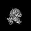
| 添付画像 |
|---|

- ダウンロードとリンク
-EMDBアーカイブ
| マップデータ | emd_38539.map.gz | 23.3 MB | EMDBマップデータ形式 | |
|---|---|---|---|---|
| ヘッダ (付随情報) | emd-38539-v30.xmlemd-38539.xml | 22.5 KB 22.5 KB | 表示 表示 | EMDBヘッダ |
| 画像 |  emd_38539.png emd_38539.png | 50.6 KB | ||
| Filedesc metadata | emd-38539.cif.gz | 7.3 KB | ||
| その他 | emd_38539_half_map_1.map.gzemd_38539_half_map_2.map.gz | 23.4 MB 23.4 MB | ||
| アーカイブディレクトリ |  http://ftp.pdbj.org/pub/emdb/structures/EMD-38539ftp://ftp.pdbj.org/pub/emdb/structures/EMD-38539 http://ftp.pdbj.org/pub/emdb/structures/EMD-38539ftp://ftp.pdbj.org/pub/emdb/structures/EMD-38539 | HTTPS FTP |
-検証レポート
| 文書・要旨 | emd_38539_validation.pdf.gz | 838 KB | 表示 | EMDB検証レポート |
|---|---|---|---|---|
| 文書・詳細版 | emd_38539_full_validation.pdf.gz | 837.4 KB | 表示 | |
| XML形式データ | emd_38539_validation.xml.gz | 10.6 KB | 表示 | |
| CIF形式データ | emd_38539_validation.cif.gz | 12.5 KB | 表示 | |
| アーカイブディレクトリ | https://ftp.pdbj.org/pub/emdb/validation_reports/EMD-38539ftp://ftp.pdbj.org/pub/emdb/validation_reports/EMD-38539 | HTTPS FTP |
-関連構造データ


-リンク
| EMDBのページ | EMDB (EBI/PDBe) / EMDataResource |
|---|---|
| 「今月の分子」の関連する項目 |


-マップ
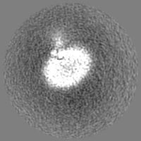
| ファイル | ダウンロード / ファイル: emd_38539.map.gz / 形式: CCP4 / 大きさ: 30.5 MB / タイプ: IMAGE STORED AS FLOATING POINT NUMBER (4 BYTES) | ||||||||||||||||||||||||||||||||||||
|---|---|---|---|---|---|---|---|---|---|---|---|---|---|---|---|---|---|---|---|---|---|---|---|---|---|---|---|---|---|---|---|---|---|---|---|---|---|
| 投影像・断面図 | 画像のコントロール
画像は Spider により作成 | ||||||||||||||||||||||||||||||||||||
| ボクセルのサイズ | X=Y=Z: 1.071 Å | ||||||||||||||||||||||||||||||||||||
| 密度 |
| ||||||||||||||||||||||||||||||||||||
| 対称性 | 空間群: 1 | ||||||||||||||||||||||||||||||||||||
| 詳細 | EMDB XML:
|
 Z (Sec.)
 Y (Row.)
 X (Col.)


-添付データ
-ハーフマップ: #1
| ファイル | emd_38539_half_map_1.map | ||||||||||||
|---|---|---|---|---|---|---|---|---|---|---|---|---|---|
| 投影像・断面図 |
| ||||||||||||
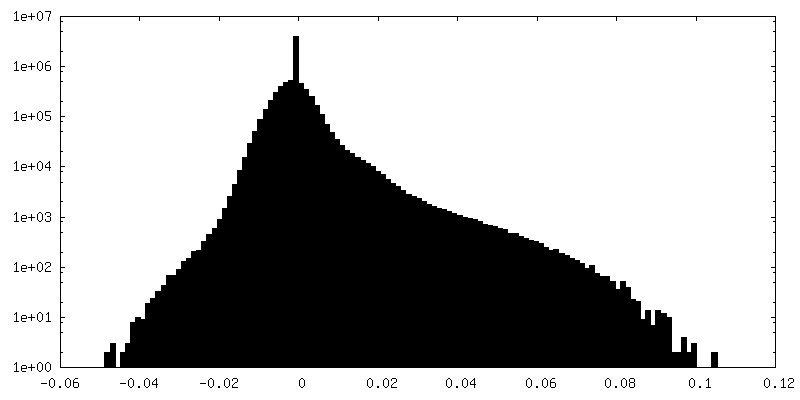
| 密度ヒストグラム |


-ハーフマップ: #2
| ファイル | emd_38539_half_map_2.map | ||||||||||||
|---|---|---|---|---|---|---|---|---|---|---|---|---|---|
| 投影像・断面図 |
| ||||||||||||
| 密度ヒストグラム |


- 試料の構成要素
-全体 : tethered agonist-bound human PAR1-Gi complex
| 全体 | 名称: tethered agonist-bound human PAR1-Gi complex |
|---|---|
| 要素 |
|
-超分子 #1: tethered agonist-bound human PAR1-Gi complex
| 超分子 | 名称: tethered agonist-bound human PAR1-Gi complex / タイプ: complex / ID: 1 / 親要素: 0 / 含まれる分子: #1-#5 |
|---|---|
| 由来(天然) | 生物種: Homo sapiens (ヒト) |
-分子 #1: Guanine nucleotide-binding protein G(i) subunit alpha-1
| 分子 | 名称: Guanine nucleotide-binding protein G(i) subunit alpha-1 タイプ: protein_or_peptide / ID: 1 / コピー数: 1 / 光学異性体: LEVO |
|---|---|
| 由来(天然) | 生物種: Homo sapiens (ヒト) |
| 分子量 | 理論値: 40.313863 KDa |
| 組換発現 | 生物種:   Spodoptera frugiperda (ツマジロクサヨトウ) Spodoptera frugiperda (ツマジロクサヨトウ) |
| 配列 | 文字列: GCTLSAEDKA AVERSKMIDR NLREDGEKAA REVKLLLLGA GESGKSTIVK QMKIIHEAGY SEEECKQYKA VVYSNTIQSI IAIIRAMGR LKIDFGDSAR ADDARQLFVL AGAAEEGFMT AELAGVIKRL WKDSGVQACF NRSREYQLND SAAYYLNDLD R IAQPNYIP ...文字列: GCTLSAEDKA AVERSKMIDR NLREDGEKAA REVKLLLLGA GESGKSTIVK QMKIIHEAGY SEEECKQYKA VVYSNTIQSI IAIIRAMGR LKIDFGDSAR ADDARQLFVL AGAAEEGFMT AELAGVIKRL WKDSGVQACF NRSREYQLND SAAYYLNDLD R IAQPNYIP TQQDVLRTRV KTTGIVETHF TFKDLHFKMF DVGAQRSERK KWIHCFEGVT AIIFCVALSD YDLVLAEDEE MN RMHESMK LFDSICNNKW FTDTSIILFL NKKDLFEEKI KKSPLTICYP EYAGSNTYEE AAAYIQCQFE DLNKRKDTKE IYT HFTCST DTKNVQFVFD AVTDVIIKNN LKDCGLF UniProtKB: Guanine nucleotide-binding protein G(i) subunit alpha-1 |
-分子 #2: Guanine nucleotide-binding protein G(I)/G(S)/G(T) subunit beta-1
| 分子 | 名称: Guanine nucleotide-binding protein G(I)/G(S)/G(T) subunit beta-1 タイプ: protein_or_peptide / ID: 2 / コピー数: 1 / 光学異性体: LEVO |
|---|---|
| 由来(天然) | 生物種: |
| 分子量 | 理論値: 41.332137 KDa |
| 組換発現 | 生物種: Spodoptera frugiperda (ツマジロクサヨトウ) |
| 配列 | 文字列: MVSGWRLFKK ISGSSGGGGS GGGGSSGGSL LQSELDQLRQ EAEQLKNQIR DARKACADAT LSQITNNIDP VGRIQMRTRR TLRGHLAKI YAMHWGTDSR LLVSASQDGK LIIWDSYTTN KVHAIPLRSS WVMTCAYAPS GNYVACGGLD NICSIYNLKT R EGNVRVSR ...文字列: MVSGWRLFKK ISGSSGGGGS GGGGSSGGSL LQSELDQLRQ EAEQLKNQIR DARKACADAT LSQITNNIDP VGRIQMRTRR TLRGHLAKI YAMHWGTDSR LLVSASQDGK LIIWDSYTTN KVHAIPLRSS WVMTCAYAPS GNYVACGGLD NICSIYNLKT R EGNVRVSR ELAGHTGYLS CCRFLDDNQI VTSSGDTTCA LWDIETGQQT TTFTGHTGDV MSLSLAPDTR LFVSGACDAS AK LWDVREG MCRQTFTGHE SDINAICFFP NGNAFATGSD DATCRLFDLR ADQELMTYSH DNIICGITSV SFSKSGRLLL AGY DDFNCN VWDALKADRA GVLAGHDNRV SCLGVTDDGM AVATGSWDSF LKIWNHHHHH HHH UniProtKB: Guanine nucleotide-binding protein G(I)/G(S)/G(T) subunit beta-1 |
-分子 #3: Guanine nucleotide-binding protein G(I)/G(S)/G(O) subunit gamma-2
| 分子 | 名称: Guanine nucleotide-binding protein G(I)/G(S)/G(O) subunit gamma-2 タイプ: protein_or_peptide / ID: 3 / コピー数: 1 / 光学異性体: LEVO |
|---|---|
| 由来(天然) | 生物種: |
| 分子量 | 理論値: 7.432554 KDa |
| 組換発現 | 生物種: Spodoptera frugiperda (ツマジロクサヨトウ) |
| 配列 | 文字列: ASNNTASIAQ ARKLVEQLKM EANIDRIKVS KAAADLMAYC EAHAKEDPLL TPVPASENPF REKKFFC UniProtKB: Guanine nucleotide-binding protein G(I)/G(S)/G(O) subunit gamma-2 |
-分子 #4: scFv16
| 分子 | 名称: scFv16 / タイプ: protein_or_peptide / ID: 4 / コピー数: 1 / 光学異性体: LEVO |
|---|---|
| 由来(天然) | 生物種: |
| 分子量 | 理論値: 40.523266 KDa |
| 組換発現 | 生物種: Spodoptera frugiperda (ツマジロクサヨトウ) |
| 配列 | 文字列: LLVNQSHQGF NKEHTSKMVS AIVLYVLLAA AAHSAFAVQL VESGGGLVQP GGSRKLSCSA SGFAFSSFGM HWVRQAPEKG LEWVAYISS GSGTIYYADT VKGRFTISRD DPKNTLFLQM TSLRSEDTAM YYCVRSIYYY GSSPFDFWGQ GTTLTVSAGG G GSGGGGSG ...文字列: LLVNQSHQGF NKEHTSKMVS AIVLYVLLAA AAHSAFAVQL VESGGGLVQP GGSRKLSCSA SGFAFSSFGM HWVRQAPEKG LEWVAYISS GSGTIYYADT VKGRFTISRD DPKNTLFLQM TSLRSEDTAM YYCVRSIYYY GSSPFDFWGQ GTTLTVSAGG G GSGGGGSG GGGSADIVMT QATSSVPVTP GESVSISCRS SKSLLHSNGN TYLYWFLQRP GQSPQLLIYR MSNLASGVPD RF SGSGSGT AFTLTISRLE AEDVGVYYCM QHLEYPLTFG AGTKLELVDE NLYFQGASHH HHHHHHWFLQ RPGQSPQLLI YRM SNLASG VPDRFSGSGS GTAFTLTISR LEAEDVGVYY CMQHLEYPLT FGAGTKLEL |
-分子 #5: Proteinase-activated receptor 1 LgBiT
| 分子 | 名称: Proteinase-activated receptor 1 LgBiT / タイプ: protein_or_peptide / ID: 5 / コピー数: 1 / 光学異性体: LEVO |
|---|---|
| 由来(天然) | 生物種: Homo sapiens (ヒト) |
| 分子量 | 理論値: 58.787539 KDa |
| 組換発現 | 生物種: Spodoptera frugiperda (ツマジロクサヨトウ) |
| 配列 | 文字列: SFLLRNPNDK YEPFWEDEEK NESGLTEYRL VSINKSSPLQ KQLPAFISED ASGYLTSSWL TLFVPSVYTG VFVVSLPLNI MAIVVFILK MKVKKPAVVY MLHLATADVL FVSVLPFKIS YYFSGSDWQF GSELCRFVTA AFYCNMYASI LLMTVISIDR F LAVVYPMQ ...文字列: SFLLRNPNDK YEPFWEDEEK NESGLTEYRL VSINKSSPLQ KQLPAFISED ASGYLTSSWL TLFVPSVYTG VFVVSLPLNI MAIVVFILK MKVKKPAVVY MLHLATADVL FVSVLPFKIS YYFSGSDWQF GSELCRFVTA AFYCNMYASI LLMTVISIDR F LAVVYPMQ SLSWRTLGRA SFTCLAIWAL AIAGVVPLLL KEQTIQVPGL NITTCHDVLN ETLLEGYYAY YFSAFSAVFF FV PLIISTV CYVSIIRCLS SSAVANRSKK SRALFLSAAV FCIFIICFGP TNVLLIAHYS FLSHTSTTEA AYFAYLLCVC VSS ISCCID PLIYYYASSE CQRYVYSILC CKESSDPSSY GSSGGGGSGG GGSSGVFTLE DFVGDWEQTA AYNLDQVLEQ GGVS SLLQN LAVSVTPIQR IVRSGENALK IDIHVIIPYE GLSADQMAQI EEVFKVVYPV DDHHFKVILP YGTLVIDGVT PNMLN YFGR PYEGIAVFDG KKITVTGTLW NGNKIIDERL ITPDGSMLFR VTINSGGS UniProtKB: Proteinase-activated receptor 1 |
-分子 #6: CHOLESTEROL
| 分子 | 名称: CHOLESTEROL / タイプ: ligand / ID: 6 / コピー数: 2 / 式: CLR |
|---|---|
| 分子量 | 理論値: 386.654 Da |
| Chemical component information |  ChemComp-CLR: |
-実験情報
-構造解析
| 手法 | クライオ電子顕微鏡法 |
|---|---|
 解析 解析 | 単粒子再構成法 |
| 試料の集合状態 | particle |
-試料調製
| 緩衝液 | pH: 7.5 |
|---|---|
| グリッド | モデル: Quantifoil R1.2/1.3 / 材質: GOLD / メッシュ: 300 / 支持フィルム - 材質: CARBON / 支持フィルム - トポロジー: HOLEY |
| 凍結 | 凍結剤: ETHANE / チャンバー内湿度: 100 % / チャンバー内温度: 277 K |
- 電子顕微鏡法
| 顕微鏡 | FEI TITAN KRIOS |
|---|---|
| 撮影 | フィルム・検出器のモデル: GATAN K3 (6k x 4k) / 平均電子線量: 70.0 e/Å2 |
| 電子線 | 加速電圧: 300 kV / 電子線源:  FIELD EMISSION GUN FIELD EMISSION GUN |
| 電子光学系 | 照射モード: FLOOD BEAM / 撮影モード: BRIGHT FIELD / 最大 デフォーカス(公称値): 2.0 µm / 最小 デフォーカス(公称値): 0.5 µm |
| 実験機器 |  モデル: Titan Krios / 画像提供: FEI Company |