[English] 日本語
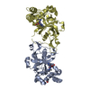
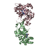
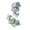
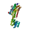
Yorodumi- PDB-3vw7: Crystal structure of human protease-activated receptor 1 (PAR1) b... -
+Open data
-Basic information
Entry | Database: PDB / ID: 3vw7 | ||||||
---|---|---|---|---|---|---|---|
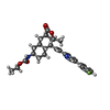
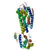
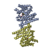
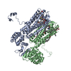
Title | Crystal structure of human protease-activated receptor 1 (PAR1) bound with antagonist vorapaxar at 2.2 angstrom | ||||||
Components | Proteinase-activated receptor 1, Lysozyme | ||||||
Keywords | Signaling protein/antagonist / High resolution structure / protease-activated receptor 1 / inactive conformation / antagonist vorapaxar / G protein-coupled receptor / signaling protein / membrane protein / thrombin receptor-antagonist complex / Signaling protein-antagonist complex | ||||||
Function / homology | Function and homology information negative regulation of renin secretion into blood stream / negative regulation of glomerular filtration / dendritic cell homeostasis / establishment of synaptic specificity at neuromuscular junction / thrombin-activated receptor signaling pathway / thrombin-activated receptor activity / platelet dense tubular network / cell-cell junction maintenance / regulation of interleukin-1 beta production / platelet dense granule organization ...negative regulation of renin secretion into blood stream / negative regulation of glomerular filtration / dendritic cell homeostasis / establishment of synaptic specificity at neuromuscular junction / thrombin-activated receptor signaling pathway / thrombin-activated receptor activity / platelet dense tubular network / cell-cell junction maintenance / regulation of interleukin-1 beta production / platelet dense granule organization / connective tissue replacement involved in inflammatory response wound healing / trans-synaptic signaling by endocannabinoid, modulating synaptic transmission / regulation of sensory perception of pain / positive regulation of smooth muscle contraction / positive regulation of calcium ion transport / : / positive regulation of Rho protein signal transduction / : / regulation of blood coagulation / positive regulation of collagen biosynthetic process / G-protein alpha-subunit binding / anatomical structure morphogenesis / positive regulation of blood coagulation / Common Pathway of Fibrin Clot Formation / viral release from host cell by cytolysis / positive regulation of vasoconstriction / homeostasis of number of cells within a tissue / release of sequestered calcium ion into cytosol / peptidoglycan catabolic process / Peptide ligand-binding receptors / positive regulation of GTPase activity / positive regulation of release of sequestered calcium ion into cytosol / positive regulation of interleukin-8 production / G protein-coupled receptor activity / positive regulation of receptor signaling pathway via JAK-STAT / neuromuscular junction / regulation of synaptic plasticity / caveola / platelet activation / : / response to wounding / positive regulation of interleukin-6 production / G-protein beta-subunit binding / cell wall macromolecule catabolic process / late endosome / lysozyme / Thrombin signalling through proteinase activated receptors (PARs) / lysozyme activity / phospholipase C-activating G protein-coupled receptor signaling pathway / positive regulation of cytosolic calcium ion concentration / G alpha (q) signalling events / postsynaptic membrane / positive regulation of canonical NF-kappaB signal transduction / negative regulation of neuron apoptotic process / host cell cytoplasm / response to lipopolysaccharide / positive regulation of MAPK cascade / positive regulation of ERK1 and ERK2 cascade / positive regulation of phosphatidylinositol 3-kinase/protein kinase B signal transduction / early endosome / defense response to bacterium / positive regulation of cell migration / inflammatory response / G protein-coupled receptor signaling pathway / negative regulation of cell population proliferation / signaling receptor binding / positive regulation of cell population proliferation / positive regulation of DNA-templated transcription / Golgi apparatus / cell surface / extracellular region / plasma membrane Similarity search - Function | ||||||
Biological species | Homo sapiens (human) Enterobacteria phage T4 (virus) | ||||||
Method | X-RAY DIFFRACTION / SYNCHROTRON / MOLECULAR REPLACEMENT / Resolution: 2.2 Å | ||||||
Authors | Zhang, C. / Srinivasan, Y. / Arlow, D.H. / Fung, J.J. / Palmer, D. / Zheng, Y. / Green, H.F. / Pandey, A. / Dror, R.O. / Shaw, D.E. ...Zhang, C. / Srinivasan, Y. / Arlow, D.H. / Fung, J.J. / Palmer, D. / Zheng, Y. / Green, H.F. / Pandey, A. / Dror, R.O. / Shaw, D.E. / Weis, W.I. / Coughlin, S.R. / Kobilka, B.K. | ||||||
Citation | Journal: Nature / Year: 2012 Title: High-resolution crystal structure of human protease-activated receptor 1 Authors: Zhang, C. / Srinivasan, Y. / Arlow, D.H. / Fung, J.J. / Palmer, D. / Zheng, Y. / Green, H.F. / Pandey, A. / Dror, R.O. / Shaw, D.E. / Weis, W.I. / Coughlin, S.R. / Kobilka, B.K. | ||||||
History |
|
-Structure visualization
Structure viewer | Molecule: MolmilJmol/JSmol |
---|
-Downloads & links
-Download
PDBx/mmCIF format | 3vw7.cif.gz | 203.3 KB | Display | PDBx/mmCIF format |
---|---|---|---|---|
PDB format | pdb3vw7.ent.gz | 159.6 KB | Display | PDB format |
PDBx/mmJSON format | 3vw7.json.gz | Tree view | PDBx/mmJSON format | |
Others | Other downloads |
-Validation report
Summary document | 3vw7_validation.pdf.gz | 1.8 MB | Display | wwPDB validaton report |
---|---|---|---|---|
Full document | 3vw7_full_validation.pdf.gz | 1.8 MB | Display | |
Data in XML | 3vw7_validation.xml.gz | 21.8 KB | Display | |
Data in CIF | 3vw7_validation.cif.gz | 29.6 KB | Display | |
Arichive directory | https://data.pdbj.org/pub/pdb/validation_reports/vw/3vw7 ftp://data.pdbj.org/pub/pdb/validation_reports/vw/3vw7 | HTTPS FTP |
-Related structure data
Related structure data | 3oduS S: Starting model for refinement |
---|---|
Similar structure data |
-Links
-Assembly
Deposited unit |
| ||||||||
---|---|---|---|---|---|---|---|---|---|
1 |
| ||||||||
Unit cell |
|
-Components
-Protein , 1 types, 1 molecules A
#1: Protein | Mass: 54338.980 Da / Num. of mol.: 1 / Mutation: N250G, N259S, D1020N, C1054T, C1097A Source method: isolated from a genetically manipulated source Details: Chimera protein of residues 86-395 form Proteinase-activated receptor 1 (P25116, PAR1_HUMAN), Lyzosyme from Enterobacteria phage T4 (P00720, LYS_BPT4) and residues 303-395 from Proteinase- ...Details: Chimera protein of residues 86-395 form Proteinase-activated receptor 1 (P25116, PAR1_HUMAN), Lyzosyme from Enterobacteria phage T4 (P00720, LYS_BPT4) and residues 303-395 from Proteinase-activated receptor 1 (P25116, PAR1_HUMAN) Source: (gene. exp.) Homo sapiens (human), (gene. exp.) Enterobacteria phage T4 (virus) Production host: Spodoptera frugiperda (fall armyworm) / References: UniProt: P25116, UniProt: P00720, lysozyme |
---|
-Non-polymers , 5 types, 103 molecules
#2: Chemical | ChemComp-VPX / | ||||||
---|---|---|---|---|---|---|---|
#3: Chemical | ChemComp-OLC / ( #4: Chemical | ChemComp-CL / | #5: Chemical | ChemComp-NA / | #6: Water | ChemComp-HOH / | |
-Details
Has protein modification | Y |
---|
-Experimental details
-Experiment
Experiment | Method: X-RAY DIFFRACTION / Number of used crystals: 18 |
---|
-Sample preparation
Crystal | Density Matthews: 2.49 Å3/Da / Density % sol: 50.67 % |
---|---|
Crystal grow | Temperature: 293 K Details: 0.1-0.2M sodium chloride, 100mM sodium phosphate pH 6.0-6.5, 25%-35% PEG 300, Lipidic cubic phase (in meso phase), temperature 293K PH range: 6.0-6.5 |
-Data collection
Diffraction | Mean temperature: 93 K | ||||||||||||||||||||||||||||||||||||||||||||||||||||||||||||||||||||||||||||||||||||||||||||||||||||||||||||||||
---|---|---|---|---|---|---|---|---|---|---|---|---|---|---|---|---|---|---|---|---|---|---|---|---|---|---|---|---|---|---|---|---|---|---|---|---|---|---|---|---|---|---|---|---|---|---|---|---|---|---|---|---|---|---|---|---|---|---|---|---|---|---|---|---|---|---|---|---|---|---|---|---|---|---|---|---|---|---|---|---|---|---|---|---|---|---|---|---|---|---|---|---|---|---|---|---|---|---|---|---|---|---|---|---|---|---|---|---|---|---|---|---|---|
Diffraction source | Source: SYNCHROTRON / Site: APS / Beamline: 23-ID-B / Wavelength: 1.033 Å | ||||||||||||||||||||||||||||||||||||||||||||||||||||||||||||||||||||||||||||||||||||||||||||||||||||||||||||||||
Detector | Type: MARMOSAIC 300 mm CCD / Detector: CCD / Date: Feb 18, 2012 | ||||||||||||||||||||||||||||||||||||||||||||||||||||||||||||||||||||||||||||||||||||||||||||||||||||||||||||||||
Radiation | Monochromator: Double crystal cryo-cooled Si(111) / Protocol: SINGLE WAVELENGTH / Monochromatic (M) / Laue (L): M / Scattering type: x-ray | ||||||||||||||||||||||||||||||||||||||||||||||||||||||||||||||||||||||||||||||||||||||||||||||||||||||||||||||||
Radiation wavelength | Wavelength: 1.033 Å / Relative weight: 1 | ||||||||||||||||||||||||||||||||||||||||||||||||||||||||||||||||||||||||||||||||||||||||||||||||||||||||||||||||
Reflection | Resolution: 2.2→30 Å / Num. all: 28455 / Num. obs: 27181 / % possible obs: 95.6 % / Observed criterion σ(F): 1 / Observed criterion σ(I): 1 / Redundancy: 4.8 % / Rmerge(I) obs: 0.135 / Χ2: 1.284 / Net I/σ(I): 7.2 | ||||||||||||||||||||||||||||||||||||||||||||||||||||||||||||||||||||||||||||||||||||||||||||||||||||||||||||||||
Reflection shell |
|
-Processing
Software |
| ||||||||||||||||||||||||||||||||||||||||||||||||||||||||||||||||||||||
---|---|---|---|---|---|---|---|---|---|---|---|---|---|---|---|---|---|---|---|---|---|---|---|---|---|---|---|---|---|---|---|---|---|---|---|---|---|---|---|---|---|---|---|---|---|---|---|---|---|---|---|---|---|---|---|---|---|---|---|---|---|---|---|---|---|---|---|---|---|---|---|
Refinement | Method to determine structure: MOLECULAR REPLACEMENT Starting model: PDB code 3ODU Resolution: 2.2→28.273 Å / Occupancy max: 1 / Occupancy min: 0 / FOM work R set: 0.8116 / SU ML: 0.26 / σ(F): 0 / Phase error: 24.96 / Stereochemistry target values: ML
| ||||||||||||||||||||||||||||||||||||||||||||||||||||||||||||||||||||||
Solvent computation | Shrinkage radii: 0.83 Å / VDW probe radii: 1.1 Å / Solvent model: FLAT BULK SOLVENT MODEL / Bsol: 59.432 Å2 / ksol: 0.344 e/Å3 | ||||||||||||||||||||||||||||||||||||||||||||||||||||||||||||||||||||||
Displacement parameters | Biso max: 224.91 Å2 / Biso mean: 46.5549 Å2 / Biso min: 19.34 Å2
| ||||||||||||||||||||||||||||||||||||||||||||||||||||||||||||||||||||||
Refinement step | Cycle: LAST / Resolution: 2.2→28.273 Å
| ||||||||||||||||||||||||||||||||||||||||||||||||||||||||||||||||||||||
Refine LS restraints |
| ||||||||||||||||||||||||||||||||||||||||||||||||||||||||||||||||||||||
LS refinement shell | Refine-ID: X-RAY DIFFRACTION / Total num. of bins used: 9
| ||||||||||||||||||||||||||||||||||||||||||||||||||||||||||||||||||||||
Refinement TLS params. | Method: refined / Origin x: -4.0967 Å / Origin y: -8.5829 Å / Origin z: 27.7598 Å
| ||||||||||||||||||||||||||||||||||||||||||||||||||||||||||||||||||||||
Refinement TLS group | Selection details: all |