National Natural Science Foundation of China (NSFC)
China
Citation
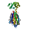
Journal: Cell Res / Year: 2024 Title: Structural basis of tethered agonism and G protein coupling of protease-activated receptors. Authors: Jia Guo / Yun-Li Zhou / Yixin Yang / Shimeng Guo / Erli You / Xin Xie / Yi Jiang / Chunyou Mao / H Eric Xu / Yan Zhang / Abstract: Protease-activated receptors (PARs) are a unique group within the G protein-coupled receptor superfamily, orchestrating cellular responses to extracellular proteases via enzymatic cleavage, which ...Protease-activated receptors (PARs) are a unique group within the G protein-coupled receptor superfamily, orchestrating cellular responses to extracellular proteases via enzymatic cleavage, which triggers intracellular signaling pathways. Protease-activated receptor 1 (PAR1) is a key member of this family and is recognized as a critical pharmacological target for managing thrombotic disorders. In this study, we present cryo-electron microscopy structures of PAR1 in its activated state, induced by its natural tethered agonist (TA), in complex with two distinct downstream proteins, the G and G heterotrimers, respectively. The TA peptide is positioned within a surface pocket, prompting PAR1 activation through notable conformational shifts. Contrary to the typical receptor activation that involves the outward movement of transmembrane helix 6 (TM6), PAR1 activation is characterized by the simultaneous downward shift of TM6 and TM7, coupled with the rotation of a group of aromatic residues. This results in the displacement of an intracellular anion, creating space for downstream G protein binding. Our findings delineate the TA recognition pattern and highlight a distinct role of the second extracellular loop in forming β-sheets with TA within the PAR family, a feature not observed in other TA-activated receptors. Moreover, the nuanced differences in the interactions between intracellular loops 2/3 and the Gα subunit of different G proteins are crucial for determining the specificity of G protein coupling. These insights contribute to our understanding of the ligand binding and activation mechanisms of PARs, illuminating the basis for PAR1's versatility in G protein coupling.
Entire : Cryo-EM structure of ligand-bound form of the receptor in complex...
Entire
Name: Cryo-EM structure of ligand-bound form of the receptor in complex with the transducer
Components
Complex: Cryo-EM structure of ligand-bound form of the receptor in complex with the transducer
Complex: G(I)/G(S)/G(T) subunit beta-1
Protein or peptide: Guanine nucleotide-binding protein G(I)/G(S)/G(T) subunit beta-1
Complex: G(I)/G(S)/G(O) subunit gamma-2
Protein or peptide: Guanine nucleotide-binding protein G(I)/G(S)/G(O) subunit gamma-2
Complex: Gi1-Gq chimeric
Protein or peptide: G subunit q (Gi1-Gq chimeric)
Complex: scFv16
Protein or peptide: scFv16
Complex: Proteinase-activated receptor 1
Protein or peptide: Proteinase-activated receptor 1 LgBiT
Ligand: CHOLESTEROL
+
Supramolecule #1: Cryo-EM structure of ligand-bound form of the receptor in complex...
Supramolecule
Name: Cryo-EM structure of ligand-bound form of the receptor in complex with the transducer type: complex / ID: 1 / Parent: 0 / Macromolecule list: #1-#5
In the structure databanks used in Yorodumi, some data are registered as the other names, "COVID-19 virus" and "2019-nCoV". Here are the details of the virus and the list of structure data.
Jan 31, 2019. EMDB accession codes are about to change! (news from PDBe EMDB page)
EMDB accession codes are about to change! (news from PDBe EMDB page)
The allocation of 4 digits for EMDB accession codes will soon come to an end. Whilst these codes will remain in use, new EMDB accession codes will include an additional digit and will expand incrementally as the available range of codes is exhausted. The current 4-digit format prefixed with “EMD-” (i.e. EMD-XXXX) will advance to a 5-digit format (i.e. EMD-XXXXX), and so on. It is currently estimated that the 4-digit codes will be depleted around Spring 2019, at which point the 5-digit format will come into force.
The EM Navigator/Yorodumi systems omit the EMD- prefix.
Related info.:Q: What is EMD? / ID/Accession-code notation in Yorodumi/EM Navigator
Yorodumi is a browser for structure data from EMDB, PDB, SASBDB, etc.
This page is also the successor to EM Navigator detail page, and also detail information page/front-end page for Omokage search.
The word "yorodu" (or yorozu) is an old Japanese word meaning "ten thousand". "mi" (miru) is to see.
Related info.:EMDB / PDB / SASBDB / Comparison of 3 databanks / Yorodumi Search / Aug 31, 2016. New EM Navigator & Yorodumi / Yorodumi Papers / Jmol/JSmol / Function and homology information / Changes in new EM Navigator and Yorodumi
 Movie
Movie Controller

 Yorodumi
 Open data
 Basic information
 Map data
 Sample
 Keywords
 Function and homology information
 Homo sapiens (human) /
  Authors
 China, 1 items
  Citation
 Structure visualization
 Downloads & links
 emd_38538.png
 http://ftp.pdbj.org/pub/emdb/structures/EMD-38538


 Z (Sec.)
 Y (Row.)
 X (Col.)


 Sample components
 Spodoptera frugiperda (fall armyworm)
 Processing
 Electron microscopy
 FIELD EMISSION GUN