Movie
Movie Controller

[English] 日本語
 Yorodumi
- EMDB-38539: Cryo-EM structure of the tethered agonist-bound human PAR1-Gi complex -
+ Open data

- Basic information
| Entry |  | |||||||||
|---|---|---|---|---|---|---|---|---|---|---|
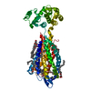
| Title | Cryo-EM structure of the tethered agonist-bound human PAR1-Gi complex | |||||||||
 Map data Map data | ||||||||||
 Sample Sample |
| |||||||||
 Keywords Keywords | GPCR / MEMBRANE PROTEIN / protease-activated receptor | |||||||||
| Function / homology |  Function and homology information Function and homology informationnegative regulation of renin secretion into blood stream / dendritic cell homeostasis / trans-synaptic signaling by endocannabinoid, modulating synaptic transmission / platelet dense tubular network / establishment of synaptic specificity at neuromuscular junction / thrombin-activated receptor activity / connective tissue replacement involved in inflammatory response wound healing / regulation of interleukin-1 beta production / platelet dense granule organization / cell-cell junction maintenance ...negative regulation of renin secretion into blood stream / dendritic cell homeostasis / trans-synaptic signaling by endocannabinoid, modulating synaptic transmission / platelet dense tubular network / establishment of synaptic specificity at neuromuscular junction / thrombin-activated receptor activity / connective tissue replacement involved in inflammatory response wound healing / regulation of interleukin-1 beta production / platelet dense granule organization / cell-cell junction maintenance / positive regulation of smooth muscle contraction / positive regulation of calcium ion transport / G-protein activation / Activation of the phototransduction cascade / Glucagon-type ligand receptors / Thromboxane signalling through TP receptor / Sensory perception of sweet, bitter, and umami (glutamate) taste / G beta:gamma signalling through PI3Kgamma / G beta:gamma signalling through CDC42 / Cooperation of PDCL (PhLP1) and TRiC/CCT in G-protein beta folding / Activation of G protein gated Potassium channels / Inhibition of voltage gated Ca2+ channels via Gbeta/gamma subunits / Ca2+ pathway / G alpha (z) signalling events / High laminar flow shear stress activates signaling by PIEZO1 and PECAM1:CDH5:KDR in endothelial cells / Glucagon-like Peptide-1 (GLP1) regulates insulin secretion / Vasopressin regulates renal water homeostasis via Aquaporins / Adrenaline,noradrenaline inhibits insulin secretion / ADP signalling through P2Y purinoceptor 12 / G alpha (q) signalling events / G alpha (i) signalling events / Thrombin signalling through proteinase activated receptors (PARs) / Activation of G protein gated Potassium channels / G-protein activation / G beta:gamma signalling through PI3Kgamma / Prostacyclin signalling through prostacyclin receptor / G beta:gamma signalling through PLC beta / ADP signalling through P2Y purinoceptor 1 / Thromboxane signalling through TP receptor / Presynaptic function of Kainate receptors / G beta:gamma signalling through CDC42 / Inhibition of voltage gated Ca2+ channels via Gbeta/gamma subunits / G alpha (12/13) signalling events / Glucagon-type ligand receptors / G beta:gamma signalling through BTK / ADP signalling through P2Y purinoceptor 12 / Adrenaline,noradrenaline inhibits insulin secretion / Cooperation of PDCL (PhLP1) and TRiC/CCT in G-protein beta folding / Ca2+ pathway / G alpha (z) signalling events / Thrombin signalling through proteinase activated receptors (PARs) / Extra-nuclear estrogen signaling / G alpha (s) signalling events / G alpha (q) signalling events / thrombin-activated receptor signaling pathway / photoreceptor outer segment membrane / negative regulation of glomerular filtration / spectrin binding / G alpha (i) signalling events / Glucagon-like Peptide-1 (GLP1) regulates insulin secretion / regulation of blood coagulation / High laminar flow shear stress activates signaling by PIEZO1 and PECAM1:CDH5:KDR in endothelial cells / Vasopressin regulates renal water homeostasis via Aquaporins / alkylglycerophosphoethanolamine phosphodiesterase activity / positive regulation of Rho protein signal transduction / positive regulation of collagen biosynthetic process / positive regulation of blood coagulation / positive regulation of vasoconstriction / anatomical structure morphogenesis / photoreceptor outer segment / : / G-protein alpha-subunit binding / cardiac muscle cell apoptotic process / release of sequestered calcium ion into cytosol / adenylate cyclase inhibitor activity / positive regulation of protein localization to cell cortex / photoreceptor inner segment / T cell migration / positive regulation of relaxation of smooth muscle / Adenylate cyclase inhibitory pathway / D2 dopamine receptor binding / homeostasis of number of cells within a tissue / adenylate cyclase-inhibiting serotonin receptor signaling pathway / G protein-coupled serotonin receptor binding / cellular response to forskolin / Peptide ligand-binding receptors / positive regulation of release of sequestered calcium ion into cytosol / regulation of mitotic spindle organization / positive regulation of interleukin-8 production / chemokine-mediated signaling pathway / positive regulation of receptor signaling pathway via JAK-STAT / neuromuscular junction / Regulation of insulin secretion / neuropeptide signaling pathway / response to prostaglandin E / platelet activation / positive regulation of cholesterol biosynthetic process / negative regulation of insulin secretion / caveola / response to wounding Similarity search - Function | |||||||||
| Biological species |  Homo sapiens (human) / Homo sapiens (human) /  | |||||||||
| Method | single particle reconstruction / cryo EM / Resolution: 3.2 Å | |||||||||
 Authors Authors | Guo J / Zhang Y | |||||||||
| Funding support |  China, 1 items China, 1 items
| |||||||||
 Citation Citation | Journal: Cell Res / Year: 2024 Title: Structural basis of tethered agonism and G protein coupling of protease-activated receptors. Authors: Jia Guo / Yun-Li Zhou / Yixin Yang / Shimeng Guo / Erli You / Xin Xie / Yi Jiang / Chunyou Mao / H Eric Xu / Yan Zhang / Abstract: Protease-activated receptors (PARs) are a unique group within the G protein-coupled receptor superfamily, orchestrating cellular responses to extracellular proteases via enzymatic cleavage, which ...Protease-activated receptors (PARs) are a unique group within the G protein-coupled receptor superfamily, orchestrating cellular responses to extracellular proteases via enzymatic cleavage, which triggers intracellular signaling pathways. Protease-activated receptor 1 (PAR1) is a key member of this family and is recognized as a critical pharmacological target for managing thrombotic disorders. In this study, we present cryo-electron microscopy structures of PAR1 in its activated state, induced by its natural tethered agonist (TA), in complex with two distinct downstream proteins, the G and G heterotrimers, respectively. The TA peptide is positioned within a surface pocket, prompting PAR1 activation through notable conformational shifts. Contrary to the typical receptor activation that involves the outward movement of transmembrane helix 6 (TM6), PAR1 activation is characterized by the simultaneous downward shift of TM6 and TM7, coupled with the rotation of a group of aromatic residues. This results in the displacement of an intracellular anion, creating space for downstream G protein binding. Our findings delineate the TA recognition pattern and highlight a distinct role of the second extracellular loop in forming β-sheets with TA within the PAR family, a feature not observed in other TA-activated receptors. Moreover, the nuanced differences in the interactions between intracellular loops 2/3 and the Gα subunit of different G proteins are crucial for determining the specificity of G protein coupling. These insights contribute to our understanding of the ligand binding and activation mechanisms of PARs, illuminating the basis for PAR1's versatility in G protein coupling. | |||||||||
| History |
|
- Structure visualization
| Supplemental images |
|---|

- Downloads & links
-EMDB archive
| Map data | emd_38539.map.gz | 23.3 MB | EMDB map data format | |
|---|---|---|---|---|
| Header (meta data) | emd-38539-v30.xmlemd-38539.xml | 22.5 KB 22.5 KB | Display Display | EMDB header |
| Images |  emd_38539.png emd_38539.png | 50.6 KB | ||
| Filedesc metadata | emd-38539.cif.gz | 7.3 KB | ||
| Others | emd_38539_half_map_1.map.gzemd_38539_half_map_2.map.gz | 23.4 MB 23.4 MB | ||
| Archive directory |  http://ftp.pdbj.org/pub/emdb/structures/EMD-38539ftp://ftp.pdbj.org/pub/emdb/structures/EMD-38539 http://ftp.pdbj.org/pub/emdb/structures/EMD-38539ftp://ftp.pdbj.org/pub/emdb/structures/EMD-38539 | HTTPS FTP |
-Related structure data
| Related structure data |  8xosMC  8xorC M: atomic model generated by this map C: citing same article ( |
|---|---|
| Similar structure data |
-Links
| EMDB pages | EMDB (EBI/PDBe) / EMDataResource |
|---|---|
| Related items in Molecule of the Month |


-Map
| File | Download / File: emd_38539.map.gz / Format: CCP4 / Size: 30.5 MB / Type: IMAGE STORED AS FLOATING POINT NUMBER (4 BYTES) | ||||||||||||||||||||||||||||||||||||
|---|---|---|---|---|---|---|---|---|---|---|---|---|---|---|---|---|---|---|---|---|---|---|---|---|---|---|---|---|---|---|---|---|---|---|---|---|---|
| Projections & slices | Image control
Images are generated by Spider. | ||||||||||||||||||||||||||||||||||||
| Voxel size | X=Y=Z: 1.071 Å | ||||||||||||||||||||||||||||||||||||
| Density |
| ||||||||||||||||||||||||||||||||||||
| Symmetry | Space group: 1 | ||||||||||||||||||||||||||||||||||||
| Details | EMDB XML:
|
 Z (Sec.)
 Y (Row.)
 X (Col.)


-Supplemental data
-Half map: #1
| File | emd_38539_half_map_1.map | ||||||||||||
|---|---|---|---|---|---|---|---|---|---|---|---|---|---|
| Projections & Slices |
| ||||||||||||
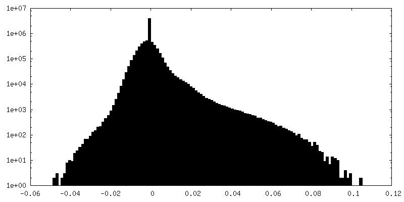
| Density Histograms |


-Half map: #2
| File | emd_38539_half_map_2.map | ||||||||||||
|---|---|---|---|---|---|---|---|---|---|---|---|---|---|
| Projections & Slices |
| ||||||||||||
| Density Histograms |


- Sample components
-Entire : tethered agonist-bound human PAR1-Gi complex
| Entire | Name: tethered agonist-bound human PAR1-Gi complex |
|---|---|
| Components |
|
-Supramolecule #1: tethered agonist-bound human PAR1-Gi complex
| Supramolecule | Name: tethered agonist-bound human PAR1-Gi complex / type: complex / ID: 1 / Parent: 0 / Macromolecule list: #1-#5 |
|---|---|
| Source (natural) | Organism: Homo sapiens (human) |
-Macromolecule #1: Guanine nucleotide-binding protein G(i) subunit alpha-1
| Macromolecule | Name: Guanine nucleotide-binding protein G(i) subunit alpha-1 type: protein_or_peptide / ID: 1 / Number of copies: 1 / Enantiomer: LEVO |
|---|---|
| Source (natural) | Organism: Homo sapiens (human) |
| Molecular weight | Theoretical: 40.313863 KDa |
| Recombinant expression | Organism:   Spodoptera frugiperda (fall armyworm) Spodoptera frugiperda (fall armyworm) |
| Sequence | String: GCTLSAEDKA AVERSKMIDR NLREDGEKAA REVKLLLLGA GESGKSTIVK QMKIIHEAGY SEEECKQYKA VVYSNTIQSI IAIIRAMGR LKIDFGDSAR ADDARQLFVL AGAAEEGFMT AELAGVIKRL WKDSGVQACF NRSREYQLND SAAYYLNDLD R IAQPNYIP ...String: GCTLSAEDKA AVERSKMIDR NLREDGEKAA REVKLLLLGA GESGKSTIVK QMKIIHEAGY SEEECKQYKA VVYSNTIQSI IAIIRAMGR LKIDFGDSAR ADDARQLFVL AGAAEEGFMT AELAGVIKRL WKDSGVQACF NRSREYQLND SAAYYLNDLD R IAQPNYIP TQQDVLRTRV KTTGIVETHF TFKDLHFKMF DVGAQRSERK KWIHCFEGVT AIIFCVALSD YDLVLAEDEE MN RMHESMK LFDSICNNKW FTDTSIILFL NKKDLFEEKI KKSPLTICYP EYAGSNTYEE AAAYIQCQFE DLNKRKDTKE IYT HFTCST DTKNVQFVFD AVTDVIIKNN LKDCGLF UniProtKB: Guanine nucleotide-binding protein G(i) subunit alpha-1 |
-Macromolecule #2: Guanine nucleotide-binding protein G(I)/G(S)/G(T) subunit beta-1
| Macromolecule | Name: Guanine nucleotide-binding protein G(I)/G(S)/G(T) subunit beta-1 type: protein_or_peptide / ID: 2 / Number of copies: 1 / Enantiomer: LEVO |
|---|---|
| Source (natural) | Organism: |
| Molecular weight | Theoretical: 41.332137 KDa |
| Recombinant expression | Organism: Spodoptera frugiperda (fall armyworm) |
| Sequence | String: MVSGWRLFKK ISGSSGGGGS GGGGSSGGSL LQSELDQLRQ EAEQLKNQIR DARKACADAT LSQITNNIDP VGRIQMRTRR TLRGHLAKI YAMHWGTDSR LLVSASQDGK LIIWDSYTTN KVHAIPLRSS WVMTCAYAPS GNYVACGGLD NICSIYNLKT R EGNVRVSR ...String: MVSGWRLFKK ISGSSGGGGS GGGGSSGGSL LQSELDQLRQ EAEQLKNQIR DARKACADAT LSQITNNIDP VGRIQMRTRR TLRGHLAKI YAMHWGTDSR LLVSASQDGK LIIWDSYTTN KVHAIPLRSS WVMTCAYAPS GNYVACGGLD NICSIYNLKT R EGNVRVSR ELAGHTGYLS CCRFLDDNQI VTSSGDTTCA LWDIETGQQT TTFTGHTGDV MSLSLAPDTR LFVSGACDAS AK LWDVREG MCRQTFTGHE SDINAICFFP NGNAFATGSD DATCRLFDLR ADQELMTYSH DNIICGITSV SFSKSGRLLL AGY DDFNCN VWDALKADRA GVLAGHDNRV SCLGVTDDGM AVATGSWDSF LKIWNHHHHH HHH UniProtKB: Guanine nucleotide-binding protein G(I)/G(S)/G(T) subunit beta-1 |
-Macromolecule #3: Guanine nucleotide-binding protein G(I)/G(S)/G(O) subunit gamma-2
| Macromolecule | Name: Guanine nucleotide-binding protein G(I)/G(S)/G(O) subunit gamma-2 type: protein_or_peptide / ID: 3 / Number of copies: 1 / Enantiomer: LEVO |
|---|---|
| Source (natural) | Organism: |
| Molecular weight | Theoretical: 7.432554 KDa |
| Recombinant expression | Organism: Spodoptera frugiperda (fall armyworm) |
| Sequence | String: ASNNTASIAQ ARKLVEQLKM EANIDRIKVS KAAADLMAYC EAHAKEDPLL TPVPASENPF REKKFFC UniProtKB: Guanine nucleotide-binding protein G(I)/G(S)/G(O) subunit gamma-2 |
-Macromolecule #4: scFv16
| Macromolecule | Name: scFv16 / type: protein_or_peptide / ID: 4 / Number of copies: 1 / Enantiomer: LEVO |
|---|---|
| Source (natural) | Organism: |
| Molecular weight | Theoretical: 40.523266 KDa |
| Recombinant expression | Organism: Spodoptera frugiperda (fall armyworm) |
| Sequence | String: LLVNQSHQGF NKEHTSKMVS AIVLYVLLAA AAHSAFAVQL VESGGGLVQP GGSRKLSCSA SGFAFSSFGM HWVRQAPEKG LEWVAYISS GSGTIYYADT VKGRFTISRD DPKNTLFLQM TSLRSEDTAM YYCVRSIYYY GSSPFDFWGQ GTTLTVSAGG G GSGGGGSG ...String: LLVNQSHQGF NKEHTSKMVS AIVLYVLLAA AAHSAFAVQL VESGGGLVQP GGSRKLSCSA SGFAFSSFGM HWVRQAPEKG LEWVAYISS GSGTIYYADT VKGRFTISRD DPKNTLFLQM TSLRSEDTAM YYCVRSIYYY GSSPFDFWGQ GTTLTVSAGG G GSGGGGSG GGGSADIVMT QATSSVPVTP GESVSISCRS SKSLLHSNGN TYLYWFLQRP GQSPQLLIYR MSNLASGVPD RF SGSGSGT AFTLTISRLE AEDVGVYYCM QHLEYPLTFG AGTKLELVDE NLYFQGASHH HHHHHHWFLQ RPGQSPQLLI YRM SNLASG VPDRFSGSGS GTAFTLTISR LEAEDVGVYY CMQHLEYPLT FGAGTKLEL |
-Macromolecule #5: Proteinase-activated receptor 1 LgBiT
| Macromolecule | Name: Proteinase-activated receptor 1 LgBiT / type: protein_or_peptide / ID: 5 / Number of copies: 1 / Enantiomer: LEVO |
|---|---|
| Source (natural) | Organism: Homo sapiens (human) |
| Molecular weight | Theoretical: 58.787539 KDa |
| Recombinant expression | Organism: Spodoptera frugiperda (fall armyworm) |
| Sequence | String: SFLLRNPNDK YEPFWEDEEK NESGLTEYRL VSINKSSPLQ KQLPAFISED ASGYLTSSWL TLFVPSVYTG VFVVSLPLNI MAIVVFILK MKVKKPAVVY MLHLATADVL FVSVLPFKIS YYFSGSDWQF GSELCRFVTA AFYCNMYASI LLMTVISIDR F LAVVYPMQ ...String: SFLLRNPNDK YEPFWEDEEK NESGLTEYRL VSINKSSPLQ KQLPAFISED ASGYLTSSWL TLFVPSVYTG VFVVSLPLNI MAIVVFILK MKVKKPAVVY MLHLATADVL FVSVLPFKIS YYFSGSDWQF GSELCRFVTA AFYCNMYASI LLMTVISIDR F LAVVYPMQ SLSWRTLGRA SFTCLAIWAL AIAGVVPLLL KEQTIQVPGL NITTCHDVLN ETLLEGYYAY YFSAFSAVFF FV PLIISTV CYVSIIRCLS SSAVANRSKK SRALFLSAAV FCIFIICFGP TNVLLIAHYS FLSHTSTTEA AYFAYLLCVC VSS ISCCID PLIYYYASSE CQRYVYSILC CKESSDPSSY GSSGGGGSGG GGSSGVFTLE DFVGDWEQTA AYNLDQVLEQ GGVS SLLQN LAVSVTPIQR IVRSGENALK IDIHVIIPYE GLSADQMAQI EEVFKVVYPV DDHHFKVILP YGTLVIDGVT PNMLN YFGR PYEGIAVFDG KKITVTGTLW NGNKIIDERL ITPDGSMLFR VTINSGGS UniProtKB: Proteinase-activated receptor 1 |
-Macromolecule #6: CHOLESTEROL
| Macromolecule | Name: CHOLESTEROL / type: ligand / ID: 6 / Number of copies: 2 / Formula: CLR |
|---|---|
| Molecular weight | Theoretical: 386.654 Da |
| Chemical component information |  ChemComp-CLR: |
-Experimental details
-Structure determination
| Method | cryo EM |
|---|---|
 Processing Processing | single particle reconstruction |
| Aggregation state | particle |
-Sample preparation
| Buffer | pH: 7.5 |
|---|---|
| Grid | Model: Quantifoil R1.2/1.3 / Material: GOLD / Mesh: 300 / Support film - Material: CARBON / Support film - topology: HOLEY |
| Vitrification | Cryogen name: ETHANE / Chamber humidity: 100 % / Chamber temperature: 277 K |
- Electron microscopy
| Microscope | FEI TITAN KRIOS |
|---|---|
| Image recording | Film or detector model: GATAN K3 (6k x 4k) / Average electron dose: 70.0 e/Å2 |
| Electron beam | Acceleration voltage: 300 kV / Electron source:  FIELD EMISSION GUN FIELD EMISSION GUN |
| Electron optics | Illumination mode: FLOOD BEAM / Imaging mode: BRIGHT FIELD / Nominal defocus max: 2.0 µm / Nominal defocus min: 0.5 µm |
| Experimental equipment |  Model: Titan Krios / Image courtesy: FEI Company |