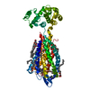
[日本語] English

- EMDB-38538: Cryo-EM structure of the tethered agonist-bound human PAR1-Gq complex -
+
データを開く
-
基本情報
登録情報 | ![]() | |||||||||
---|---|---|---|---|---|---|---|---|---|---|
タイトル | Cryo-EM structure of the tethered agonist-bound human PAR1-Gq complex | |||||||||
![]() | ||||||||||
![]() |
| |||||||||
![]() | GPCR / MEMBRANE PROTEIN / protease-activated receptor | |||||||||
機能・相同性 | ![]() negative regulation of renin secretion into blood stream / dendritic cell homeostasis / trans-synaptic signaling by endocannabinoid, modulating synaptic transmission / establishment of synaptic specificity at neuromuscular junction / platelet dense tubular network / thrombin-activated receptor activity / regulation of interleukin-1 beta production / connective tissue replacement involved in inflammatory response wound healing / platelet dense granule organization / cell-cell junction maintenance ...negative regulation of renin secretion into blood stream / dendritic cell homeostasis / trans-synaptic signaling by endocannabinoid, modulating synaptic transmission / establishment of synaptic specificity at neuromuscular junction / platelet dense tubular network / thrombin-activated receptor activity / regulation of interleukin-1 beta production / connective tissue replacement involved in inflammatory response wound healing / platelet dense granule organization / cell-cell junction maintenance / regulation of sensory perception of pain / positive regulation of smooth muscle contraction / positive regulation of calcium ion transport / G-protein activation / Activation of the phototransduction cascade / Glucagon-type ligand receptors / Thromboxane signalling through TP receptor / Sensory perception of sweet, bitter, and umami (glutamate) taste / G beta:gamma signalling through PI3Kgamma / G beta:gamma signalling through CDC42 / Cooperation of PDCL (PhLP1) and TRiC/CCT in G-protein beta folding / Activation of G protein gated Potassium channels / Inhibition of voltage gated Ca2+ channels via Gbeta/gamma subunits / Ca2+ pathway / G alpha (z) signalling events / High laminar flow shear stress activates signaling by PIEZO1 and PECAM1:CDH5:KDR in endothelial cells / Glucagon-like Peptide-1 (GLP1) regulates insulin secretion / Vasopressin regulates renal water homeostasis via Aquaporins / Adrenaline,noradrenaline inhibits insulin secretion / ADP signalling through P2Y purinoceptor 12 / G alpha (q) signalling events / G alpha (i) signalling events / Activation of G protein gated Potassium channels / G-protein activation / G beta:gamma signalling through PI3Kgamma / Prostacyclin signalling through prostacyclin receptor / G beta:gamma signalling through PLC beta / ADP signalling through P2Y purinoceptor 1 / Thromboxane signalling through TP receptor / Presynaptic function of Kainate receptors / G beta:gamma signalling through CDC42 / Inhibition of voltage gated Ca2+ channels via Gbeta/gamma subunits / Thrombin signalling through proteinase activated receptors (PARs) / G alpha (12/13) signalling events / Glucagon-type ligand receptors / G beta:gamma signalling through BTK / ADP signalling through P2Y purinoceptor 12 / Adrenaline,noradrenaline inhibits insulin secretion / Cooperation of PDCL (PhLP1) and TRiC/CCT in G-protein beta folding / Ca2+ pathway / Thrombin signalling through proteinase activated receptors (PARs) / G alpha (z) signalling events / Extra-nuclear estrogen signaling / G alpha (s) signalling events / photoreceptor outer segment membrane / G alpha (q) signalling events / G alpha (i) signalling events / spectrin binding / negative regulation of glomerular filtration / Glucagon-like Peptide-1 (GLP1) regulates insulin secretion / thrombin-activated receptor signaling pathway / High laminar flow shear stress activates signaling by PIEZO1 and PECAM1:CDH5:KDR in endothelial cells / Vasopressin regulates renal water homeostasis via Aquaporins / positive regulation of Rho protein signal transduction / regulation of blood coagulation / alkylglycerophosphoethanolamine phosphodiesterase activity / positive regulation of collagen biosynthetic process / anatomical structure morphogenesis / photoreceptor outer segment / positive regulation of blood coagulation / G-protein alpha-subunit binding / Common Pathway of Fibrin Clot Formation / homeostasis of number of cells within a tissue / photoreceptor inner segment / positive regulation of vasoconstriction / release of sequestered calcium ion into cytosol / cardiac muscle cell apoptotic process / positive regulation of GTPase activity / positive regulation of release of sequestered calcium ion into cytosol / Peptide ligand-binding receptors / positive regulation of interleukin-8 production / positive regulation of receptor signaling pathway via JAK-STAT / G protein-coupled receptor activity / neuromuscular junction / regulation of synaptic plasticity / caveola / platelet activation / response to wounding / positive regulation of interleukin-6 production / cellular response to catecholamine stimulus / adenylate cyclase-activating dopamine receptor signaling pathway / late endosome / cellular response to prostaglandin E stimulus / G-protein beta-subunit binding / heterotrimeric G-protein complex / sensory perception of taste / signaling receptor complex adaptor activity / Thrombin signalling through proteinase activated receptors (PARs) / cell body / positive regulation of cytosolic calcium ion concentration 類似検索 - 分子機能 | |||||||||
生物種 | ![]() ![]() ![]() ![]() ![]() ![]() ![]() | |||||||||
手法 | 単粒子再構成法 / クライオ電子顕微鏡法 / 解像度: 3.0 Å | |||||||||
![]() | Guo J / Zhang Y / Shen Q / Zang S / Shen D / Zhang H / Chen Z / Wang G / Zhang C / Liu Z | |||||||||
資金援助 | ![]()
| |||||||||
![]() | ![]() タイトル: Structural basis of tethered agonism and G protein coupling of protease-activated receptors. 著者: Jia Guo / Yun-Li Zhou / Yixin Yang / Shimeng Guo / Erli You / Xin Xie / Yi Jiang / Chunyou Mao / H Eric Xu / Yan Zhang / ![]() 要旨: Protease-activated receptors (PARs) are a unique group within the G protein-coupled receptor superfamily, orchestrating cellular responses to extracellular proteases via enzymatic cleavage, which ...Protease-activated receptors (PARs) are a unique group within the G protein-coupled receptor superfamily, orchestrating cellular responses to extracellular proteases via enzymatic cleavage, which triggers intracellular signaling pathways. Protease-activated receptor 1 (PAR1) is a key member of this family and is recognized as a critical pharmacological target for managing thrombotic disorders. In this study, we present cryo-electron microscopy structures of PAR1 in its activated state, induced by its natural tethered agonist (TA), in complex with two distinct downstream proteins, the G and G heterotrimers, respectively. The TA peptide is positioned within a surface pocket, prompting PAR1 activation through notable conformational shifts. Contrary to the typical receptor activation that involves the outward movement of transmembrane helix 6 (TM6), PAR1 activation is characterized by the simultaneous downward shift of TM6 and TM7, coupled with the rotation of a group of aromatic residues. This results in the displacement of an intracellular anion, creating space for downstream G protein binding. Our findings delineate the TA recognition pattern and highlight a distinct role of the second extracellular loop in forming β-sheets with TA within the PAR family, a feature not observed in other TA-activated receptors. Moreover, the nuanced differences in the interactions between intracellular loops 2/3 and the Gα subunit of different G proteins are crucial for determining the specificity of G protein coupling. These insights contribute to our understanding of the ligand binding and activation mechanisms of PARs, illuminating the basis for PAR1's versatility in G protein coupling. | |||||||||
履歴 |
|
-
構造の表示
添付画像 |
---|
-
ダウンロードとリンク
-EMDBアーカイブ
マップデータ | ![]() | 23.2 MB | ![]() | |
---|---|---|---|---|
ヘッダ (付随情報) | ![]() ![]() | 22.9 KB 22.9 KB | 表示 表示 | ![]() |
画像 | ![]() | 44.9 KB | ||
Filedesc metadata | ![]() | 7 KB | ||
その他 | ![]() ![]() | 23.3 MB 23.3 MB | ||
アーカイブディレクトリ | ![]() ![]() | HTTPS FTP |
-検証レポート
文書・要旨 | ![]() | 720.8 KB | 表示 | ![]() |
---|---|---|---|---|
文書・詳細版 | ![]() | 720.4 KB | 表示 | |
XML形式データ | ![]() | 10.6 KB | 表示 | |
CIF形式データ | ![]() | 12.4 KB | 表示 | |
アーカイブディレクトリ | ![]() ![]() | HTTPS FTP |
-関連構造データ
関連構造データ | ![]() 8xorMC ![]() 8xosC M: このマップから作成された原子モデル C: 同じ文献を引用 ( |
---|---|
類似構造データ | 類似検索 - 機能・相同性 ![]() |
-
リンク
EMDBのページ | ![]() ![]() |
---|---|
「今月の分子」の関連する項目 |
-
マップ
ファイル | ![]() | ||||||||||||||||||||||||||||||||||||
---|---|---|---|---|---|---|---|---|---|---|---|---|---|---|---|---|---|---|---|---|---|---|---|---|---|---|---|---|---|---|---|---|---|---|---|---|---|
投影像・断面図 | 画像のコントロール
画像は Spider により作成 | ||||||||||||||||||||||||||||||||||||
ボクセルのサイズ | X=Y=Z: 1.071 Å | ||||||||||||||||||||||||||||||||||||
密度 |
| ||||||||||||||||||||||||||||||||||||
対称性 | 空間群: 1 | ||||||||||||||||||||||||||||||||||||
詳細 | EMDB XML:
|
-添付データ
-ハーフマップ: #2
ファイル | emd_38538_half_map_1.map | ||||||||||||
---|---|---|---|---|---|---|---|---|---|---|---|---|---|
投影像・断面図 |
| ||||||||||||
密度ヒストグラム |
-ハーフマップ: #1
ファイル | emd_38538_half_map_2.map | ||||||||||||
---|---|---|---|---|---|---|---|---|---|---|---|---|---|
投影像・断面図 |
| ||||||||||||
密度ヒストグラム |
-
試料の構成要素
+全体 : Cryo-EM structure of ligand-bound form of the receptor in complex...
+超分子 #1: Cryo-EM structure of ligand-bound form of the receptor in complex...
+超分子 #2: G(I)/G(S)/G(T) subunit beta-1
+超分子 #3: G(I)/G(S)/G(O) subunit gamma-2
+超分子 #4: Gi1-Gq chimeric
+超分子 #5: scFv16
+超分子 #6: Proteinase-activated receptor 1
+分子 #1: Guanine nucleotide-binding protein G(I)/G(S)/G(T) subunit beta-1
+分子 #2: Guanine nucleotide-binding protein G(I)/G(S)/G(O) subunit gamma-2
+分子 #3: G subunit q (Gi1-Gq chimeric)
+分子 #4: scFv16
+分子 #5: Proteinase-activated receptor 1 LgBiT
+分子 #6: CHOLESTEROL
-実験情報
-構造解析
手法 | クライオ電子顕微鏡法 |
---|---|
![]() | 単粒子再構成法 |
試料の集合状態 | particle |
-
試料調製
緩衝液 | pH: 7.5 |
---|---|
グリッド | モデル: Quantifoil R1.2/1.3 / 材質: GOLD / メッシュ: 300 / 支持フィルム - 材質: CARBON / 支持フィルム - トポロジー: HOLEY |
凍結 | 凍結剤: ETHANE / チャンバー内湿度: 100 % / チャンバー内温度: 277 K |
-
電子顕微鏡法
顕微鏡 | FEI TITAN KRIOS |
---|---|
撮影 | フィルム・検出器のモデル: GATAN K3 (6k x 4k) / デジタル化 - 画像ごとのフレーム数: 1-40 / 実像数: 5048 / 平均電子線量: 70.0 e/Å2 |
電子線 | 加速電圧: 300 kV / 電子線源: ![]() |
電子光学系 | 倍率(補正後): 49310 / 照射モード: FLOOD BEAM / 撮影モード: BRIGHT FIELD / 最大 デフォーカス(公称値): 2.0 µm / 最小 デフォーカス(公称値): 0.5 µm / 倍率(公称値): 29000 |
試料ステージ | 試料ホルダーモデル: FEI TITAN KRIOS AUTOGRID HOLDER ホルダー冷却材: NITROGEN |
実験機器 | ![]() モデル: Titan Krios / 画像提供: FEI Company |
-
画像解析
初期モデル | モデルのタイプ: PDB ENTRY PDBモデル - PDB ID: |
---|---|
最終 再構成 | 解像度のタイプ: BY AUTHOR / 解像度: 3.0 Å / 解像度の算出法: FSC 0.143 CUT-OFF / 使用した粒子像数: 723778 |
初期 角度割当 | タイプ: RANDOM ASSIGNMENT |
最終 角度割当 | タイプ: MAXIMUM LIKELIHOOD |