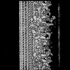
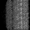
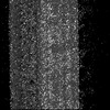
 Movie
Movie Controller

[English] 日本語
 Yorodumi
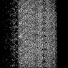
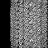
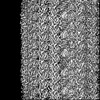
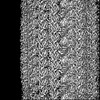
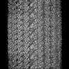
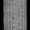
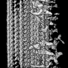
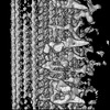
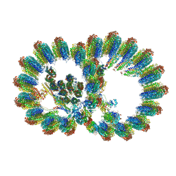
- EMDB-35225: A3 part of axonemal doublet microtubules in mouse sperm with 48-n... -
+ Open data

- Basic information
| Entry | 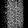 | |||||||||
|---|---|---|---|---|---|---|---|---|---|---|
| Title | A3 part of axonemal doublet microtubules in mouse sperm with 48-nm repeat | |||||||||
 Map data Map data | A3 part of axonemal doublet microtubules in mouse sperm with 48-nm repeat | |||||||||
 Sample Sample |
| |||||||||
 Keywords Keywords | microtubules / axoneme / sperm / filament / PROTEIN TRANSPORT | |||||||||
| Biological species |  | |||||||||
| Method | subtomogram averaging / cryo EM / Resolution: 6.6 Å | |||||||||
 Authors Authors | Zhu Y / Tai LH / Yin GL / Sun F | |||||||||
| Funding support |  China, 1 items China, 1 items
| |||||||||
 Citation Citation | Journal: Cell Discov / Year: 2023 Title: In-cell structural insight into the stability of sperm microtubule doublet. Authors: Tai L / Yin G / Huang X / Sun F / Zhu Y | |||||||||
| History |
|
- Structure visualization
- Downloads & links
-EMDB archive
| Map data | emd_35225.map.gz | 58.8 MB |  EMDB map data format EMDB map data format | |
|---|---|---|---|---|
| Header (meta data) | emd-35225-v30.xmlemd-35225.xml | 12 KB 12 KB | Display Display | EMDB header |
| FSC (resolution estimation) | emd_35225_fsc.xml | 9.1 KB | Display | FSC data file |
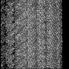
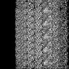
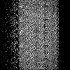
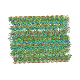
| Images |  emd_35225.png emd_35225.png | 147.9 KB | ||
| Masks | emd_35225_msk_1.map | 64 MB | Mask map | |
| Filedesc metadata | emd-35225.cif.gz | 3.8 KB | ||
| Others | emd_35225_half_map_1.map.gzemd_35225_half_map_2.map.gz | 59.4 MB 59.4 MB | ||
| Archive directory |  http://ftp.pdbj.org/pub/emdb/structures/EMD-35225ftp://ftp.pdbj.org/pub/emdb/structures/EMD-35225 http://ftp.pdbj.org/pub/emdb/structures/EMD-35225ftp://ftp.pdbj.org/pub/emdb/structures/EMD-35225 | HTTPS FTP |
-Validation report
| Summary document | emd_35225_validation.pdf.gz | 1.6 MB | Display | EMDB validaton report |
|---|---|---|---|---|
| Full document | emd_35225_full_validation.pdf.gz | 1.6 MB | Display | |
| Data in XML | emd_35225_validation.xml.gz | 15.8 KB | Display | |
| Data in CIF | emd_35225_validation.cif.gz | 20.7 KB | Display | |
| Arichive directory | https://ftp.pdbj.org/pub/emdb/validation_reports/EMD-35225ftp://ftp.pdbj.org/pub/emdb/validation_reports/EMD-35225 | HTTPS FTP |
-Related structure data

-Links
| EMDB pages | EMDB (EBI/PDBe) / EMDataResource |
|---|
-Map
| File | Download / File: emd_35225.map.gz / Format: CCP4 / Size: 64 MB / Type: IMAGE STORED AS FLOATING POINT NUMBER (4 BYTES) | ||||||||||||||||||||
|---|---|---|---|---|---|---|---|---|---|---|---|---|---|---|---|---|---|---|---|---|---|
| Annotation | A3 part of axonemal doublet microtubules in mouse sperm with 48-nm repeat | ||||||||||||||||||||
| Voxel size | X=Y=Z: 1.76 Å | ||||||||||||||||||||
| Density |
| ||||||||||||||||||||
| Symmetry | Space group: 1 | ||||||||||||||||||||
| Details | EMDB XML:
|
-Supplemental data
- Sample components
-Entire : mouse sperm
| Entire | Name: mouse sperm |
|---|---|
| Components |
|
-Supramolecule #1: mouse sperm
| Supramolecule | Name: mouse sperm / type: cell / ID: 1 / Parent: 0 |
|---|---|
| Source (natural) | Organism: |
-Experimental details
-Structure determination
| Method | cryo EM |
|---|---|
 Processing Processing | subtomogram averaging |
| Aggregation state | cell |
-Sample preparation
| Buffer | pH: 7 |
|---|---|
| Vitrification | Cryogen name: ETHANE |
- Electron microscopy
| Microscope | FEI TITAN KRIOS |
|---|---|
| Image recording | Film or detector model: GATAN K2 QUANTUM (4k x 4k) / Detector mode: SUPER-RESOLUTION / Average electron dose: 3.0 e/Å2 |
| Electron beam | Acceleration voltage: 300 kV / Electron source:  FIELD EMISSION GUN FIELD EMISSION GUN |
| Electron optics | Illumination mode: SPOT SCAN / Imaging mode: BRIGHT FIELD / Cs: 2.7 mm / Nominal defocus max: 5.0 µm / Nominal defocus min: 1.0 µm |
| Experimental equipment |  Model: Titan Krios / Image courtesy: FEI Company |
-Image processing
| Final reconstruction | Applied symmetry - Point group: C1 (asymmetric) / Resolution.type: BY AUTHOR / Resolution: 6.6 Å / Resolution method: FSC 0.143 CUT-OFF / Number subtomograms used: 17450 |
|---|---|
| Extraction | Number tomograms: 689 / Number images used: 17450 |
| Final angle assignment | Type: MAXIMUM LIKELIHOOD |
