 Movie
Movie Controller

[English] 日本語
 Yorodumi
- EMDB-35230: In situ structure of axonemal doublet microtubules in mouse sperm... -
+ Open data

- Basic information
| Entry | 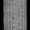 | |||||||||
|---|---|---|---|---|---|---|---|---|---|---|
| Title | In situ structure of axonemal doublet microtubules in mouse sperm with 48-nm repeat | |||||||||
 Map data Map data | In situ structure of axonemal doublet microtubules in mouse sperm with 48-nm repeat | |||||||||
 Sample Sample |
| |||||||||
 Keywords Keywords | microtubules / axoneme / sperm / filament / STRUCTURAL PROTEIN | |||||||||
| Function / homology |  Function and homology information Function and homology informationprotein localization to motile cilium / outer acrosomal membrane / epithelial cilium movement involved in determination of left/right asymmetry / regulation of brood size / establishment of left/right asymmetry / 9+0 motile cilium / axonemal microtubule doublet inner sheath / manchette assembly / sperm flagellum assembly / axonemal B tubule inner sheath ...protein localization to motile cilium / outer acrosomal membrane / epithelial cilium movement involved in determination of left/right asymmetry / regulation of brood size / establishment of left/right asymmetry / 9+0 motile cilium / axonemal microtubule doublet inner sheath / manchette assembly / sperm flagellum assembly / axonemal B tubule inner sheath / axonemal A tubule inner sheath / Microtubule-dependent trafficking of connexons from Golgi to the plasma membrane / Cilium Assembly / Sealing of the nuclear envelope (NE) by ESCRT-III / protein polyglutamylation / left/right pattern formation / sperm axoneme assembly / regulation of calcineurin-NFAT signaling cascade / regulation of microtubule nucleation / Intraflagellar transport / Carboxyterminal post-translational modifications of tubulin / positive regulation of feeding behavior / COPI-independent Golgi-to-ER retrograde traffic / cerebrospinal fluid circulation / MAP kinase tyrosine/serine/threonine phosphatase activity / inner dynein arm assembly / cilium-dependent cell motility / epithelial cilium movement involved in extracellular fluid movement / regulation of cilium beat frequency involved in ciliary motility / HSP90 chaperone cycle for steroid hormone receptors (SHR) in the presence of ligand / COPI-mediated anterograde transport / cilium movement involved in cell motility / 9+2 motile cilium / intraciliary transport / Kinesins / regulation of store-operated calcium entry / acrosomal membrane / cilium movement / axoneme assembly / PKR-mediated signaling / left/right axis specification / Aggrephagy / RHO GTPases activate IQGAPs / microtubule sliding / Mitotic Prometaphase / EML4 and NUDC in mitotic spindle formation / Resolution of Sister Chromatid Cohesion / Recycling pathway of L1 / calcium ion sensor activity / The role of GTSE1 in G2/M progression after G2 checkpoint / COPI-dependent Golgi-to-ER retrograde traffic / axonemal microtubule / Hedgehog 'off' state / RHO GTPases Activate Formins / Loss of Nlp from mitotic centrosomes / Recruitment of mitotic centrosome proteins and complexes / Loss of proteins required for interphase microtubule organization from the centrosome / Anchoring of the basal body to the plasma membrane / Separation of Sister Chromatids / cilium organization / Recruitment of NuMA to mitotic centrosomes / AURKA Activation by TPX2 / gamma-tubulin ring complex / manchette / Regulation of PLK1 Activity at G2/M Transition / positive regulation of cilium assembly / MHC class II antigen presentation / 3'-5'-DNA exonuclease activity / flagellated sperm motility / UTP biosynthetic process / CTP biosynthetic process / motile cilium / determination of left/right symmetry / DNA catabolic process / extrinsic component of membrane / nucleoside diphosphate kinase activity / protein targeting to membrane / positive regulation of cell motility / : / GTP biosynthetic process / intermediate filament / Hydrolases; Acting on ester bonds; Exodeoxyribonucleases producing 5'-phosphomonoesters / tubulin complex / protein-serine/threonine phosphatase / AMP binding / regulation of neuron projection development / ciliary base / receptor clustering / protein serine/threonine phosphatase activity / cerebral cortex cell migration / ciliary transition zone / phosphatase activity / mitotic cytokinesis / microtubule organizing center / cellular response to UV-C / cilium assembly / cellular response to unfolded protein / spermatid development / regulation of cell division / negative regulation of protein binding Similarity search - Function | |||||||||
| Biological species |  | |||||||||
| Method | subtomogram averaging / cryo EM / Resolution: 6.5 Å | |||||||||
 Authors Authors | Zhu Y / Yin GL / Tai LH / Sun F | |||||||||
| Funding support |  China, 1 items China, 1 items
| |||||||||
 Citation Citation | Journal: Cell Discov / Year: 2023 Title: In-cell structural insight into the stability of sperm microtubule doublet. Authors: Linhua Tai / Guoliang Yin / Xiaojun Huang / Fei Sun / Yun Zhu / Abstract: The propulsion for mammalian sperm swimming is generated by flagella beating. Microtubule doublets (DMTs) along with microtubule inner proteins (MIPs) are essential structural blocks of flagella. ...The propulsion for mammalian sperm swimming is generated by flagella beating. Microtubule doublets (DMTs) along with microtubule inner proteins (MIPs) are essential structural blocks of flagella. However, the intricate molecular architecture of intact sperm DMT remains elusive. Here, by in situ cryo-electron tomography, we solved the in-cell structure of mouse sperm DMT at 4.5-7.5 Å resolutions, and built its model with 36 kinds of MIPs in 48 nm periodicity. We identified multiple copies of Tektin5 that reinforce Tektin bundle, and multiple MIPs with different periodicities that anchor the Tektin bundle to tubulin wall. This architecture contributes to a superior stability of A-tubule than B-tubule of DMT, which was revealed by structural comparison of DMTs from the intact and deformed axonemes. Our work provides an overall molecular picture of intact sperm DMT in 48 nm periodicity that is essential to understand the molecular mechanism of sperm motility as well as the related ciliopathies. | |||||||||
| History |
|
- Structure visualization
| Supplemental images |
|---|

- Downloads & links
-EMDB archive
| Map data | emd_35230.map.gz | 56.3 MB | EMDB map data format | |
|---|---|---|---|---|
| Header (meta data) | emd-35230-v30.xmlemd-35230.xml | 61.9 KB 61.9 KB | Display Display | EMDB header |
| Images |  emd_35230.png emd_35230.png | 165.9 KB | ||
| Filedesc metadata | emd-35230.cif.gz | 17.2 KB | ||
| Archive directory |  http://ftp.pdbj.org/pub/emdb/structures/EMD-35230ftp://ftp.pdbj.org/pub/emdb/structures/EMD-35230 http://ftp.pdbj.org/pub/emdb/structures/EMD-35230ftp://ftp.pdbj.org/pub/emdb/structures/EMD-35230 | HTTPS FTP |
-Related structure data
| Related structure data | 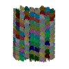 8i7rMC  8i7oC C: citing same article ( M: atomic model generated by this map |
|---|---|
| Similar structure data |

-Links
| EMDB pages | EMDB (EBI/PDBe) / EMDataResource |
|---|---|
| Related items in Molecule of the Month |


-Map
| File | Download / File: emd_35230.map.gz / Format: CCP4 / Size: 125 MB / Type: IMAGE STORED AS FLOATING POINT NUMBER (4 BYTES) | ||||||||||||||||||||||||||||||||||||
|---|---|---|---|---|---|---|---|---|---|---|---|---|---|---|---|---|---|---|---|---|---|---|---|---|---|---|---|---|---|---|---|---|---|---|---|---|---|
| Annotation | In situ structure of axonemal doublet microtubules in mouse sperm with 48-nm repeat | ||||||||||||||||||||||||||||||||||||
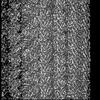
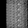
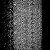
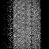
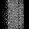
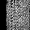
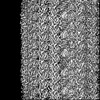
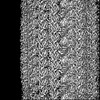
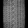
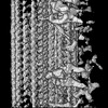
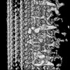
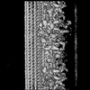
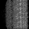
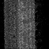
| Projections & slices | Image control
Images are generated by Spider. | ||||||||||||||||||||||||||||||||||||
| Voxel size | X=Y=Z: 1.76 Å | ||||||||||||||||||||||||||||||||||||
| Density |
| ||||||||||||||||||||||||||||||||||||
| Symmetry | Space group: 1 | ||||||||||||||||||||||||||||||||||||
| Details | EMDB XML:
|
 Z (Sec.)
 Y (Row.)
 X (Col.)


-Supplemental data
- Sample components
+Entire : mouse sperm
+Supramolecule #1: mouse sperm
+Macromolecule #1: Meiosis-specific nuclear structural protein 1
+Macromolecule #2: Tektin-1
+Macromolecule #3: Tubulin beta-4B chain
+Macromolecule #4: Detyrosinated tubulin alpha-3 chain
+Macromolecule #5: Tektin-2
+Macromolecule #6: Nucleoside diphosphate kinase 7
+Macromolecule #7: Tektin-3
+Macromolecule #8: Tektin-4
+Macromolecule #9: EF-hand domain-containing family member B
+Macromolecule #10: Tektin bundle-interacting protein 1
+Macromolecule #11: Tektin-5
+Macromolecule #12: Cilia- and flagella-associated protein 53
+Macromolecule #13: EF-hand domain-containing protein 1
+Macromolecule #14: EF-hand domain-containing family member C2
+Macromolecule #15: Protein FAM166A
+Macromolecule #16: Cilia- and flagella-associated protein 95
+Macromolecule #17: Protein FAM166C
+Macromolecule #18: Cilia- and flagella-associated protein 107
+Macromolecule #19: Dual specificity phosphatase 21
+Macromolecule #20: Cilia- and flagella-associated protein 161
+Macromolecule #21: Coiled-coil domain-containing protein 105
+Macromolecule #22: Enkurin
+Macromolecule #23: Piercer of microtubule wall 1 protein
+Macromolecule #24: Testis-expressed protein 43
+Macromolecule #25: Piercer of microtubule wall 2 protein
+Macromolecule #26: Cilia- and flagella-associated protein 276
+Macromolecule #27: RIB43A-like with coiled-coils protein 2
+Macromolecule #28: Protein Flattop
+Macromolecule #29: Cilia- and flagella-associated protein 52
+Macromolecule #30: EF-hand calcium-binding domain-containing protein 6
+Macromolecule #31: Cilia and flagella-associated protein 77
+Macromolecule #32: Sperm-associated antigen 8
+Macromolecule #33: Cilia- and flagella-associated protein 45
+Macromolecule #34: Cilia- and flagella-associated protein 20
+Macromolecule #35: Parkin coregulated gene protein homolog
+Macromolecule #36: Cilia- and flagella-associated protein 210
+Macromolecule #37: Sperm acrosome-associated protein 9
+Macromolecule #38: Cilia- and flagella-associated protein 141
+Macromolecule #39: GUANOSINE-5'-TRIPHOSPHATE

-Experimental details
-Structure determination
| Method | cryo EM |
|---|---|
 Processing Processing | subtomogram averaging |
| Aggregation state | cell |
-Sample preparation
| Buffer | pH: 7 |
|---|---|
| Vitrification | Cryogen name: ETHANE |
- Electron microscopy
| Microscope | FEI TITAN KRIOS |
|---|---|
| Image recording | Film or detector model: GATAN K2 QUANTUM (4k x 4k) / Detector mode: SUPER-RESOLUTION / Average electron dose: 3.0 e/Å2 |
| Electron beam | Acceleration voltage: 300 kV / Electron source:  FIELD EMISSION GUN FIELD EMISSION GUN |
| Electron optics | Illumination mode: SPOT SCAN / Imaging mode: BRIGHT FIELD / Cs: 2.7 mm / Nominal defocus max: 5.0 µm / Nominal defocus min: 1.0 µm |
| Experimental equipment |  Model: Titan Krios / Image courtesy: FEI Company |
-Image processing
| Final reconstruction | Applied symmetry - Point group: C1 (asymmetric) / Resolution.type: BY AUTHOR / Resolution: 6.5 Å / Resolution method: FSC 0.143 CUT-OFF / Number subtomograms used: 17450 |
|---|---|
| Extraction | Number tomograms: 689 / Number images used: 17450 |
| CTF correction | Type: PHASE FLIPPING AND AMPLITUDE CORRECTION |
| Final angle assignment | Type: MAXIMUM LIKELIHOOD |
