[English] 日本語
Yorodumi
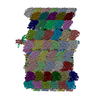
- PDB-8i7o: In situ structure of axonemal doublet microtubules in mouse sperm... -

+
Open data


ID or keywords:

Loading...

-
Basic information

Entry
Database: PDB / ID: 8i7o
TitleIn situ structure of axonemal doublet microtubules in mouse sperm with 16-nm repeat
Components
  • (Cilia- and flagella-associated protein ...) x 3
  • (EF-hand domain-containing ...) x 2
  • Cilia and flagella-associated protein 77
  • Coiled-coil domain-containing protein 105
  • Detyrosinated tubulin alpha-3 chain
  • Dual specificity phosphatase 21
  • EF-hand calcium-binding domain-containing protein 6
  • Enkurin
  • Parkin coregulated gene protein homolog
  • Protein FAM166A
  • Protein FAM166C
  • Protein Flattop
  • Sperm acrosome-associated protein 9
  • Tektin bundle-interacting protein 1
  • Tektin-1
  • Tektin-2
  • Tektin-3
  • Tektin-4
  • Tektin-5
  • Testis-expressed protein 43
  • Tubulin beta-4B chain
KeywordsSTRUCTURAL PROTEIN / microtubules / axoneme / sperm / filament
Function / homology
Function and homology information


male germ-line stem cell population maintenance / outer acrosomal membrane / regulation of brood size / establishment of left/right asymmetry / 9+0 motile cilium / Microtubule-dependent trafficking of connexons from Golgi to the plasma membrane / Cilium Assembly / Sealing of the nuclear envelope (NE) by ESCRT-III / Carboxyterminal post-translational modifications of tubulin / protein polyglutamylation ...male germ-line stem cell population maintenance / outer acrosomal membrane / regulation of brood size / establishment of left/right asymmetry / 9+0 motile cilium / Microtubule-dependent trafficking of connexons from Golgi to the plasma membrane / Cilium Assembly / Sealing of the nuclear envelope (NE) by ESCRT-III / Carboxyterminal post-translational modifications of tubulin / protein polyglutamylation / Intraflagellar transport / inner dynein arm assembly / cilium-dependent cell motility / positive regulation of feeding behavior / regulation of cilium beat frequency involved in ciliary motility / sperm principal piece / COPI-independent Golgi-to-ER retrograde traffic / MAP kinase tyrosine/serine/threonine phosphatase activity / HSP90 chaperone cycle for steroid hormone receptors (SHR) in the presence of ligand / cilium movement involved in cell motility / 9+2 motile cilium / PKR-mediated signaling / COPI-mediated anterograde transport / protein localization to organelle / Aggrephagy / acrosomal membrane / Kinesins / Mitotic Prometaphase / EML4 and NUDC in mitotic spindle formation / Resolution of Sister Chromatid Cohesion / RHO GTPases activate IQGAPs / axoneme assembly / The role of GTSE1 in G2/M progression after G2 checkpoint / flagellated sperm motility / axonemal microtubule / cilium organization / Recycling pathway of L1 / RHO GTPases Activate Formins / COPI-dependent Golgi-to-ER retrograde traffic / Separation of Sister Chromatids / Hedgehog 'off' state / Loss of Nlp from mitotic centrosomes / Recruitment of mitotic centrosome proteins and complexes / Loss of proteins required for interphase microtubule organization from the centrosome / Recruitment of NuMA to mitotic centrosomes / Anchoring of the basal body to the plasma membrane / AURKA Activation by TPX2 / manchette / Regulation of PLK1 Activity at G2/M Transition / MHC class II antigen presentation / protein targeting to mitochondrion / motile cilium / positive regulation of cell motility / protein targeting to membrane / microtubule organizing center / tubulin complex / ciliary base / myosin phosphatase activity / extrinsic component of membrane / intercellular bridge / beta-tubulin binding / protein-serine/threonine phosphatase / regulation of neuron projection development / regulation of cell division / spermatid development / axoneme / phosphatase activity / cerebral cortex cell migration / alpha-tubulin binding / mitotic cytokinesis / cilium assembly / sperm flagellum / cellular response to unfolded protein / cytoplasmic microtubule / microtubule-based process / dephosphorylation / sperm midpiece / Hsp70 protein binding / heat shock protein binding / centriole / cellular response to leukemia inhibitory factor / Neutrophil degranulation / mitotic spindle organization / protein-tyrosine-phosphatase / acrosomal vesicle / ciliary basal body / protein tyrosine phosphatase activity / cell projection / G protein-coupled receptor binding / Hydrolases; Acting on acid anhydrides; Acting on GTP to facilitate cellular and subcellular movement / Hsp90 protein binding / protein localization / cilium / mitochondrial intermembrane space / structural constituent of cytoskeleton / intracellular calcium ion homeostasis / mitotic spindle / microtubule cytoskeleton organization / SH3 domain binding / spindle pole
Similarity search - Function
Atypical dual specificity phosphatase, subfamily B / Protein of unknown function DUF4513 / Domain of unknown function (DUF4513) / DJBP, EF-hand domain / Tektin bundle interacting protein 1 / Coiled-coil domain-containing protein 105 / EF-hand domain / Tektin bundle interacting protein 1 / Sperm acrosome-associated protein 9 / : ...Atypical dual specificity phosphatase, subfamily B / Protein of unknown function DUF4513 / Domain of unknown function (DUF4513) / DJBP, EF-hand domain / Tektin bundle interacting protein 1 / Coiled-coil domain-containing protein 105 / EF-hand domain / Tektin bundle interacting protein 1 / Sperm acrosome-associated protein 9 / : / Sperm acrosome-associated protein 9 / Tektin / Cilia- and flagella-associated protein 276 / Tektin family / Protein of unknown function (DUF3695) / Cilia- and flagella-associated protein 77 / Cilia- and flagella-associated protein 77 / Uncharacterised protein FAM166/UPF0605 / Protein of unknown function (DUF2475) / Protein Flattop / DM10 domain / EF-hand domain-containing protein EFHC1/EFHC2/EFHB / DM10 domain / DM10 domain profile. / Domains in hypothetical proteins in Drosophila, C. elegans and mammals. Occurs singly in some nucleoside diphosphate kinases. / Enkurin domain / Calmodulin-binding / Enkurin domain profile. / CFA20 domain / Cilia- and flagella-associated protein 20/CFAP20DC / CFA20 domain / Parkin co-regulated protein / Parkin co-regulated protein / Dual specificity phosphatase, catalytic domain / EF hand / Dual specificity phosphatase, catalytic domain / Dual specificity phosphatase, catalytic domain / Dual specificity protein phosphatase domain / Dual specificity protein phosphatase domain profile. / EF-hand domain pair / Tubulin-beta mRNA autoregulation signal. / Alpha tubulin / Beta tubulin, autoregulation binding site / Beta tubulin / Tubulin / Tubulin, C-terminal / Tubulin C-terminal domain / Tubulin, conserved site / Tubulin subunits alpha, beta, and gamma signature. / Tyrosine specific protein phosphatases active site. / Protein-tyrosine phosphatase, active site / Tubulin/FtsZ family, C-terminal domain / Tubulin/FtsZ-like, C-terminal domain / Tubulin/FtsZ, C-terminal / Tubulin/FtsZ, 2-layer sandwich domain / Tubulin/FtsZ family, GTPase domain / Tubulin/FtsZ family, GTPase domain / Tubulin/FtsZ, GTPase domain / Tubulin/FtsZ, GTPase domain superfamily / Tyrosine-specific protein phosphatases domain / Tyrosine specific protein phosphatases domain profile. / Protein-tyrosine phosphatase-like / EF-hand domain pair / EF-hand, calcium binding motif / EF-Hand 1, calcium-binding site / EF-hand calcium-binding domain. / EF-hand calcium-binding domain profile. / EF-hand domain / EF-hand domain pair / WD40 repeat, conserved site / Trp-Asp (WD) repeats signature. / WD domain, G-beta repeat / WD40 repeats / WD40 repeat / Trp-Asp (WD) repeats profile. / Trp-Asp (WD) repeats circular profile. / WD40-repeat-containing domain superfamily / WD40/YVTN repeat-like-containing domain superfamily
Similarity search - Domain/homology
GUANOSINE-5'-TRIPHOSPHATE / Cilia and flagella associated protein 77 / Tektin bundle-interacting protein 1 / Tektin-5 / Tubulin alpha-3 chain / Tubulin beta-4B chain / Tektin-4 / Cilia- and flagella-associated protein 52 / EF-hand calcium-binding domain-containing protein 6 / Protein Flattop ...GUANOSINE-5'-TRIPHOSPHATE / Cilia and flagella associated protein 77 / Tektin bundle-interacting protein 1 / Tektin-5 / Tubulin alpha-3 chain / Tubulin beta-4B chain / Tektin-4 / Cilia- and flagella-associated protein 52 / EF-hand calcium-binding domain-containing protein 6 / Protein Flattop / Enkurin / Tektin-3 / Sperm acrosome-associated protein 9 / Cilia- and flagella-associated protein 20 / Tektin-2 / EF-hand domain-containing family member C2 / Ciliary microtubule inner protein 2A / Tektin-like protein 1 / Dual specificity phosphatase 21 / Sperm-associated microtubule inner protein 10 / EF-hand domain-containing protein 1 / Cilia- and flagella-associated protein 276 / Tektin-1 / Parkin coregulated gene protein homolog / Ciliary microtubule inner protein 2C
Similarity search - Component
Biological speciesMus musculus (house mouse)
MethodELECTRON MICROSCOPY / subtomogram averaging / cryo EM / Resolution: 4.5 Å
AuthorsZhu, Y. / Yin, G.L. / Tai, L.H. / Sun, F.
Funding support China, 1items
OrganizationGrant numberCountry
National Natural Science Foundation of China (NSFC)31925026 China
CitationJournal: Cell Discov / Year: 2023
Title: In-cell structural insight into the stability of sperm microtubule doublet.
Authors: Linhua Tai / Guoliang Yin / Xiaojun Huang / Fei Sun / Yun Zhu /
Abstract: The propulsion for mammalian sperm swimming is generated by flagella beating. Microtubule doublets (DMTs) along with microtubule inner proteins (MIPs) are essential structural blocks of flagella. ...The propulsion for mammalian sperm swimming is generated by flagella beating. Microtubule doublets (DMTs) along with microtubule inner proteins (MIPs) are essential structural blocks of flagella. However, the intricate molecular architecture of intact sperm DMT remains elusive. Here, by in situ cryo-electron tomography, we solved the in-cell structure of mouse sperm DMT at 4.5-7.5 Å resolutions, and built its model with 36 kinds of MIPs in 48 nm periodicity. We identified multiple copies of Tektin5 that reinforce Tektin bundle, and multiple MIPs with different periodicities that anchor the Tektin bundle to tubulin wall. This architecture contributes to a superior stability of A-tubule than B-tubule of DMT, which was revealed by structural comparison of DMTs from the intact and deformed axonemes. Our work provides an overall molecular picture of intact sperm DMT in 48 nm periodicity that is essential to understand the molecular mechanism of sperm motility as well as the related ciliopathies.
History
DepositionFeb 1, 2023Deposition site: PDBJ / Processing site: PDBJ
Revision 1.0Oct 11, 2023Provider: repository / Type: Initial release
Revision 1.1Dec 6, 2023Group: Database references / Category: citation / citation_author
Item: _citation.journal_volume / _citation.page_first ..._citation.journal_volume / _citation.page_first / _citation.page_last / _citation.pdbx_database_id_DOI / _citation.pdbx_database_id_PubMed / _citation.title

-
Structure visualization

Structure viewerMolecule:
MolmilJmol/JSmol

Downloads & links

-
Assembly

Deposited unit
A2: Tektin-1
A3: Tektin-1
AE: Detyrosinated tubulin alpha-3 chain
AF: Tubulin beta-4B chain
AG: Detyrosinated tubulin alpha-3 chain
AH: Tubulin beta-4B chain
AI: Detyrosinated tubulin alpha-3 chain
B2: Tektin-2
B3: Tektin-2
B7: Tektin-2
B8: Tektin-2
BE: Detyrosinated tubulin alpha-3 chain
BF: Tubulin beta-4B chain
BG: Detyrosinated tubulin alpha-3 chain
BH: Tubulin beta-4B chain
BI: Detyrosinated tubulin alpha-3 chain
C2: Tektin-3
C3: Tektin-3
C4: Tektin-3
C7: Tektin-3
C8: Tektin-3
CF: Tubulin beta-4B chain
CG: Detyrosinated tubulin alpha-3 chain
CH: Tubulin beta-4B chain
CI: Detyrosinated tubulin alpha-3 chain
CJ: Tubulin beta-4B chain
Cb: Tektin-3
Cc: Tektin-3
D2: Tektin-4
D3: Tektin-4
D4: Tektin-4
D7: Tektin-4
D8: Tektin-4
D9: Tektin-4
DD: Tubulin beta-4B chain
DE: Detyrosinated tubulin alpha-3 chain
DF: Tubulin beta-4B chain
DG: Detyrosinated tubulin alpha-3 chain
DH: Tubulin beta-4B chain
E2: Tektin bundle-interacting protein 1
E3: Tektin bundle-interacting protein 1
ED: Tubulin beta-4B chain
EE: Detyrosinated tubulin alpha-3 chain
EF: Tubulin beta-4B chain
EG: Detyrosinated tubulin alpha-3 chain
EH: Tubulin beta-4B chain
F2: Tektin-5
F3: Tektin-5
F6: Tektin-5
F7: Tektin-5
FD: Tubulin beta-4B chain
FE: Detyrosinated tubulin alpha-3 chain
FF: Tubulin beta-4B chain
FG: Detyrosinated tubulin alpha-3 chain
FH: Tubulin beta-4B chain
Fa: Tektin-5
Fb: Tektin-5
Fc: Tektin-5
Fl: Tektin-5
G1: EF-hand domain-containing protein 1
G2: EF-hand domain-containing protein 1
G5: EF-hand domain-containing family member C2
GD: Tubulin beta-4B chain
GE: Detyrosinated tubulin alpha-3 chain
GF: Tubulin beta-4B chain
GG: Detyrosinated tubulin alpha-3 chain
GH: Tubulin beta-4B chain
GI: Detyrosinated tubulin alpha-3 chain
H3: Protein FAM166A
H4: Protein FAM166A
HD: Tubulin beta-4B chain
HE: Detyrosinated tubulin alpha-3 chain
HF: Tubulin beta-4B chain
HG: Detyrosinated tubulin alpha-3 chain
HH: Tubulin beta-4B chain
HI: Detyrosinated tubulin alpha-3 chain
I2: Protein FAM166C
I3: Protein FAM166C
IE: Detyrosinated tubulin alpha-3 chain
IF: Tubulin beta-4B chain
IG: Detyrosinated tubulin alpha-3 chain
IH: Tubulin beta-4B chain
II: Detyrosinated tubulin alpha-3 chain
J2: Dual specificity phosphatase 21
J3: Dual specificity phosphatase 21
JD: Tubulin beta-4B chain
JE: Detyrosinated tubulin alpha-3 chain
JF: Tubulin beta-4B chain
JG: Detyrosinated tubulin alpha-3 chain
JH: Tubulin beta-4B chain
K2: Coiled-coil domain-containing protein 105
K3: Coiled-coil domain-containing protein 105
K4: Coiled-coil domain-containing protein 105
KD: Tubulin beta-4B chain
KE: Detyrosinated tubulin alpha-3 chain
KF: Tubulin beta-4B chain
KG: Detyrosinated tubulin alpha-3 chain
KH: Tubulin beta-4B chain
KI: Detyrosinated tubulin alpha-3 chain
L2: Enkurin
L3: Enkurin
LD: Tubulin beta-4B chain
LE: Detyrosinated tubulin alpha-3 chain
LF: Tubulin beta-4B chain
LG: Detyrosinated tubulin alpha-3 chain
LH: Tubulin beta-4B chain
LI: Detyrosinated tubulin alpha-3 chain
M2: Testis-expressed protein 43
M3: Testis-expressed protein 43
ME: Detyrosinated tubulin alpha-3 chain
MF: Tubulin beta-4B chain
MG: Detyrosinated tubulin alpha-3 chain
MH: Tubulin beta-4B chain
MI: Detyrosinated tubulin alpha-3 chain
N2: Cilia- and flagella-associated protein 276
N3: Cilia- and flagella-associated protein 276
ND: Tubulin beta-4B chain
NE: Detyrosinated tubulin alpha-3 chain
NF: Tubulin beta-4B chain
NG: Detyrosinated tubulin alpha-3 chain
NH: Tubulin beta-4B chain
NI: Detyrosinated tubulin alpha-3 chain
O1: Protein Flattop
O2: Protein Flattop
OD: Tubulin beta-4B chain
OE: Detyrosinated tubulin alpha-3 chain
OF: Tubulin beta-4B chain
OG: Detyrosinated tubulin alpha-3 chain
OH: Tubulin beta-4B chain
OI: Detyrosinated tubulin alpha-3 chain
P1: Cilia- and flagella-associated protein 52
P2: Cilia- and flagella-associated protein 52
PE: Detyrosinated tubulin alpha-3 chain
PF: Tubulin beta-4B chain
PG: Detyrosinated tubulin alpha-3 chain
PH: Tubulin beta-4B chain
PI: Detyrosinated tubulin alpha-3 chain
Q1: EF-hand calcium-binding domain-containing protein 6
Q2: EF-hand calcium-binding domain-containing protein 6
QE: Detyrosinated tubulin alpha-3 chain
QF: Tubulin beta-4B chain
QG: Detyrosinated tubulin alpha-3 chain
QH: Tubulin beta-4B chain
QI: Detyrosinated tubulin alpha-3 chain
R2: Cilia and flagella-associated protein 77
RE: Detyrosinated tubulin alpha-3 chain
RF: Tubulin beta-4B chain
RG: Detyrosinated tubulin alpha-3 chain
RH: Tubulin beta-4B chain
RI: Detyrosinated tubulin alpha-3 chain
RJ: Tubulin beta-4B chain
SE: Detyrosinated tubulin alpha-3 chain
SF: Tubulin beta-4B chain
SG: Detyrosinated tubulin alpha-3 chain
SH: Tubulin beta-4B chain
SI: Detyrosinated tubulin alpha-3 chain
SJ: Tubulin beta-4B chain
TF: Tubulin beta-4B chain
TG: Detyrosinated tubulin alpha-3 chain
TH: Tubulin beta-4B chain
TI: Detyrosinated tubulin alpha-3 chain
TJ: Tubulin beta-4B chain
UD: Tubulin beta-4B chain
UE: Detyrosinated tubulin alpha-3 chain
UF: Tubulin beta-4B chain
UG: Detyrosinated tubulin alpha-3 chain
UH: Tubulin beta-4B chain
VD: Tubulin beta-4B chain
VE: Detyrosinated tubulin alpha-3 chain
VF: Tubulin beta-4B chain
VG: Detyrosinated tubulin alpha-3 chain
VH: Tubulin beta-4B chain
WE: Detyrosinated tubulin alpha-3 chain
WF: Tubulin beta-4B chain
WG: Detyrosinated tubulin alpha-3 chain
WH: Tubulin beta-4B chain
WI: Detyrosinated tubulin alpha-3 chain
XC: Cilia- and flagella-associated protein 20
XD: Cilia- and flagella-associated protein 20
XE: Cilia- and flagella-associated protein 20
XJ: Parkin coregulated gene protein homolog
XK: Parkin coregulated gene protein homolog
XL: Parkin coregulated gene protein homolog
YI: Sperm acrosome-associated protein 9
YJ: Sperm acrosome-associated protein 9
YK: Sperm acrosome-associated protein 9
YL: Sperm acrosome-associated protein 9
YM: Sperm acrosome-associated protein 9
YN: Sperm acrosome-associated protein 9
hetero molecules


Theoretical massNumber of molelcules
Total (without water)9,097,620312
Polymers9,033,269189
Non-polymers64,351123
Water0
1


  • Idetical with deposited unit
  • defined by author
  • Evidence: electron microscopy
TypeNameSymmetry operationNumber
identity operation1_5551

-
Components

-
Protein , 19 types, 179 molecules A2A3AEAGAIBEBGBICGCIDEDGEEEGFEFGGEGGGIHEHGHIIEIGIIJEJGKEKGKI...

#1: Protein Tektin-1 /


Mass: 48713.035 Da / Num. of mol.: 2 / Source method: isolated from a natural source / Source: (natural) Mus musculus (house mouse) / References: UniProt: Q9DAJ2
#2: Protein ...
Detyrosinated tubulin alpha-3 chain


Mass: 48689.090 Da / Num. of mol.: 61 / Source method: isolated from a natural source / Source: (natural) Mus musculus (house mouse) / References: UniProt: P05214
#3: Protein ...
Tubulin beta-4B chain / / Tubulin beta-2C chain


Mass: 47897.918 Da / Num. of mol.: 62 / Source method: isolated from a natural source / Source: (natural) Mus musculus (house mouse) / References: UniProt: P68372
#4: Protein
Tektin-2 / / Tektin-t / Testicular tektin


Mass: 50385.066 Da / Num. of mol.: 4 / Source method: isolated from a natural source / Source: (natural) Mus musculus (house mouse) / References: UniProt: Q922G7
#5: Protein
Tektin-3 /


Mass: 56754.777 Da / Num. of mol.: 7 / Source method: isolated from a natural source / Source: (natural) Mus musculus (house mouse) / References: UniProt: Q6X6Z7
#6: Protein
Tektin-4 / / Testicular microtubules-related protein 4


Mass: 52121.449 Da / Num. of mol.: 6 / Source method: isolated from a natural source / Source: (natural) Mus musculus (house mouse) / References: UniProt: Q149S1
#7: Protein Tektin bundle-interacting protein 1


Mass: 23987.262 Da / Num. of mol.: 2 / Source method: isolated from a natural source / Source: (natural) Mus musculus (house mouse) / References: UniProt: A6H6Q4
#8: Protein
Tektin-5 /


Mass: 62817.535 Da / Num. of mol.: 8 / Source method: isolated from a natural source / Source: (natural) Mus musculus (house mouse) / References: UniProt: G5E8A8
#11: Protein Protein FAM166A


Mass: 36659.566 Da / Num. of mol.: 2 / Source method: isolated from a natural source / Source: (natural) Mus musculus (house mouse) / References: UniProt: Q9D4K5
#12: Protein Protein FAM166C


Mass: 23097.957 Da / Num. of mol.: 2 / Source method: isolated from a natural source / Source: (natural) Mus musculus (house mouse) / References: UniProt: Q9DAS2
#13: Protein Dual specificity phosphatase 21


Mass: 21527.574 Da / Num. of mol.: 2 / Source method: isolated from a natural source / Source: (natural) Mus musculus (house mouse)
References: UniProt: Q9D9D8, protein-serine/threonine phosphatase, protein-tyrosine-phosphatase
#14: Protein Coiled-coil domain-containing protein 105


Mass: 57406.484 Da / Num. of mol.: 3 / Source method: isolated from a natural source / Source: (natural) Mus musculus (house mouse) / References: UniProt: Q9D4K7
#15: Protein Enkurin /


Mass: 29587.521 Da / Num. of mol.: 2 / Source method: isolated from a natural source / Source: (natural) Mus musculus (house mouse) / References: UniProt: Q6SP97
#16: Protein Testis-expressed protein 43


Mass: 16305.608 Da / Num. of mol.: 2 / Source method: isolated from a natural source / Source: (natural) Mus musculus (house mouse) / References: UniProt: Q9D9I1
#18: Protein Protein Flattop / Cilia- and flagella-associated protein 126


Mass: 20575.123 Da / Num. of mol.: 2 / Source method: isolated from a natural source / Source: (natural) Mus musculus (house mouse) / References: UniProt: Q6P8X9
#20: Protein EF-hand calcium-binding domain-containing protein 6 / DJ-1-binding protein / DJBP


Mass: 176028.312 Da / Num. of mol.: 2 / Source method: isolated from a natural source / Source: (natural) Mus musculus (house mouse) / References: UniProt: Q6P1E8
#21: Protein Cilia and flagella-associated protein 77


Mass: 32786.020 Da / Num. of mol.: 1 / Source method: isolated from a natural source / Source: (natural) Mus musculus (house mouse) / References: UniProt: A0A087WRI3
#23: Protein Parkin coregulated gene protein homolog / Hypertension-related protein 1-like protein / PARK2 coregulated gene protein


Mass: 27763.268 Da / Num. of mol.: 3 / Source method: isolated from a natural source / Source: (natural) Mus musculus (house mouse) / References: UniProt: Q9DAK2
#24: Protein
Sperm acrosome-associated protein 9 / Acrosome and sperm tail protein / MAST


Mass: 19512.373 Da / Num. of mol.: 6 / Source method: isolated from a natural source / Source: (natural) Mus musculus (house mouse) / References: UniProt: Q7TPM5

-
EF-hand domain-containing ... , 2 types, 3 molecules G1G2G5

#9: Protein EF-hand domain-containing protein 1 / Myoclonin-1


Mass: 75235.422 Da / Num. of mol.: 2 / Source method: isolated from a natural source / Source: (natural) Mus musculus (house mouse) / References: UniProt: Q9D9T8
#10: Protein EF-hand domain-containing family member C2


Mass: 87758.023 Da / Num. of mol.: 1 / Source method: isolated from a natural source / Source: (natural) Mus musculus (house mouse) / References: UniProt: Q9D485

-
Cilia- and flagella-associated protein ... , 3 types, 7 molecules N2N3P1P2XCXDXE

#17: Protein Cilia- and flagella-associated protein 276


Mass: 18960.092 Da / Num. of mol.: 2 / Source method: isolated from a natural source / Source: (natural) Mus musculus (house mouse) / References: UniProt: Q9DAD0
#19: Protein Cilia- and flagella-associated protein 52 / WD repeat-containing protein 16


Mass: 68322.164 Da / Num. of mol.: 2 / Source method: isolated from a natural source / Source: (natural) Mus musculus (house mouse) / References: UniProt: Q5F201
#22: Protein Cilia- and flagella-associated protein 20 / Gene trap locus 3 protein


Mass: 22781.389 Da / Num. of mol.: 3 / Source method: isolated from a natural source / Source: (natural) Mus musculus (house mouse) / References: UniProt: Q8BTU1

-
Non-polymers , 1 types, 123 molecules

#25: Chemical...
ChemComp-GTP / GUANOSINE-5'-TRIPHOSPHATE / Guanosine triphosphate


Mass: 523.180 Da / Num. of mol.: 123 / Source method: obtained synthetically / Formula: C10H16N5O14P3 / Comment: GTP, energy-carrying molecule*YM

-
Details

Has ligand of interestN

-
Experimental details

-
Experiment

ExperimentMethod: ELECTRON MICROSCOPY
EM experimentAggregation state: CELL / 3D reconstruction method: subtomogram averaging

-
Sample preparation

ComponentName: mouse sperm / Type: CELL / Entity ID: #1-#24 / Source: NATURAL
Source (natural)Organism: Mus musculus (house mouse)
Buffer solutionpH: 7
SpecimenEmbedding applied: NO / Shadowing applied: NO / Staining applied: NO / Vitrification applied: YES
VitrificationCryogen name: ETHANE

-
Electron microscopy imaging

Experimental equipment
Model: Titan Krios / Image courtesy: FEI Company
MicroscopyModel: FEI TITAN KRIOS
Electron gunElectron source: FIELD EMISSION GUN / Accelerating voltage: 300 kV / Illumination mode: SPOT SCAN
Electron lensMode: BRIGHT FIELDBright-field microscopy / Nominal defocus max: 5000 nm / Nominal defocus min: 1000 nm / Cs: 2.7 mm
Image recordingElectron dose: 3 e/Å2 / Avg electron dose per subtomogram: 117 e/Å2 / Detector mode: SUPER-RESOLUTION / Film or detector model: GATAN K2 QUANTUM (4k x 4k)

-
Processing

SoftwareName: UCSF ChimeraX / Version: 1.6/v9 / Classification: model building / URL: https://www.rbvi.ucsf.edu/chimerax/ / Os: Windows / Type: package
CTF correctionType: PHASE FLIPPING AND AMPLITUDE CORRECTION
SymmetryPoint symmetry: C1 (asymmetric)
3D reconstructionResolution: 4.5 Å / Resolution method: FSC 0.143 CUT-OFF / Num. of particles: 37018 / Symmetry type: POINT
EM volume selectionNum. of tomograms: 689 / Num. of volumes extracted: 37018

+
About Yorodumi

-
News

-
Feb 9, 2022. New format data for meta-information of EMDB entries

New format data for meta-information of EMDB entries

  • Version 3 of the EMDB header file is now the official format.
  • The previous official version 1.9 will be removed from the archive.

Related info.:EMDB header

External links:wwPDB to switch to version 3 of the EMDB data model

-
Aug 12, 2020. Covid-19 info

Covid-19 info

URL: https://pdbj.org/emnavi/covid19.php

New page: Covid-19 featured information page in EM Navigator.

Related info.:Covid-19 info / Mar 5, 2020. Novel coronavirus structure data

+
Mar 5, 2020. Novel coronavirus structure data

Novel coronavirus structure data

Related info.:Yorodumi Speices / Aug 12, 2020. Covid-19 info

External links:COVID-19 featured content - PDBj / Molecule of the Month (242):Coronavirus Proteases

+
Jan 31, 2019. EMDB accession codes are about to change! (news from PDBe EMDB page)

EMDB accession codes are about to change! (news from PDBe EMDB page)

  • The allocation of 4 digits for EMDB accession codes will soon come to an end. Whilst these codes will remain in use, new EMDB accession codes will include an additional digit and will expand incrementally as the available range of codes is exhausted. The current 4-digit format prefixed with “EMD-” (i.e. EMD-XXXX) will advance to a 5-digit format (i.e. EMD-XXXXX), and so on. It is currently estimated that the 4-digit codes will be depleted around Spring 2019, at which point the 5-digit format will come into force.
  • The EM Navigator/Yorodumi systems omit the EMD- prefix.

Related info.:Q: What is EMD? / ID/Accession-code notation in Yorodumi/EM Navigator

External links:EMDB Accession Codes are Changing Soon! / Contact to PDBj

+
Jul 12, 2017. Major update of PDB

Major update of PDB

  • wwPDB released updated PDB data conforming to the new PDBx/mmCIF dictionary.
  • This is a major update changing the version number from 4 to 5, and with Remediation, in which all the entries are updated.
  • In this update, many items about electron microscopy experimental information are reorganized (e.g. em_software).
  • Now, EM Navigator and Yorodumi are based on the updated data.

External links:wwPDB Remediation / Enriched Model Files Conforming to OneDep Data Standards Now Available in the PDB FTP Archive

-
Yorodumi

Thousand views of thousand structures

  • Yorodumi is a browser for structure data from EMDB, PDB, SASBDB, etc.
  • This page is also the successor to EM Navigator detail page, and also detail information page/front-end page for Omokage search.
  • The word "yorodu" (or yorozu) is an old Japanese word meaning "ten thousand". "mi" (miru) is to see.

Related info.:EMDB / PDB / SASBDB / Comparison of 3 databanks / Yorodumi Search / Aug 31, 2016. New EM Navigator & Yorodumi / Yorodumi Papers / Jmol/JSmol / Function and homology information / Changes in new EM Navigator and Yorodumi

Read more