 Movie
Movie Controller

[English] 日本語
 Yorodumi
- EMDB-35210: In situ structure of A-tubule of axonemal doublet microtubules in... -
+ Open data

- Basic information
| Entry |  | |||||||||
|---|---|---|---|---|---|---|---|---|---|---|
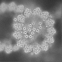
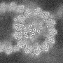
| Title | In situ structure of A-tubule of axonemal doublet microtubules in mouse sperm with 16-nm repeat | |||||||||
 Map data Map data | A-tubule of axonemal doublet microtubules in mouse sperm with 16-nm repeat | |||||||||
 Sample Sample |
| |||||||||
 Keywords Keywords | microtubules / axoneme / sperm / filament / PROTEIN TRANSPORT | |||||||||
| Biological species |  | |||||||||
| Method | subtomogram averaging / cryo EM / Resolution: 4.5 Å | |||||||||
 Authors Authors | Zhu Y / Tai LH / Yin GL / Sun F | |||||||||
| Funding support |  China, 1 items China, 1 items
| |||||||||
 Citation Citation | Journal: Cell Discov / Year: 2023 Title: In-cell structural insight into the stability of sperm microtubule doublet. Authors: Linhua Tai / Guoliang Yin / Xiaojun Huang / Fei Sun / Yun Zhu / Abstract: The propulsion for mammalian sperm swimming is generated by flagella beating. Microtubule doublets (DMTs) along with microtubule inner proteins (MIPs) are essential structural blocks of flagella. ...The propulsion for mammalian sperm swimming is generated by flagella beating. Microtubule doublets (DMTs) along with microtubule inner proteins (MIPs) are essential structural blocks of flagella. However, the intricate molecular architecture of intact sperm DMT remains elusive. Here, by in situ cryo-electron tomography, we solved the in-cell structure of mouse sperm DMT at 4.5-7.5 Å resolutions, and built its model with 36 kinds of MIPs in 48 nm periodicity. We identified multiple copies of Tektin5 that reinforce Tektin bundle, and multiple MIPs with different periodicities that anchor the Tektin bundle to tubulin wall. This architecture contributes to a superior stability of A-tubule than B-tubule of DMT, which was revealed by structural comparison of DMTs from the intact and deformed axonemes. Our work provides an overall molecular picture of intact sperm DMT in 48 nm periodicity that is essential to understand the molecular mechanism of sperm motility as well as the related ciliopathies. | |||||||||
| History |
|
- Structure visualization
| Supplemental images |
|---|

- Downloads & links
-EMDB archive
| Map data | emd_35210.map.gz | 35.4 MB |  EMDB map data format EMDB map data format | |
|---|---|---|---|---|
| Header (meta data) | emd-35210-v30.xmlemd-35210.xml | 12.1 KB 12.1 KB | Display Display | EMDB header |
| FSC (resolution estimation) | emd_35210_fsc.xml | 7.7 KB | Display | FSC data file |
| Images | 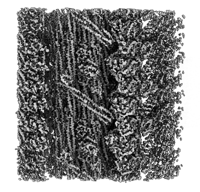 emd_35210.png emd_35210.png | 224.6 KB | ||
| Masks | emd_35210_msk_1.map | 38.4 MB | Mask map | |
| Filedesc metadata | emd-35210.cif.gz | 3.8 KB | ||
| Others | emd_35210_half_map_1.map.gzemd_35210_half_map_2.map.gz | 35.6 MB 35.6 MB | ||
| Archive directory |  http://ftp.pdbj.org/pub/emdb/structures/EMD-35210ftp://ftp.pdbj.org/pub/emdb/structures/EMD-35210 http://ftp.pdbj.org/pub/emdb/structures/EMD-35210ftp://ftp.pdbj.org/pub/emdb/structures/EMD-35210 | HTTPS FTP |
-Related structure data


-Links
| EMDB pages | EMDB (EBI/PDBe) / EMDataResource |
|---|
-Map
| File | Download / File: emd_35210.map.gz / Format: CCP4 / Size: 38.4 MB / Type: IMAGE STORED AS FLOATING POINT NUMBER (4 BYTES) | ||||||||||||||||||||||||||||||||||||
|---|---|---|---|---|---|---|---|---|---|---|---|---|---|---|---|---|---|---|---|---|---|---|---|---|---|---|---|---|---|---|---|---|---|---|---|---|---|
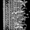
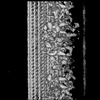
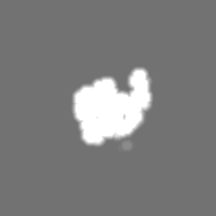
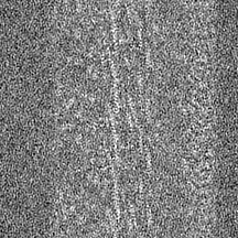
| Annotation | A-tubule of axonemal doublet microtubules in mouse sperm with 16-nm repeat | ||||||||||||||||||||||||||||||||||||
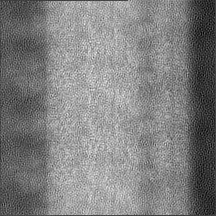
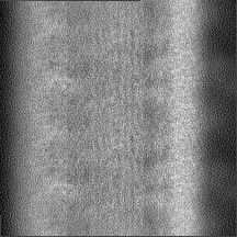
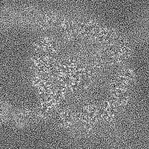
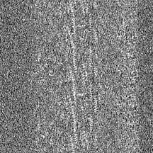
| Projections & slices | Image control
Images are generated by Spider. | ||||||||||||||||||||||||||||||||||||
| Voxel size | X=Y=Z: 1.76 Å | ||||||||||||||||||||||||||||||||||||
| Density |
| ||||||||||||||||||||||||||||||||||||
| Symmetry | Space group: 1 | ||||||||||||||||||||||||||||||||||||
| Details | EMDB XML:
|
 Z (Sec.)
 Y (Row.)
 X (Col.)


-Supplemental data
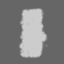
-Mask #1
| File | emd_35210_msk_1.map | ||||||||||||
|---|---|---|---|---|---|---|---|---|---|---|---|---|---|
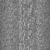
| Projections & Slices |
| ||||||||||||
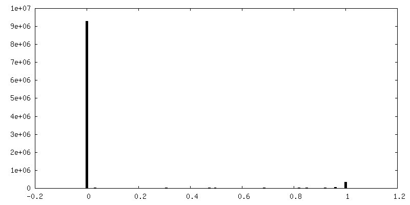
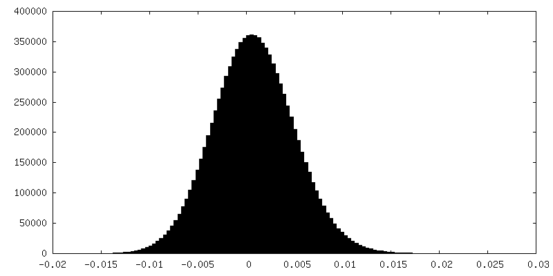
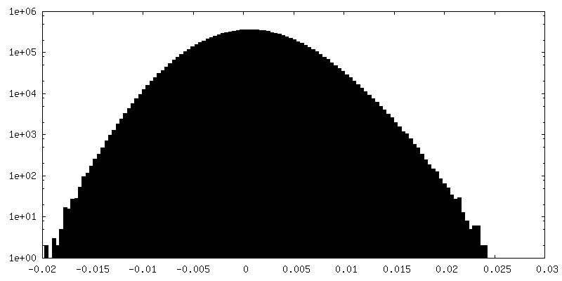
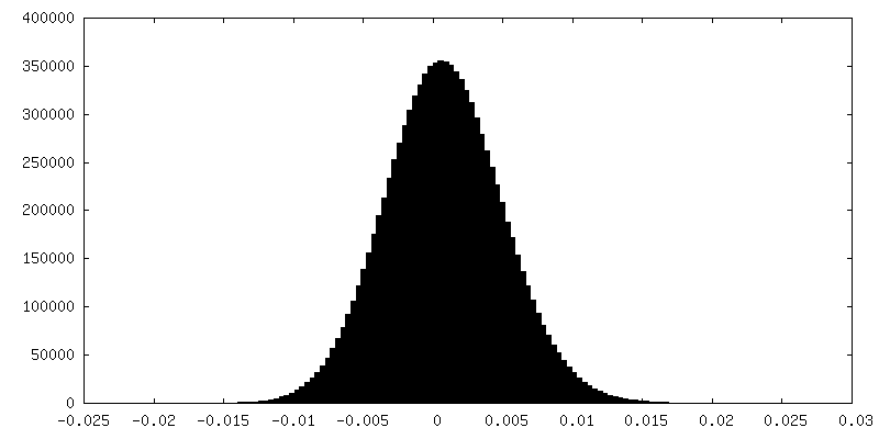
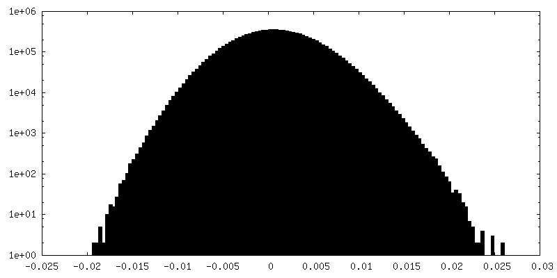
| Density Histograms |
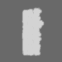

-Half map: Half 1 map of mouse sperm DMT A-tubule
| File | emd_35210_half_map_1.map | ||||||||||||
|---|---|---|---|---|---|---|---|---|---|---|---|---|---|
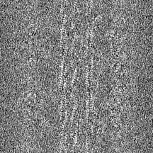
| Annotation | Half 1 map of mouse sperm DMT A-tubule | ||||||||||||
| Projections & Slices |
| ||||||||||||
| Density Histograms |
-Half map: Half 2 map of mouse sperm DMT A-tubule
| File | emd_35210_half_map_2.map | ||||||||||||
|---|---|---|---|---|---|---|---|---|---|---|---|---|---|
| Annotation | Half 2 map of mouse sperm DMT A-tubule | ||||||||||||
| Projections & Slices |
| ||||||||||||
| Density Histograms |
- Sample components
-Entire : mouse sperm
| Entire | Name: mouse sperm |
|---|---|
| Components |
|
-Supramolecule #1: mouse sperm
| Supramolecule | Name: mouse sperm / type: cell / ID: 1 / Parent: 0 |
|---|---|
| Source (natural) | Organism: |
-Experimental details
-Structure determination
| Method | cryo EM |
|---|---|
 Processing Processing | subtomogram averaging |
| Aggregation state | cell |
-Sample preparation
| Buffer | pH: 7 |
|---|---|
| Vitrification | Cryogen name: ETHANE |
- Electron microscopy
| Microscope | FEI TITAN KRIOS |
|---|---|
| Image recording | Film or detector model: GATAN K2 QUANTUM (4k x 4k) / Detector mode: SUPER-RESOLUTION / Average electron dose: 3.0 e/Å2 |
| Electron beam | Acceleration voltage: 300 kV / Electron source:  FIELD EMISSION GUN FIELD EMISSION GUN |
| Electron optics | Illumination mode: SPOT SCAN / Imaging mode: BRIGHT FIELD / Cs: 2.7 mm / Nominal defocus max: 5.0 µm / Nominal defocus min: 1.0 µm |
| Experimental equipment |  Model: Titan Krios / Image courtesy: FEI Company |
-Image processing
| Final reconstruction | Applied symmetry - Point group: C1 (asymmetric) / Resolution.type: BY AUTHOR / Resolution: 4.5 Å / Resolution method: FSC 0.143 CUT-OFF / Number subtomograms used: 37018 |
|---|---|
| Extraction | Number tomograms: 689 / Number images used: 37018 |
| Final angle assignment | Type: MAXIMUM LIKELIHOOD |
| FSC plot (resolution estimation) | 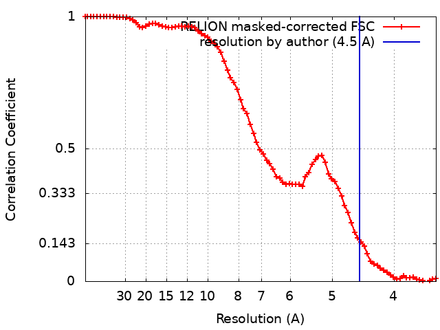 |
