Movie
Movie Controller


+ Open data

- Basic information
| Entry | 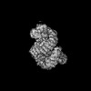 | |||||||||
|---|---|---|---|---|---|---|---|---|---|---|
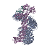
| Title | Cryo-EM Structure of the CAND1-Cul3-Rbx1 complex | |||||||||
 Map data Map data | ||||||||||
 Sample Sample |
| |||||||||
 Keywords Keywords | ligase / complex | |||||||||
| Function / homology |  Function and homology information Function and homology informationSCF complex assembly / positive regulation of mitotic cell cycle phase transition / trophectodermal cellular morphogenesis / liver morphogenesis / POZ domain binding / negative regulation of catalytic activity / polar microtubule / regulation protein catabolic process at postsynapse / anaphase-promoting complex-dependent catabolic process / nuclear protein quality control by the ubiquitin-proteasome system ...SCF complex assembly / positive regulation of mitotic cell cycle phase transition / trophectodermal cellular morphogenesis / liver morphogenesis / POZ domain binding / negative regulation of catalytic activity / polar microtubule / regulation protein catabolic process at postsynapse / anaphase-promoting complex-dependent catabolic process / nuclear protein quality control by the ubiquitin-proteasome system / COPII vesicle coat assembly / negative regulation of beige fat cell differentiation / RHOBTB3 ATPase cycle / cullin-RING-type E3 NEDD8 transferase / NEDD8 transferase activity / embryonic cleavage / cullin-RING ubiquitin ligase complex / Cul7-RING ubiquitin ligase complex / cellular response to chemical stress / Loss of Function of FBXW7 in Cancer and NOTCH1 Signaling / positive regulation of mitotic metaphase/anaphase transition / positive regulation of protein autoubiquitination / fibroblast apoptotic process / Notch binding / RNA polymerase II transcription initiation surveillance / protein neddylation / cell projection organization / negative regulation of Rho protein signal transduction / NEDD8 ligase activity / RHOBTB1 GTPase cycle / protein K27-linked ubiquitination / negative regulation of response to oxidative stress / VCB complex / Cul5-RING ubiquitin ligase complex / stem cell division / ubiquitin-ubiquitin ligase activity / mitotic metaphase chromosome alignment / ubiquitin-dependent protein catabolic process via the C-end degron rule pathway / SCF ubiquitin ligase complex / Cul2-RING ubiquitin ligase complex / stress fiber assembly / Cul3-RING ubiquitin ligase complex / negative regulation of type I interferon production / positive regulation of cytokinesis / SCF-dependent proteasomal ubiquitin-dependent protein catabolic process / Prolactin receptor signaling / Cul4A-RING E3 ubiquitin ligase complex / negative regulation of mitophagy / Cul4-RING E3 ubiquitin ligase complex / Cul4B-RING E3 ubiquitin ligase complex / ubiquitin ligase complex scaffold activity / positive regulation of RNA polymerase II transcription preinitiation complex assembly / cullin family protein binding / endoplasmic reticulum to Golgi vesicle-mediated transport / protein monoubiquitination / RHOBTB2 GTPase cycle / sperm flagellum / ubiquitin ligase complex / protein autoubiquitination / site of DNA damage / signal transduction in response to DNA damage / Nuclear events stimulated by ALK signaling in cancer / protein K48-linked ubiquitination / negative regulation of insulin receptor signaling pathway / regulation of cellular response to insulin stimulus / transcription-coupled nucleotide-excision repair / gastrulation / positive regulation of TORC1 signaling / post-translational protein modification / intrinsic apoptotic signaling pathway / TBP-class protein binding / cyclin binding / T cell activation / positive regulation of protein ubiquitination / Regulation of BACH1 activity / integrin-mediated signaling pathway / negative regulation of canonical NF-kappaB signal transduction / kidney development / cellular response to amino acid stimulus / negative regulation of canonical Wnt signaling pathway / G1/S transition of mitotic cell cycle / Degradation of DVL / Degradation of CRY and PER proteins / Iron uptake and transport / Degradation of GLI1 by the proteasome / Recognition of DNA damage by PCNA-containing replication complex / RING-type E3 ubiquitin transferase / Negative regulation of NOTCH4 signaling / GSK3B and BTRC:CUL1-mediated-degradation of NFE2L2 / Hedgehog 'on' state / Vif-mediated degradation of APOBEC3G / FBXL7 down-regulates AURKA during mitotic entry and in early mitosis / Degradation of GLI2 by the proteasome / GLI3 is processed to GLI3R by the proteasome / Ubiquitin-Mediated Degradation of Phosphorylated Cdc25A / protein destabilization / Evasion by RSV of host interferon responses / NOTCH1 Intracellular Domain Regulates Transcription / Degradation of beta-catenin by the destruction complex / DNA Damage Recognition in GG-NER Similarity search - Function | |||||||||
| Biological species |  Homo sapiens (human) Homo sapiens (human) | |||||||||
| Method | single particle reconstruction / cryo EM / Resolution: 3.76 Å | |||||||||
 Authors Authors | Hu Y / Mao Q / Chen Z / Sun L | |||||||||
| Funding support |  China, 1 items China, 1 items
| |||||||||
 Citation Citation | Journal: Nat Struct Mol Biol / Year: 2024 Title: Dynamic molecular architecture and substrate recruitment of cullin3-RING E3 ligase CRL3. Authors: Yuxia Hu / Zhao Zhang / Qiyu Mao / Xiang Zhang / Aihua Hao / Yu Xun / Yeda Wang / Lin Han / Wuqiang Zhan / Qianying Liu / Yue Yin / Chao Peng / Eva Marie Y Moresco / Zhenguo Chen / Bruce Beutler / Lei Sun /  Abstract: Phosphatidylinositol 3-kinase α, a heterodimer of catalytic p110α and one of five regulatory subunits, mediates insulin- and insulin like growth factor-signaling and, frequently, oncogenesis. ...Phosphatidylinositol 3-kinase α, a heterodimer of catalytic p110α and one of five regulatory subunits, mediates insulin- and insulin like growth factor-signaling and, frequently, oncogenesis. Cellular levels of the regulatory p85α subunit are tightly controlled by regulated proteasomal degradation. In adipose tissue and growth plates, failure of K48-linked p85α ubiquitination causes diabetes, lipodystrophy and dwarfism in mice, as in humans with SHORT syndrome. Here we elucidated the structures of the key ubiquitin ligase complexes regulating p85α availability. Specificity is provided by the substrate receptor KBTBD2, which recruits p85α to the cullin3-RING E3 ubiquitin ligase (CRL3). CRL3 forms multimers, which disassemble into dimers upon substrate binding (CRL3-p85α) and/or neddylation by the activator NEDD8 (CRL3~N8), leading to p85α ubiquitination and degradation. Deactivation involves dissociation of NEDD8 mediated by the COP9 signalosome and displacement of KBTBD2 by the inhibitor CAND1. The hereby identified structural basis of p85α regulation opens the way to better understanding disturbances of glucose regulation, growth and cancer. | |||||||||
| History |
|
- Structure visualization
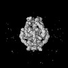
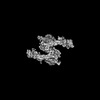
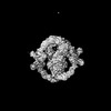
| Supplemental images |
|---|

- Downloads & links
-EMDB archive
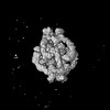
| Map data | emd_34473.map.gz | 57.1 MB | EMDB map data format | |
|---|---|---|---|---|
| Header (meta data) | emd-34473-v30.xmlemd-34473.xml | 21.9 KB 21.9 KB | Display Display | EMDB header |
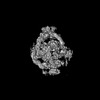
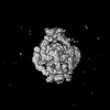
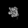
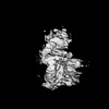
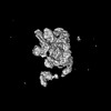
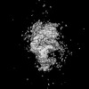
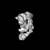
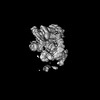
| Images | 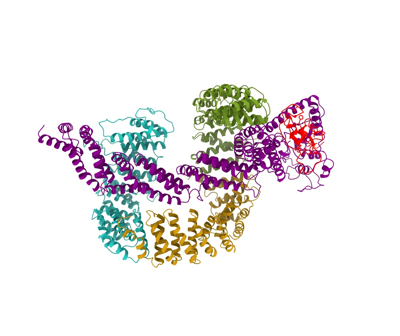 emd_34473.png emd_34473.png | 81 KB | ||
| Filedesc metadata | emd-34473.cif.gz | 7.6 KB | ||
| Others | emd_34473_half_map_1.map.gzemd_34473_half_map_2.map.gz | 49.7 MB 49.7 MB | ||
| Archive directory |  http://ftp.pdbj.org/pub/emdb/structures/EMD-34473ftp://ftp.pdbj.org/pub/emdb/structures/EMD-34473 http://ftp.pdbj.org/pub/emdb/structures/EMD-34473ftp://ftp.pdbj.org/pub/emdb/structures/EMD-34473 | HTTPS FTP |
-Related structure data
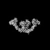
| Related structure data | 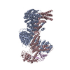 8h3qMC 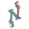 8gq6C 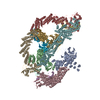 8h33C 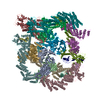 8h34C 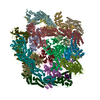 8h35C 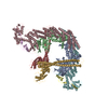 8h36C  8h37C  8h38C  8h3aC  8h3fC 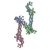 8h3rC M: atomic model generated by this map C: citing same article ( |
|---|---|
| Similar structure data |


-Links
| EMDB pages | EMDB (EBI/PDBe) / EMDataResource |
|---|---|
| Related items in Molecule of the Month |


-Map
| File | Download / File: emd_34473.map.gz / Format: CCP4 / Size: 64 MB / Type: IMAGE STORED AS FLOATING POINT NUMBER (4 BYTES) | ||||||||||||||||||||||||||||||||||||
|---|---|---|---|---|---|---|---|---|---|---|---|---|---|---|---|---|---|---|---|---|---|---|---|---|---|---|---|---|---|---|---|---|---|---|---|---|---|
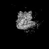
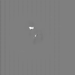
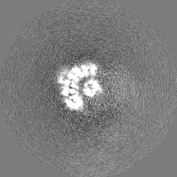
| Projections & slices | Image control
Images are generated by Spider. | ||||||||||||||||||||||||||||||||||||
| Voxel size | X=Y=Z: 1.044 Å | ||||||||||||||||||||||||||||||||||||
| Density |
| ||||||||||||||||||||||||||||||||||||
| Symmetry | Space group: 1 | ||||||||||||||||||||||||||||||||||||
| Details | EMDB XML:
|
 Z (Sec.)
 Y (Row.)
 X (Col.)


-Supplemental data
-Half map: #2
| File | emd_34473_half_map_1.map | ||||||||||||
|---|---|---|---|---|---|---|---|---|---|---|---|---|---|
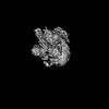
| Projections & Slices |
| ||||||||||||
| Density Histograms |


-Half map: #1
| File | emd_34473_half_map_2.map | ||||||||||||
|---|---|---|---|---|---|---|---|---|---|---|---|---|---|
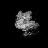
| Projections & Slices |
| ||||||||||||
| Density Histograms |


- Sample components
-Entire : CAND1-Cul3-Rbx1 complex
| Entire | Name: CAND1-Cul3-Rbx1 complex |
|---|---|
| Components |
|
-Supramolecule #1: CAND1-Cul3-Rbx1 complex
| Supramolecule | Name: CAND1-Cul3-Rbx1 complex / type: complex / ID: 1 / Parent: 0 / Macromolecule list: #1-#3 |
|---|---|
| Source (natural) | Organism: Homo sapiens (human) |
-Supramolecule #2: CAND1
| Supramolecule | Name: CAND1 / type: complex / ID: 2 / Parent: 1 / Macromolecule list: #1 |
|---|
-Supramolecule #3: Cul3, Rbx1
| Supramolecule | Name: Cul3, Rbx1 / type: complex / ID: 3 / Parent: 1 / Macromolecule list: #2-#3 |
|---|
-Macromolecule #1: Cullin-associated NEDD8-dissociated protein 1
| Macromolecule | Name: Cullin-associated NEDD8-dissociated protein 1 / type: protein_or_peptide / ID: 1 / Number of copies: 1 / Enantiomer: LEVO |
|---|---|
| Source (natural) | Organism: Homo sapiens (human) |
| Molecular weight | Theoretical: 136.529297 KDa |
| Recombinant expression | Organism:  |
| Sequence | String: MASASYHISN LLEKMTSSDK DFRFMATNDL MTELQKDSIK LDDDSERKVV KMILKLLEDK NGEVQNLAVK CLGPLVSKVK EYQVETIVD TLCTNMLSDK EQLRDISSIG LKTVIGELPP ASSGSALAAN VCKKITGRLT SAIAKQEDVS VQLEALDIMA D MLSRQGGL ...String: MASASYHISN LLEKMTSSDK DFRFMATNDL MTELQKDSIK LDDDSERKVV KMILKLLEDK NGEVQNLAVK CLGPLVSKVK EYQVETIVD TLCTNMLSDK EQLRDISSIG LKTVIGELPP ASSGSALAAN VCKKITGRLT SAIAKQEDVS VQLEALDIMA D MLSRQGGL LVNFHPSILT CLLPQLTSPR LAVRKRTIIA LGHLVMSCGN IVFVDLIEHL LSELSKNDSM STTRTYIQCI AA ISRQAGH RIGEYLEKII PLVVKFCNVD DDELREYCIQ AFESFVRRCP KEVYPHVSTI INICLKYLTY DPNYNYDDED EDE NAMDAD GGDDDDQGSD DEYSDDDDMS WKVRRAAAKC LDAVVSTRHE MLPEFYKTVS PALISRFKER EENVKADVFH AYLS LLKQT RPVQSWLCDP DAMEQGETPL TMLQSQVPNI VKALHKQMKE KSVKTRQCCF NMLTELVNVL PGALTQHIPV LVPGI IFSL NDKSSSSNLK IDALSCLYVI LCNHSPQVFH PHVQALVPPV VACVGDPFYK ITSEALLVTQ QLVKVIRPLD QPSSFD ATP YIKDLFTCTI KRLKAADIDQ EVKERAISCM GQIICNLGDN LGSDLPNTLQ IFLERLKNEI TRLTTVKALT LIAGSPL KI DLRPVLGEGV PILASFLRKN QRALKLGTLS ALDILIKNYS DSLTAAMIDA VLDELPPLIS ESDMHVSQMA ISFLTTLA K VYPSSLSKIS GSILNELIGL VRSPLLQGGA LSAMLDFFQA LVVTGTNNLG YMDLLRMLTG PVYSQSTALT HKQSYYSIA KCVAALTRAC PKEGPAVVGQ FIQDVKNSRS TDSIRLLALL SLGEVGHHID LSGQLELKSV ILEAFSSPSE EVKSAASYAL GSISVGNLP EYLPFVLQEI TSQPKRQYLL LHSLKEIISS ASVVGLKPYV ENIWALLLKH CECAEEGTRN VVAECLGKLT L IDPETLLP RLKGYLISGS SYARSSVVTA VKFTISDHPQ PIDPLLKNCI GDFLKTLEDP DLNVRRVALV TFNSAAHNKP SL IRDLLDT VLPHLYNETK VRKELIREVE MGPFKHTVDD GLDIRKAAFE CMYTLLDSCL DRLDIFEFLN HVEDGLKDHY DIK MLTFLM LVRLSTLCPS AVLQRLDRLV EPLRATCTTK VKANSVKQEF EKQDELKRSA MRAVAALLTI PEAEKSPLMS EFQS QISSN PELAAIFESI QKDSSSTNLE SMDTS UniProtKB: Cullin-associated NEDD8-dissociated protein 1 |
-Macromolecule #2: Cullin-3
| Macromolecule | Name: Cullin-3 / type: protein_or_peptide / ID: 2 / Number of copies: 1 / Enantiomer: LEVO |
|---|---|
| Source (natural) | Organism: Homo sapiens (human) |
| Molecular weight | Theoretical: 89.063328 KDa |
| Recombinant expression | Organism:   Spodoptera frugiperda (fall armyworm) Spodoptera frugiperda (fall armyworm) |
| Sequence | String: MSNLSKGTGS RKDTKMRIRA FPMTMDEKYV NSIWDLLKNA IQEIQRKNNS GLSFEELYRN AYTMVLHKHG EKLYTGLREV VTEHLINKV REDVLNSLNN NFLQTLNQAW NDHQTAMVMI RDILMYMDRV YVQQNNVENV YNLGLIIFRD QVVRYGCIRD H LRQTLLDM ...String: MSNLSKGTGS RKDTKMRIRA FPMTMDEKYV NSIWDLLKNA IQEIQRKNNS GLSFEELYRN AYTMVLHKHG EKLYTGLREV VTEHLINKV REDVLNSLNN NFLQTLNQAW NDHQTAMVMI RDILMYMDRV YVQQNNVENV YNLGLIIFRD QVVRYGCIRD H LRQTLLDM IARERKGEVV DRGAIRNACQ MLMILGLEGR SVYEEDFEAP FLEMSAEFFQ MESQKFLAEN SASVYIKKVE AR INEEIER VMHCLDKSTE EPIVKVVERE LISKHMKTIV EMENSGLVHM LKNGKTEDLG CMYKLFSRVP NGLKTMCECM SSY LREQGK ALVSEEGEGK NPVDYIQGLL DLKSRFDRFL LESFNNDRLF KQTIAGDFEY FLNLNSRSPE YLSLFIDDKL KKGV KGLTE QEVETILDKA MVLFRFMQEK DVFERYYKQH LARRLLTNKS VSDDSEKNMI SKLKTECGCQ FTSKLEGMFR DMSIS NTTM DEFRQHLQAT GVSLGGVDLT VRVLTTGYWP TQSATPKCNI PPAPRHAFEI FRRFYLAKHS GRQLTLQHHM GSADLN ATF YGPVKKEDGS EVGVGGAQVT GSNTRKHILQ VSTFQMTILM LFNNREKYTF EEIQQETDIP ERELVRALQS LACGKPT QR VLTKEPKSKE IENGHIFTVN DQFTSKLHRV KIQTVAAKQG ESDPERKETR QKVDDDRKHE IEAAIVRIMK SRKKMQHN V LVAEVTQQLK ARFLPSPVVI KKRIEGLIER EYLARTPEDR KVYTYVA UniProtKB: Cullin-3 |
-Macromolecule #3: E3 ubiquitin-protein ligase RBX1
| Macromolecule | Name: E3 ubiquitin-protein ligase RBX1 / type: protein_or_peptide / ID: 3 / Number of copies: 1 / Enantiomer: LEVO / EC number: RING-type E3 ubiquitin transferase |
|---|---|
| Source (natural) | Organism: Homo sapiens (human) |
| Molecular weight | Theoretical: 12.289977 KDa |
| Recombinant expression | Organism: Spodoptera frugiperda (fall armyworm) |
| Sequence | String: MAAAMDVDTP SGTNSGAGKK RFEVKKWNAV ALWAWDIVVD NCAICRNHIM DLCIECQANQ ASATSEECTV AWGVCNHAFH FHCISRWLK TRQVCPLDNR EWEFQKYGH UniProtKB: E3 ubiquitin-protein ligase RBX1 |
-Macromolecule #4: ZINC ION
| Macromolecule | Name: ZINC ION / type: ligand / ID: 4 / Number of copies: 3 / Formula: ZN |
|---|---|
| Molecular weight | Theoretical: 65.409 Da |
-Experimental details
-Structure determination
| Method | cryo EM |
|---|---|
 Processing Processing | single particle reconstruction |
| Aggregation state | particle |
-Sample preparation
| Buffer | pH: 7.8 |
|---|---|
| Vitrification | Cryogen name: ETHANE |
- Electron microscopy
| Microscope | FEI TITAN KRIOS |
|---|---|
| Image recording | Film or detector model: GATAN K2 SUMMIT (4k x 4k) / Detector mode: SUPER-RESOLUTION / Average electron dose: 53.0 e/Å2 |
| Electron beam | Acceleration voltage: 300 kV / Electron source:  FIELD EMISSION GUN FIELD EMISSION GUN |
| Electron optics | Illumination mode: FLOOD BEAM / Imaging mode: BRIGHT FIELD / Nominal defocus max: 2.2 µm / Nominal defocus min: 1.2 µm |
| Experimental equipment |  Model: Titan Krios / Image courtesy: FEI Company |