 Movie
Movie Controller


+ Open data

- Basic information
| Entry | Database: PDB / ID: 8gq6 | |||||||||||||||||||||||||||||||||
|---|---|---|---|---|---|---|---|---|---|---|---|---|---|---|---|---|---|---|---|---|---|---|---|---|---|---|---|---|---|---|---|---|---|---|
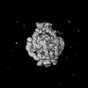
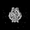
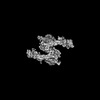
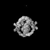
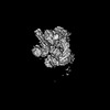
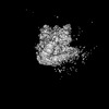
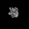
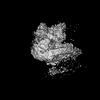
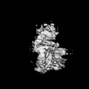
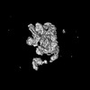
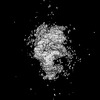
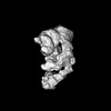
| Title | Cryo-EM Structure of the KBTBD2-CUL3-Rbx1 dimeric complex | |||||||||||||||||||||||||||||||||
 Components Components |
| |||||||||||||||||||||||||||||||||
 Keywords Keywords | LIGASE / complex | |||||||||||||||||||||||||||||||||
| Function / homology |  Function and homology information Function and homology informationpositive regulation of mitotic cell cycle phase transition / trophectodermal cellular morphogenesis / liver morphogenesis / POZ domain binding / polar microtubule / regulation protein catabolic process at postsynapse / anaphase-promoting complex-dependent catabolic process / nuclear protein quality control by the ubiquitin-proteasome system / COPII vesicle coat assembly / negative regulation of beige fat cell differentiation ...positive regulation of mitotic cell cycle phase transition / trophectodermal cellular morphogenesis / liver morphogenesis / POZ domain binding / polar microtubule / regulation protein catabolic process at postsynapse / anaphase-promoting complex-dependent catabolic process / nuclear protein quality control by the ubiquitin-proteasome system / COPII vesicle coat assembly / negative regulation of beige fat cell differentiation / RHOBTB3 ATPase cycle / cullin-RING-type E3 NEDD8 transferase / NEDD8 transferase activity / embryonic cleavage / cullin-RING ubiquitin ligase complex / Cul7-RING ubiquitin ligase complex / cellular response to chemical stress / Loss of Function of FBXW7 in Cancer and NOTCH1 Signaling / positive regulation of mitotic metaphase/anaphase transition / positive regulation of protein autoubiquitination / fibroblast apoptotic process / Notch binding / RNA polymerase II transcription initiation surveillance / protein neddylation / cell projection organization / negative regulation of Rho protein signal transduction / NEDD8 ligase activity / RHOBTB1 GTPase cycle / protein K27-linked ubiquitination / negative regulation of response to oxidative stress / VCB complex / Cul5-RING ubiquitin ligase complex / stem cell division / ubiquitin-ubiquitin ligase activity / mitotic metaphase chromosome alignment / ubiquitin-dependent protein catabolic process via the C-end degron rule pathway / SCF ubiquitin ligase complex / Cul2-RING ubiquitin ligase complex / stress fiber assembly / Cul3-RING ubiquitin ligase complex / negative regulation of type I interferon production / positive regulation of cytokinesis / SCF-dependent proteasomal ubiquitin-dependent protein catabolic process / Prolactin receptor signaling / Cul4A-RING E3 ubiquitin ligase complex / negative regulation of mitophagy / Cul4-RING E3 ubiquitin ligase complex / Cul4B-RING E3 ubiquitin ligase complex / ubiquitin ligase complex scaffold activity / cullin family protein binding / endoplasmic reticulum to Golgi vesicle-mediated transport / protein monoubiquitination / RHOBTB2 GTPase cycle / sperm flagellum / protein autoubiquitination / ubiquitin-like ligase-substrate adaptor activity / site of DNA damage / signal transduction in response to DNA damage / Nuclear events stimulated by ALK signaling in cancer / protein K48-linked ubiquitination / negative regulation of insulin receptor signaling pathway / regulation of cellular response to insulin stimulus / transcription-coupled nucleotide-excision repair / gastrulation / positive regulation of TORC1 signaling / post-translational protein modification / intrinsic apoptotic signaling pathway / cyclin binding / T cell activation / positive regulation of protein ubiquitination / Regulation of BACH1 activity / integrin-mediated signaling pathway / negative regulation of canonical NF-kappaB signal transduction / lipid metabolic process / kidney development / phosphatidylinositol 3-kinase/protein kinase B signal transduction / cellular response to amino acid stimulus / negative regulation of canonical Wnt signaling pathway / G1/S transition of mitotic cell cycle / Degradation of DVL / Degradation of CRY and PER proteins / Degradation of GLI1 by the proteasome / Recognition of DNA damage by PCNA-containing replication complex / response to insulin / RING-type E3 ubiquitin transferase / Negative regulation of NOTCH4 signaling / GSK3B and BTRC:CUL1-mediated-degradation of NFE2L2 / Hedgehog 'on' state / Vif-mediated degradation of APOBEC3G / FBXL7 down-regulates AURKA during mitotic entry and in early mitosis / Degradation of GLI2 by the proteasome / GLI3 is processed to GLI3R by the proteasome / Ubiquitin-Mediated Degradation of Phosphorylated Cdc25A / protein destabilization / Evasion by RSV of host interferon responses / NOTCH1 Intracellular Domain Regulates Transcription / Degradation of beta-catenin by the destruction complex / DNA Damage Recognition in GG-NER / Oxygen-dependent proline hydroxylation of Hypoxia-inducible Factor Alpha / glucose metabolic process Similarity search - Function | |||||||||||||||||||||||||||||||||
| Biological species |  Homo sapiens (human) Homo sapiens (human) | |||||||||||||||||||||||||||||||||
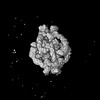
| Method | ELECTRON MICROSCOPY / single particle reconstruction / cryo EM / Resolution: 3.96 Å | |||||||||||||||||||||||||||||||||
 Authors Authors | Sun, L. / Chen, Z. / Hu, Y. / Mao, Q. | |||||||||||||||||||||||||||||||||
| Funding support |  China, 1items China, 1items
| |||||||||||||||||||||||||||||||||
 Citation Citation | Journal: Nat Struct Mol Biol / Year: 2024 Title: Dynamic molecular architecture and substrate recruitment of cullin3-RING E3 ligase CRL3. Authors: Yuxia Hu / Zhao Zhang / Qiyu Mao / Xiang Zhang / Aihua Hao / Yu Xun / Yeda Wang / Lin Han / Wuqiang Zhan / Qianying Liu / Yue Yin / Chao Peng / Eva Marie Y Moresco / Zhenguo Chen / Bruce Beutler / Lei Sun /  Abstract: Phosphatidylinositol 3-kinase α, a heterodimer of catalytic p110α and one of five regulatory subunits, mediates insulin- and insulin like growth factor-signaling and, frequently, oncogenesis. ...Phosphatidylinositol 3-kinase α, a heterodimer of catalytic p110α and one of five regulatory subunits, mediates insulin- and insulin like growth factor-signaling and, frequently, oncogenesis. Cellular levels of the regulatory p85α subunit are tightly controlled by regulated proteasomal degradation. In adipose tissue and growth plates, failure of K48-linked p85α ubiquitination causes diabetes, lipodystrophy and dwarfism in mice, as in humans with SHORT syndrome. Here we elucidated the structures of the key ubiquitin ligase complexes regulating p85α availability. Specificity is provided by the substrate receptor KBTBD2, which recruits p85α to the cullin3-RING E3 ubiquitin ligase (CRL3). CRL3 forms multimers, which disassemble into dimers upon substrate binding (CRL3-p85α) and/or neddylation by the activator NEDD8 (CRL3~N8), leading to p85α ubiquitination and degradation. Deactivation involves dissociation of NEDD8 mediated by the COP9 signalosome and displacement of KBTBD2 by the inhibitor CAND1. The hereby identified structural basis of p85α regulation opens the way to better understanding disturbances of glucose regulation, growth and cancer. | |||||||||||||||||||||||||||||||||
| History |
|
- Structure visualization
| Structure viewer | Molecule: MolmilJmol/JSmol |
|---|
- Downloads & links
-Download
| PDBx/mmCIF format | 8gq6.cif.gz | 504 KB | Display | PDBx/mmCIF format |
|---|---|---|---|---|
| PDB format | pdb8gq6.ent.gz | 404.7 KB | Display | PDB format |
| PDBx/mmJSON format | 8gq6.json.gz | Tree view | PDBx/mmJSON format | |
| Others |  Other downloads Other downloads |
-Validation report
| Arichive directory | https://data.pdbj.org/pub/pdb/validation_reports/gq/8gq6ftp://data.pdbj.org/pub/pdb/validation_reports/gq/8gq6 | HTTPS FTP |
|---|
-Related structure data
| Related structure data |  34199MC 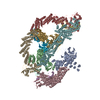 8h33C 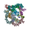 8h34C 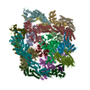 8h35C 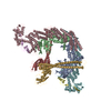 8h36C  8h37C  8h38C  8h3aC  8h3fC 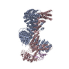 8h3qC 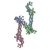 8h3rC M: map data used to model this data C: citing same article ( |
|---|---|
| Similar structure data |


-Links
 PDBj
PDBj


- Assembly
| Deposited unit | 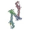
|
|---|---|
| 1 |
|
-Components
| #1: Protein | Mass: 71403.367 Da / Num. of mol.: 2 Source method: isolated from a genetically manipulated source Source: (gene. exp.) Homo sapiens (human) / Production host:   Spodoptera frugiperda (fall armyworm) / References: UniProt: Q8IY47 Spodoptera frugiperda (fall armyworm) / References: UniProt: Q8IY47#2: Protein | Mass: 13970.756 Da / Num. of mol.: 2 Source method: isolated from a genetically manipulated source Source: (gene. exp.) Homo sapiens (human) / Gene: RBX1, RNF75, ROC1 / Production host: Spodoptera frugiperda (fall armyworm)References: UniProt: P62877, RING-type E3 ubiquitin transferase, cullin-RING-type E3 NEDD8 transferase #3: Protein | Mass: 90105.469 Da / Num. of mol.: 2 Source method: isolated from a genetically manipulated source Source: (gene. exp.) Homo sapiens (human) / Production host: Spodoptera frugiperda (fall armyworm) / References: UniProt: Q13618#4: Chemical | ChemComp-ZN /   Mass: 65.409 Da / Num. of mol.: 6 / Source method: obtained synthetically / Formula: Zn Mass: 65.409 Da / Num. of mol.: 6 / Source method: obtained synthetically / Formula: ZnHas ligand of interest | N | Has protein modification | N | |
|---|
-Experimental details
-Experiment
| Experiment | Method: ELECTRON MICROSCOPY |
|---|---|
| EM experiment | Aggregation state: PARTICLE / 3D reconstruction method: single particle reconstruction |
- Sample preparation
| Component | Name: KBTBD2-CUL3-Rbx1 dimeric complex / Type: COMPLEX / Entity ID: #1-#3 / Source: RECOMBINANT |
|---|---|
| Molecular weight | Experimental value: NO |
| Source (natural) | Organism: Homo sapiens (human) |
| Source (recombinant) | Organism: Spodoptera frugiperda (fall armyworm) |
| Buffer solution | pH: 7.8 |
| Specimen | Embedding applied: NO / Shadowing applied: NO / Staining applied: NO / Vitrification applied: YES |
| Vitrification | Cryogen name: ETHANE |
- Electron microscopy imaging
| Experimental equipment |  Model: Titan Krios / Image courtesy: FEI Company |
|---|---|
| Microscopy | Model: FEI TITAN KRIOS |
| Electron gun | Electron source:  FIELD EMISSION GUN / Accelerating voltage: 300 kV / Illumination mode: FLOOD BEAM FIELD EMISSION GUN / Accelerating voltage: 300 kV / Illumination mode: FLOOD BEAM |
| Electron lens | Mode: BRIGHT FIELD / Nominal defocus max: 2200 nm / Nominal defocus min: 1200 nm |
| Image recording | Electron dose: 45 e/Å2 / Detector mode: SUPER-RESOLUTION / Film or detector model: GATAN K2 SUMMIT (4k x 4k) |
- Processing
| Software | Name: PHENIX / Version: 1.20_4459: / Classification: refinement | ||||||||||||||||||||||||
|---|---|---|---|---|---|---|---|---|---|---|---|---|---|---|---|---|---|---|---|---|---|---|---|---|---|
| EM software |
| ||||||||||||||||||||||||
| CTF correction | Type: PHASE FLIPPING AND AMPLITUDE CORRECTION | ||||||||||||||||||||||||
| Symmetry | Point symmetry: C1 (asymmetric) | ||||||||||||||||||||||||
| 3D reconstruction | Resolution: 3.96 Å / Resolution method: FSC 0.143 CUT-OFF / Num. of particles: 433329 / Symmetry type: POINT | ||||||||||||||||||||||||
| Refine LS restraints |
|
