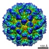
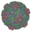
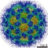
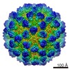
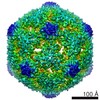
ジャーナル: J Virol / 年: 2015 タイトル: Antigenic and Cryo-Electron Microscopy Structure Analysis of a Chimeric Sapovirus Capsid. 著者: Naoyuki Miyazaki / David W Taylor / Grant S Hansman / Kazuyoshi Murata / 要旨: The capsid protein (VP1) of all caliciviruses forms an icosahedral particle with two principal domains, shell (S) and protruding (P) domains, which are connected via a flexible hinge region. The S ...The capsid protein (VP1) of all caliciviruses forms an icosahedral particle with two principal domains, shell (S) and protruding (P) domains, which are connected via a flexible hinge region. The S domain forms a scaffold surrounding the nucleic acid, while the P domains form a homodimer that interacts with receptors. The P domain is further subdivided into two subdomains, termed P1 and P2. The P2 subdomain is likely an insertion in the P1 subdomain; consequently, the P domain is divided into the P1-1, P2, and P1-2 subdomains. In order to investigate capsid antigenicity, N-terminal (N-term)/S/P1-1 and P2/P1-2 were switched between two sapovirus genotypes GI.1 and GI.5. The chimeric VP1 constructs were expressed in insect cells and were shown to self-assemble into virus-like particles (VLPs) morphologically similar to the parental VLPs. Interestingly, the chimeric VLPs had higher levels of cross-reactivities to heterogeneous antisera than the parental VLPs. In order to better understand the antigenicity from a structural perspective, we determined an intermediate-resolution (8.5-Å) cryo-electron microscopy (cryo-EM) structure of a chimeric VLP and developed a VP1 homology model. The cryo-EM structure revealed that the P domain dimers were raised slightly (∼5 Å) above the S domain. The VP1 homology model allowed us predict the S domain (67-229) and P1-1 (229-280), P2 (281-447), and P1-2 (448-567) subdomains. Our results suggested that the raised P dimers might expose immunoreactive S/P1-1 subdomain epitopes. Consequently, the higher levels of cross-reactivities with the chimeric VLPs resulted from a combination of GI.1 and GI.5 epitopes. IMPORTANCE: We developed sapovirus chimeric VP1 constructs and produced the chimeric VLPs in insect cells. We found that both chimeric VLPs had a higher level of cross-reactivity against ...IMPORTANCE: We developed sapovirus chimeric VP1 constructs and produced the chimeric VLPs in insect cells. We found that both chimeric VLPs had a higher level of cross-reactivity against heterogeneous VLP antisera than the parental VLPs. The cryo-EM structure of one chimeric VLP (Yokote/Mc114) was solved to 8.5-Å resolution. A homology model of the VP1 indicated for the first time the putative S and P (P1-1, P2, and P1-2) domains. The overall structure of Yokote/Mc114 contained features common among other caliciviruses. We showed that the P2 subdomain was mainly involved in the homodimeric interface, whereas a large gap between the P1 subdomains had fewer interactions.
 ムービー
ムービー コントローラー


 データを開く
 基本情報
基本情報 マップデータ
 試料
試料 キーワード
キーワード Sapovirus (ウイルス)
 データ登録者
 引用
引用


 構造の表示
構造の表示 ムービービューア
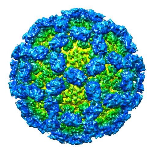
 ダウンロードとリンク
 EMD-3281.png
 http://ftp.pdbj.org/pub/emdb/structures/EMD-3281
 Z (Sec.)
 Y (Row.)
 X (Col.)


 試料の構成要素
 Homo sapiens (ヒト) / 別称: VERTEBRATES
 解析
解析 電子顕微鏡法
 FIELD EMISSION GUN