 Movie
Movie Controller


+ Open data

- Basic information
| Entry |  | |||||||||
|---|---|---|---|---|---|---|---|---|---|---|
| Title | Structure of Inhibited-EP | |||||||||
 Map data Map data | ||||||||||
 Sample Sample |
| |||||||||
 Keywords Keywords | COMPLEX / MEMBRANE PROTEIN | |||||||||
| Function / homology |  Function and homology information Function and homology informationenteropeptidase / brush border / serine-type peptidase activity / protein processing / serine-type endopeptidase activity / membrane Similarity search - Function | |||||||||
| Biological species |  Homo sapiens (human) Homo sapiens (human) | |||||||||
| Method | single particle reconstruction / cryo EM / Resolution: 3.1 Å | |||||||||
 Authors Authors | Yang XL / Ding ZY / Huang HJ | |||||||||
| Funding support | 1 items
| |||||||||
 Citation Citation | Journal: Nat Commun / Year: 2022 Title: Cryo-EM structures reveal the activation and substrate recognition mechanism of human enteropeptidase. Authors: Xiaoli Yang / Zhanyu Ding / Lisi Peng / Qiuyue Song / Deyu Zhang / Fang Cui / Chuanchao Xia / Keliang Li / Hua Yin / Shiyu Li / Zhaoshen Li / Haojie Huang /  Abstract: Enteropeptidase (EP) initiates intestinal digestion by proteolytically processing trypsinogen, generating catalytically active trypsin. EP dysfunction causes a series of pancreatic diseases including ...Enteropeptidase (EP) initiates intestinal digestion by proteolytically processing trypsinogen, generating catalytically active trypsin. EP dysfunction causes a series of pancreatic diseases including acute necrotizing pancreatitis. However, the molecular mechanisms of EP activation and substrate recognition remain elusive, due to the lack of structural information on the EP heavy chain. Here, we report cryo-EM structures of human EP in inactive, active, and substrate-bound states at resolutions from 2.7 to 4.9 Å. The EP heavy chain was observed to clamp the light chain with CUB2 domain for substrate recognition. The EP light chain N-terminus induced a rearrangement of surface-loops from inactive to active conformations, resulting in activated EP. The heavy chain then served as a hinge for light-chain conformational changes to recruit and subsequently cleave substrate. Our study provides structural insights into rearrangements of EP surface-loops and heavy chain dynamics in the EP catalytic cycle, advancing our understanding of EP-associated pancreatitis. | |||||||||
| History |
|
- Structure visualization
| Supplemental images |
|---|

- Downloads & links
-EMDB archive
| Map data | emd_32717.map.gz | 55.5 MB | EMDB map data format | |
|---|---|---|---|---|
| Header (meta data) | emd-32717-v30.xmlemd-32717.xml | 11.4 KB 11.4 KB | Display Display | EMDB header |
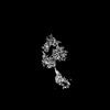
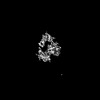
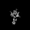
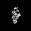
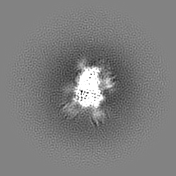
| Images | 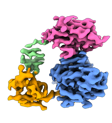 emd_32717.png emd_32717.png | 118.5 KB | ||
| Filedesc metadata | emd-32717.cif.gz | 5.6 KB | ||
| Archive directory |  http://ftp.pdbj.org/pub/emdb/structures/EMD-32717ftp://ftp.pdbj.org/pub/emdb/structures/EMD-32717 http://ftp.pdbj.org/pub/emdb/structures/EMD-32717ftp://ftp.pdbj.org/pub/emdb/structures/EMD-32717 | HTTPS FTP |
-Related structure data
| Related structure data | 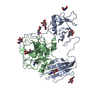 7wr7MC 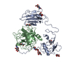 7wqwC 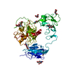 7wqxC  7wqzC 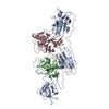 8h3sC 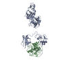 8h3uC M: atomic model generated by this map C: citing same article ( |
|---|---|
| Similar structure data |
-Links
| EMDB pages | EMDB (EBI/PDBe) / EMDataResource |
|---|---|
| Related items in Molecule of the Month |

-Map
| File | Download / File: emd_32717.map.gz / Format: CCP4 / Size: 64 MB / Type: IMAGE STORED AS FLOATING POINT NUMBER (4 BYTES) | ||||||||||||||||||||||||||||||||||||
|---|---|---|---|---|---|---|---|---|---|---|---|---|---|---|---|---|---|---|---|---|---|---|---|---|---|---|---|---|---|---|---|---|---|---|---|---|---|
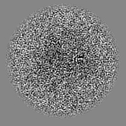
| Projections & slices | Image control
Images are generated by Spider. | ||||||||||||||||||||||||||||||||||||
| Voxel size | X=Y=Z: 1.046 Å | ||||||||||||||||||||||||||||||||||||
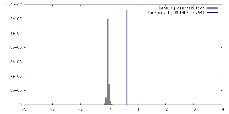
| Density |
| ||||||||||||||||||||||||||||||||||||
| Symmetry | Space group: 1 | ||||||||||||||||||||||||||||||||||||
| Details | EMDB XML:
|
 Z (Sec.)
 Y (Row.)
 X (Col.)


-Supplemental data
- Sample components
-Entire : inhibited-EP
| Entire | Name: inhibited-EP |
|---|---|
| Components |
|
-Supramolecule #1: inhibited-EP
| Supramolecule | Name: inhibited-EP / type: complex / ID: 1 / Parent: 0 / Macromolecule list: #1-#2 |
|---|---|
| Source (natural) | Organism: Homo sapiens (human) |
-Macromolecule #1: Enteropeptidase non-catalytic heavy chain
| Macromolecule | Name: Enteropeptidase non-catalytic heavy chain / type: protein_or_peptide / ID: 1 / Number of copies: 1 / Enantiomer: LEVO |
|---|---|
| Source (natural) | Organism: Homo sapiens (human) |
| Molecular weight | Theoretical: 28.703256 KDa |
| Recombinant expression | Organism: Homo sapiens (human) |
| Sequence | String: CGGPFELWEP NTTFSSTNFP NSYPNLAFCV WILNAQKGKN IQLHFQEFDL ENINDVVEIR DGEEADSLLL AVYTGPGPVK DVFSTTNRM TVLLITNDVL ARGGFKANFT TGYHLGIPEP CKADHFQCKN GECVPLVNLC DGHLHCEDGS DEADCVRFFN G TTNNNGLV ...String: CGGPFELWEP NTTFSSTNFP NSYPNLAFCV WILNAQKGKN IQLHFQEFDL ENINDVVEIR DGEEADSLLL AVYTGPGPVK DVFSTTNRM TVLLITNDVL ARGGFKANFT TGYHLGIPEP CKADHFQCKN GECVPLVNLC DGHLHCEDGS DEADCVRFFN G TTNNNGLV RFRIQSIWHT ACAENWTTQI SNDVCQLLGL GSGNSSKPIF PTDGGPFVKL NTAPDGHLIL TPSQQCLQDS LI RLQCNHK SCGKKLAAQD ITPK UniProtKB: Enteropeptidase |
-Macromolecule #2: Enteropeptidase catalytic light chain
| Macromolecule | Name: Enteropeptidase catalytic light chain / type: protein_or_peptide / ID: 2 / Number of copies: 1 / Enantiomer: LEVO |
|---|---|
| Source (natural) | Organism: Homo sapiens (human) |
| Molecular weight | Theoretical: 26.27776 KDa |
| Recombinant expression | Organism: Homo sapiens (human) |
| Sequence | String: IVGGSNAKEG AWPWVVGLYY GGRLLCGASL VSSDWLVSAA HCVYGRNLEP SKWTAILGLH MKSNLTSPQT VPRLIDEIVI NPHYNRRRK DNDIAMMHLE FKVNYTDYIQ PICLPEENQV FPPGRNCSIA GWGTVVYQGT TANILQEADV PLLSNERCQQ Q MPEYNITE ...String: IVGGSNAKEG AWPWVVGLYY GGRLLCGASL VSSDWLVSAA HCVYGRNLEP SKWTAILGLH MKSNLTSPQT VPRLIDEIVI NPHYNRRRK DNDIAMMHLE FKVNYTDYIQ PICLPEENQV FPPGRNCSIA GWGTVVYQGT TANILQEADV PLLSNERCQQ Q MPEYNITE NMICAGYEEG GIDSCQGDSG GPLMCQENNR WFLAGVTSFG YKCALPNRPG VYARVSRFTE WIQSFLH UniProtKB: Enteropeptidase |
-Macromolecule #3: 2-acetamido-2-deoxy-beta-D-glucopyranose
| Macromolecule | Name: 2-acetamido-2-deoxy-beta-D-glucopyranose / type: ligand / ID: 3 / Number of copies: 9 / Formula: NAG |
|---|---|
| Molecular weight | Theoretical: 221.208 Da |
| Chemical component information |  ChemComp-NAG: |
-Macromolecule #4: 4-carbamimidamidobenzoic acid
| Macromolecule | Name: 4-carbamimidamidobenzoic acid / type: ligand / ID: 4 / Number of copies: 1 / Formula: GBS |
|---|---|
| Molecular weight | Theoretical: 179.176 Da |
| Chemical component information | 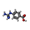 ChemComp-GBS: |
-Experimental details
-Structure determination
| Method | cryo EM |
|---|---|
 Processing Processing | single particle reconstruction |
| Aggregation state | particle |
-Sample preparation
| Buffer | pH: 7.6 |
|---|---|
| Vitrification | Cryogen name: ETHANE |
- Electron microscopy
| Microscope | FEI TITAN KRIOS |
|---|---|
| Image recording | Film or detector model: GATAN K2 SUMMIT (4k x 4k) / Average electron dose: 52.0 e/Å2 |
| Electron beam | Acceleration voltage: 300 kV / Electron source:  FIELD EMISSION GUN FIELD EMISSION GUN |
| Electron optics | Illumination mode: FLOOD BEAM / Imaging mode: BRIGHT FIELD / Nominal defocus max: 3.4 µm / Nominal defocus min: 0.7000000000000001 µm |
| Experimental equipment |  Model: Titan Krios / Image courtesy: FEI Company |
-Image processing
| Startup model | Type of model: INSILICO MODEL |
|---|---|
| Final reconstruction | Resolution.type: BY AUTHOR / Resolution: 3.1 Å / Resolution method: FSC 0.143 CUT-OFF / Number images used: 343205 |
| Initial angle assignment | Type: PROJECTION MATCHING |
| Final angle assignment | Type: ANGULAR RECONSTITUTION |
