 Movie
Movie Controller


+ Open data

- Basic information
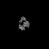
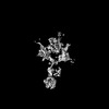
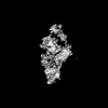
| Entry | Database: PDB / ID: 7wqz | |||||||||||||||||||||||||||
|---|---|---|---|---|---|---|---|---|---|---|---|---|---|---|---|---|---|---|---|---|---|---|---|---|---|---|---|---|
| Title | Structure of Active-mutEP | |||||||||||||||||||||||||||
 Components Components |
| |||||||||||||||||||||||||||
 Keywords Keywords | MEMBRANE PROTEIN / COMPLEX | |||||||||||||||||||||||||||
| Function / homology |  Function and homology information Function and homology informationenteropeptidase / brush border / serine-type peptidase activity / serine-type endopeptidase activity / proteolysis / membrane Similarity search - Function | |||||||||||||||||||||||||||
| Biological species |  Homo sapiens (human) Homo sapiens (human) | |||||||||||||||||||||||||||
| Method | ELECTRON MICROSCOPY / single particle reconstruction / cryo EM / Resolution: 3.7 Å | |||||||||||||||||||||||||||
 Authors Authors | Yang, X.L. / Ding, Z.Y. / Huang, H.J. | |||||||||||||||||||||||||||
| Funding support | 1items
| |||||||||||||||||||||||||||
 Citation Citation | Journal: Nat Commun / Year: 2022 Title: Cryo-EM structures reveal the activation and substrate recognition mechanism of human enteropeptidase. Authors: Xiaoli Yang / Zhanyu Ding / Lisi Peng / Qiuyue Song / Deyu Zhang / Fang Cui / Chuanchao Xia / Keliang Li / Hua Yin / Shiyu Li / Zhaoshen Li / Haojie Huang /  Abstract: Enteropeptidase (EP) initiates intestinal digestion by proteolytically processing trypsinogen, generating catalytically active trypsin. EP dysfunction causes a series of pancreatic diseases including ...Enteropeptidase (EP) initiates intestinal digestion by proteolytically processing trypsinogen, generating catalytically active trypsin. EP dysfunction causes a series of pancreatic diseases including acute necrotizing pancreatitis. However, the molecular mechanisms of EP activation and substrate recognition remain elusive, due to the lack of structural information on the EP heavy chain. Here, we report cryo-EM structures of human EP in inactive, active, and substrate-bound states at resolutions from 2.7 to 4.9 Å. The EP heavy chain was observed to clamp the light chain with CUB2 domain for substrate recognition. The EP light chain N-terminus induced a rearrangement of surface-loops from inactive to active conformations, resulting in activated EP. The heavy chain then served as a hinge for light-chain conformational changes to recruit and subsequently cleave substrate. Our study provides structural insights into rearrangements of EP surface-loops and heavy chain dynamics in the EP catalytic cycle, advancing our understanding of EP-associated pancreatitis. | |||||||||||||||||||||||||||
| History |
|
- Structure visualization
| Structure viewer | Molecule: MolmilJmol/JSmol |
|---|
- Downloads & links
-Download
| PDBx/mmCIF format | 7wqz.cif.gz | 102.1 KB | Display | PDBx/mmCIF format |
|---|---|---|---|---|
| PDB format | pdb7wqz.ent.gz | 75.8 KB | Display | PDB format |
| PDBx/mmJSON format | 7wqz.json.gz | Tree view | PDBx/mmJSON format | |
| Others |  Other downloads Other downloads |
-Validation report
| Arichive directory | https://data.pdbj.org/pub/pdb/validation_reports/wq/7wqzftp://data.pdbj.org/pub/pdb/validation_reports/wq/7wqz | HTTPS FTP |
|---|
-Related structure data
| Related structure data |  32716MC 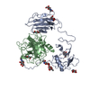 7wqwC 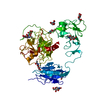 7wqxC 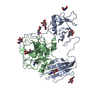 7wr7C 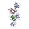 8h3sC 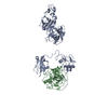 8h3uC M: map data used to model this data C: citing same article ( |
|---|---|
| Similar structure data |
-Links
 PDBj
PDBj
- Assembly
| Deposited unit | 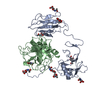
|
|---|---|
| 1 |
|
-Components
| #1: Protein | Mass: 28703.256 Da / Num. of mol.: 1 Source method: isolated from a genetically manipulated source Source: (gene. exp.) Homo sapiens (human) / Gene: TMPRSS15, ENTK, PRSS7 / Production host: Homo sapiens (human) / References: UniProt: P98073 | ||||
|---|---|---|---|---|---|
| #2: Protein | Mass: 26150.680 Da / Num. of mol.: 1 / Mutation: H825A, D876A, S971A Source method: isolated from a genetically manipulated source Source: (gene. exp.) Homo sapiens (human) / Gene: TMPRSS15, ENTK, PRSS7 / Production host: Homo sapiens (human) / References: UniProt: P98073 | ||||
| #3: Sugar | ChemComp-NAG /   Type: D-saccharide, beta linking / Mass: 221.208 Da / Num. of mol.: 9 / Source method: obtained synthetically / Formula: C8H15NO6 / Feature type: SUBJECT OF INVESTIGATION Type: D-saccharide, beta linking / Mass: 221.208 Da / Num. of mol.: 9 / Source method: obtained synthetically / Formula: C8H15NO6 / Feature type: SUBJECT OF INVESTIGATIONHas ligand of interest | Y | Has protein modification | Y | |
-Experimental details
-Experiment
| Experiment | Method: ELECTRON MICROSCOPY |
|---|---|
| EM experiment | Aggregation state: PARTICLE / 3D reconstruction method: single particle reconstruction |
- Sample preparation
| Component | Name: active-mutEP / Type: COMPLEX / Entity ID: #1-#2 / Source: RECOMBINANT |
|---|---|
| Source (natural) | Organism: Homo sapiens (human) |
| Source (recombinant) | Organism: Homo sapiens (human) |
| Buffer solution | pH: 7.6 |
| Specimen | Embedding applied: NO / Shadowing applied: NO / Staining applied: NO / Vitrification applied: YES |
| Vitrification | Cryogen name: ETHANE |
- Electron microscopy imaging
| Experimental equipment |  Model: Titan Krios / Image courtesy: FEI Company |
|---|---|
| Microscopy | Model: FEI TITAN KRIOS |
| Electron gun | Electron source:  FIELD EMISSION GUN / Accelerating voltage: 300 kV / Illumination mode: FLOOD BEAM FIELD EMISSION GUN / Accelerating voltage: 300 kV / Illumination mode: FLOOD BEAM |
| Electron lens | Mode: BRIGHT FIELD / Nominal defocus max: 3400 nm / Nominal defocus min: 700 nm |
| Image recording | Electron dose: 52 e/Å2 / Film or detector model: GATAN K2 SUMMIT (4k x 4k) |
- Processing
| Software | Name: PHENIX / Version: 1.17.1_3660: / Classification: refinement | ||||||||||||||||||||||||
|---|---|---|---|---|---|---|---|---|---|---|---|---|---|---|---|---|---|---|---|---|---|---|---|---|---|
| EM software | Name: PHENIX / Category: model refinement | ||||||||||||||||||||||||
| CTF correction | Type: PHASE FLIPPING AND AMPLITUDE CORRECTION | ||||||||||||||||||||||||
| 3D reconstruction | Resolution: 3.7 Å / Resolution method: FSC 0.143 CUT-OFF / Num. of particles: 156344 / Symmetry type: POINT | ||||||||||||||||||||||||
| Refine LS restraints |
|
