+ データを開く
- 基本情報
基本情報
| 登録情報 | データベース: PDB / ID: 7wqw | |||||||||||||||||||||||||||
|---|---|---|---|---|---|---|---|---|---|---|---|---|---|---|---|---|---|---|---|---|---|---|---|---|---|---|---|---|
| タイトル | Structure of Active-EP | |||||||||||||||||||||||||||
 要素 要素 |
| |||||||||||||||||||||||||||
 キーワード キーワード | MEMBRANE PROTEIN / COMPLEX | |||||||||||||||||||||||||||
| 機能・相同性 |  機能・相同性情報 機能・相同性情報enteropeptidase / brush border / serine-type endopeptidase activity / proteolysis / membrane 類似検索 - 分子機能 | |||||||||||||||||||||||||||
| 生物種 |  Homo sapiens (ヒト) Homo sapiens (ヒト) | |||||||||||||||||||||||||||
| 手法 | 電子顕微鏡法 / 単粒子再構成法 / クライオ電子顕微鏡法 / 解像度: 3.2 Å | |||||||||||||||||||||||||||
 データ登録者 データ登録者 | Yang, X.L. / Ding, Z.Y. / Huang, H.J. | |||||||||||||||||||||||||||
| 資金援助 | 1件
| |||||||||||||||||||||||||||
 引用 引用 |  ジャーナル: Nat Commun / 年: 2022 ジャーナル: Nat Commun / 年: 2022タイトル: Cryo-EM structures reveal the activation and substrate recognition mechanism of human enteropeptidase. 著者: Xiaoli Yang / Zhanyu Ding / Lisi Peng / Qiuyue Song / Deyu Zhang / Fang Cui / Chuanchao Xia / Keliang Li / Hua Yin / Shiyu Li / Zhaoshen Li / Haojie Huang /  要旨: Enteropeptidase (EP) initiates intestinal digestion by proteolytically processing trypsinogen, generating catalytically active trypsin. EP dysfunction causes a series of pancreatic diseases including ...Enteropeptidase (EP) initiates intestinal digestion by proteolytically processing trypsinogen, generating catalytically active trypsin. EP dysfunction causes a series of pancreatic diseases including acute necrotizing pancreatitis. However, the molecular mechanisms of EP activation and substrate recognition remain elusive, due to the lack of structural information on the EP heavy chain. Here, we report cryo-EM structures of human EP in inactive, active, and substrate-bound states at resolutions from 2.7 to 4.9 Å. The EP heavy chain was observed to clamp the light chain with CUB2 domain for substrate recognition. The EP light chain N-terminus induced a rearrangement of surface-loops from inactive to active conformations, resulting in activated EP. The heavy chain then served as a hinge for light-chain conformational changes to recruit and subsequently cleave substrate. Our study provides structural insights into rearrangements of EP surface-loops and heavy chain dynamics in the EP catalytic cycle, advancing our understanding of EP-associated pancreatitis. | |||||||||||||||||||||||||||
| 履歴 |
|
- 構造の表示
構造の表示
| 構造ビューア | 分子:  Molmil Molmil Jmol/JSmol Jmol/JSmol |
|---|
- ダウンロードとリンク
- ダウンロード
| PDBx/mmCIF形式 |  7wqw.cif.gz 7wqw.cif.gz | 101.8 KB | 表示 |  PDBx/mmCIF形式 PDBx/mmCIF形式 |
|---|---|---|---|---|
| PDB形式 |  pdb7wqw.ent.gz pdb7wqw.ent.gz | 75.6 KB | 表示 |  PDB形式 PDB形式 |
| PDBx/mmJSON形式 |  7wqw.json.gz 7wqw.json.gz | ツリー表示 |  PDBx/mmJSON形式 PDBx/mmJSON形式 | |
| その他 |  その他のダウンロード その他のダウンロード |
-検証レポート
| アーカイブディレクトリ |  https://data.pdbj.org/pub/pdb/validation_reports/wq/7wqw https://data.pdbj.org/pub/pdb/validation_reports/wq/7wqw ftp://data.pdbj.org/pub/pdb/validation_reports/wq/7wqw ftp://data.pdbj.org/pub/pdb/validation_reports/wq/7wqw | HTTPS FTP |
|---|
-関連構造データ
| 関連構造データ |  32714MC 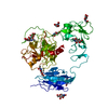 7wqxC  7wqzC 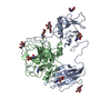 7wr7C 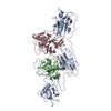 8h3sC 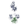 8h3uC M: このデータのモデリングに利用したマップデータ C: 同じ文献を引用 ( |
|---|---|
| 類似構造データ | 類似検索 - 機能・相同性  F&H 検索 F&H 検索 |
- リンク
リンク
- 集合体
集合体
| 登録構造単位 | 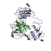
|
|---|---|
| 1 |
|
- 要素
要素
| #1: タンパク質 | 分子量: 28703.256 Da / 分子数: 1 / 由来タイプ: 組換発現 / 由来: (組換発現)  Homo sapiens (ヒト) / 遺伝子: TMPRSS15, ENTK, PRSS7 / 発現宿主: Homo sapiens (ヒト) / 遺伝子: TMPRSS15, ENTK, PRSS7 / 発現宿主:  Homo sapiens (ヒト) / 参照: UniProt: P98073 Homo sapiens (ヒト) / 参照: UniProt: P98073 | ||||
|---|---|---|---|---|---|
| #2: タンパク質 | 分子量: 26277.760 Da / 分子数: 1 / 由来タイプ: 組換発現 / 由来: (組換発現)  Homo sapiens (ヒト) / 遺伝子: TMPRSS15, ENTK, PRSS7 / 発現宿主: Homo sapiens (ヒト) / 遺伝子: TMPRSS15, ENTK, PRSS7 / 発現宿主:  Homo sapiens (ヒト) / 参照: UniProt: P98073 Homo sapiens (ヒト) / 参照: UniProt: P98073 | ||||
| #3: 糖 | ChemComp-NAG / 研究の焦点であるリガンドがあるか | Y | Has protein modification | Y | |
-実験情報
-実験
| 実験 | 手法: 電子顕微鏡法 |
|---|---|
| EM実験 | 試料の集合状態: PARTICLE / 3次元再構成法: 単粒子再構成法 |
- 試料調製
試料調製
| 構成要素 | 名称: active-EP / タイプ: COMPLEX / Entity ID: #1-#2 / 由来: RECOMBINANT |
|---|---|
| 由来(天然) | 生物種:  Homo sapiens (ヒト) Homo sapiens (ヒト) |
| 由来(組換発現) | 生物種:  Homo sapiens (ヒト) Homo sapiens (ヒト) |
| 緩衝液 | pH: 7.6 |
| 試料 | 包埋: NO / シャドウイング: NO / 染色: NO / 凍結: YES |
| 急速凍結 | 凍結剤: ETHANE |
- 電子顕微鏡撮影
| 実験機器 |  モデル: Titan Krios / 画像提供: FEI Company |
|---|---|
| 顕微鏡 | モデル: FEI TITAN KRIOS |
| 電子銃 | 電子線源:  FIELD EMISSION GUN / 加速電圧: 300 kV / 照射モード: FLOOD BEAM FIELD EMISSION GUN / 加速電圧: 300 kV / 照射モード: FLOOD BEAM |
| 電子レンズ | モード: BRIGHT FIELD / 最大 デフォーカス(公称値): 3400 nm / 最小 デフォーカス(公称値): 700 nm |
| 撮影 | 電子線照射量: 52 e/Å2 フィルム・検出器のモデル: GATAN K2 SUMMIT (4k x 4k) |
- 解析
解析
| ソフトウェア | 名称: PHENIX / バージョン: 1.17.1_3660: / 分類: 精密化 | ||||||||||||||||||||||||
|---|---|---|---|---|---|---|---|---|---|---|---|---|---|---|---|---|---|---|---|---|---|---|---|---|---|
| EMソフトウェア | 名称: PHENIX / カテゴリ: モデル精密化 | ||||||||||||||||||||||||
| CTF補正 | タイプ: PHASE FLIPPING AND AMPLITUDE CORRECTION | ||||||||||||||||||||||||
| 3次元再構成 | 解像度: 3.2 Å / 解像度の算出法: FSC 0.143 CUT-OFF / 粒子像の数: 307754 / 対称性のタイプ: POINT | ||||||||||||||||||||||||
| 拘束条件 |
|
 ムービー
ムービー コントローラー


 PDBj
PDBj


