 Movie
Movie Controller

[English] 日本語
 Yorodumi
- EMDB-3244: Electron negative-staining microscopy of an aerolysin-like protein -
+ Open data

- Basic information
| Entry | Database: EMDB / ID: EMD-3244 | |||||||||
|---|---|---|---|---|---|---|---|---|---|---|
| Title | Electron negative-staining microscopy of an aerolysin-like protein | |||||||||
 Map data Map data | Reconstruction of mutant aerolysin-like protein | |||||||||
 Sample Sample |
| |||||||||
| Function / homology |  Function and homology information Function and homology informationdisaccharide binding / pore complex / D-mannose binding / defense response to bacterium / identical protein binding / plasma membrane Similarity search - Function | |||||||||
| Biological species |  | |||||||||
| Method | electron crystallography / negative staining / Resolution: 20.0 Å | |||||||||
 Authors Authors | Jia N / Liu N / Cheng W / Jiang YL / Sun H / Chen LL / Peng JH / Zhang YH / Zhang ZH / Wang XJ ...Jia N / Liu N / Cheng W / Jiang YL / Sun H / Chen LL / Peng JH / Zhang YH / Zhang ZH / Wang XJ / Cai G / Wang JF / Zhang ZY / Wu H / Wang HW / Chen YX / Zhou CZ | |||||||||
 Citation Citation | Journal: EMBO Rep / Year: 2016 Title: Structural basis for receptor recognition and pore formation of a zebrafish aerolysin-like protein. Authors: Ning Jia / Nan Liu / Wang Cheng / Yong-Liang Jiang / Hui Sun / Lan-Lan Chen / Junhui Peng / Yonghui Zhang / Yue-He Ding / Zhi-Hui Zhang / Xuejuan Wang / Gang Cai / Junfeng Wang / Meng-Qiu ...Authors: Ning Jia / Nan Liu / Wang Cheng / Yong-Liang Jiang / Hui Sun / Lan-Lan Chen / Junhui Peng / Yonghui Zhang / Yue-He Ding / Zhi-Hui Zhang / Xuejuan Wang / Gang Cai / Junfeng Wang / Meng-Qiu Dong / Zhiyong Zhang / Hui Wu / Hong-Wei Wang / Yuxing Chen / Cong-Zhao Zhou /   Abstract: Various aerolysin-like pore-forming proteins have been identified from bacteria to vertebrates. However, the mechanism of receptor recognition and/or pore formation of the eukaryotic members remains ...Various aerolysin-like pore-forming proteins have been identified from bacteria to vertebrates. However, the mechanism of receptor recognition and/or pore formation of the eukaryotic members remains unknown. Here, we present the first crystal and electron microscopy structures of a vertebrate aerolysin-like protein from Danio rerio, termed Dln1, before and after pore formation. Each subunit of Dln1 dimer comprises a β-prism lectin module followed by an aerolysin module. Specific binding of the lectin module toward high-mannose glycans triggers drastic conformational changes of the aerolysin module in a pH-dependent manner, ultimately resulting in the formation of a membrane-bound octameric pore. Structural analyses combined with computational simulations and biochemical assays suggest a pore-forming process with an activation mechanism distinct from the previously characterized bacterial members. Moreover, Dln1 and its homologs are ubiquitously distributed in bony fishes and lamprey, suggesting a novel fish-specific defense molecule. | |||||||||
| History |
|
- Structure visualization
| Movie |
Movie viewer |
|---|---|
| Structure viewer | EM map: SurfViewMolmilJmol/JSmol |
| Supplemental images |
 UCSF Chimera
- Downloads & links
-EMDB archive
| Map data | emd_3244.map.gz | 27.8 MB | EMDB map data format | |
|---|---|---|---|---|
| Header (meta data) | emd-3244-v30.xmlemd-3244.xml | 11 KB 11 KB | Display Display | EMDB header |
| Images | 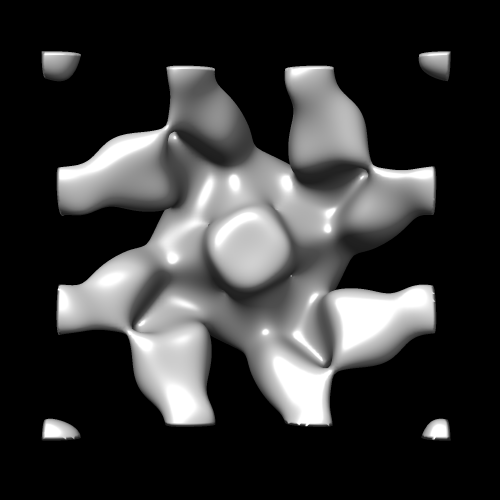 EMD-3244_Dln1_octomer.png EMD-3244_Dln1_octomer.png | 54.1 KB | ||
| Archive directory |  http://ftp.pdbj.org/pub/emdb/structures/EMD-3244ftp://ftp.pdbj.org/pub/emdb/structures/EMD-3244 http://ftp.pdbj.org/pub/emdb/structures/EMD-3244ftp://ftp.pdbj.org/pub/emdb/structures/EMD-3244 | HTTPS FTP |
-Validation report
| Summary document | emd_3244_validation.pdf.gz | 187.5 KB | Display | EMDB validaton report |
|---|---|---|---|---|
| Full document | emd_3244_full_validation.pdf.gz | 186.6 KB | Display | |
| Data in XML | emd_3244_validation.xml.gz | 4.7 KB | Display | |
| Arichive directory | https://ftp.pdbj.org/pub/emdb/validation_reports/EMD-3244ftp://ftp.pdbj.org/pub/emdb/validation_reports/EMD-3244 | HTTPS FTP |
-Related structure data


-Links
| EMDB pages | EMDB (EBI/PDBe) / EMDataResource |
|---|
-Map
| File | Download / File: emd_3244.map.gz / Format: CCP4 / Size: 30.7 MB / Type: IMAGE STORED AS FLOATING POINT NUMBER (4 BYTES) | ||||||||||||||||||||||||||||||||||||||||||||||||||||||||||||
|---|---|---|---|---|---|---|---|---|---|---|---|---|---|---|---|---|---|---|---|---|---|---|---|---|---|---|---|---|---|---|---|---|---|---|---|---|---|---|---|---|---|---|---|---|---|---|---|---|---|---|---|---|---|---|---|---|---|---|---|---|---|
| Annotation | Reconstruction of mutant aerolysin-like protein | ||||||||||||||||||||||||||||||||||||||||||||||||||||||||||||
| Projections & slices | Image control
Images are generated by Spider. generated in cubic-lattice coordinate | ||||||||||||||||||||||||||||||||||||||||||||||||||||||||||||
| Voxel size | X=Y=Z: 0.625 Å | ||||||||||||||||||||||||||||||||||||||||||||||||||||||||||||
| Density |
| ||||||||||||||||||||||||||||||||||||||||||||||||||||||||||||
| Symmetry | Space group: 1 | ||||||||||||||||||||||||||||||||||||||||||||||||||||||||||||
| Details | EMDB XML:
CCP4 map header:
| ||||||||||||||||||||||||||||||||||||||||||||||||||||||||||||
 Z (Sec.)
 Y (Row.)
 X (Col.)


-Supplemental data
- Sample components
-Entire : Dln1 (a zebrafish aerolysin-like protein) oligomer on phospholipi...
| Entire | Name: Dln1 (a zebrafish aerolysin-like protein) oligomer on phospholipid monolayer |
|---|---|
| Components |
|
-Supramolecule #1000: Dln1 (a zebrafish aerolysin-like protein) oligomer on phospholipi...
| Supramolecule | Name: Dln1 (a zebrafish aerolysin-like protein) oligomer on phospholipid monolayer type: sample / ID: 1000 Oligomeric state: Octameric Dln1 reconstituted on lipid monolayer Number unique components: 1 |
|---|---|
| Molecular weight | Experimental: 280 KDa / Theoretical: 280 KDa |
-Macromolecule #1: Dln1
| Macromolecule | Name: Dln1 / type: protein_or_peptide / ID: 1 / Number of copies: 8 / Oligomeric state: Octamer / Recombinant expression: Yes |
|---|---|
| Source (natural) | Organism: |
| Molecular weight | Experimental: 280 KDa / Theoretical: 280 KDa |
| Recombinant expression | Organism:  |
| Sequence | UniProtKB: Aerolysin-like protein |
-Experimental details
-Structure determination
| Method | negative staining |
|---|---|
 Processing Processing | electron crystallography |
| Aggregation state | 2D array |
-Sample preparation
| Concentration | 0.2 mg/mL |
|---|---|
| Buffer | pH: 5.5 / Details: 50 mM MES, 150 mM NaC |
| Staining | Type: NEGATIVE Details: Grids attached with two-dimensional protein crystal on lipid monolayer were gently washed, followed by negative staining with 2% w/v uranyl acetate for about 30 seconds. |
| Grid | Details: 200 mesh copper grid with thin holey carbon support. The grids were not glow discharged. |
| Vitrification | Cryogen name: NONE / Instrument: OTHER |
| Details | Crystals grown on a lipid-monolayer |
| Crystal formation | Details: Crystals grown on a lipid-monolayer |
- Electron microscopy
| Microscope | FEI TECNAI 12 |
|---|---|
| Date | Oct 9, 2014 |
| Image recording | Category: CCD / Film or detector model: GATAN ULTRASCAN 4000 (4k x 4k) / Number real images: 45 |
| Tilt angle min | 0 |
| Electron beam | Acceleration voltage: 120 kV / Electron source: LAB6 |
| Electron optics | Calibrated magnification: 98000 / Illumination mode: FLOOD BEAM / Imaging mode: BRIGHT FIELD / Cs: 6.7 mm / Nominal defocus max: 1.5 µm / Nominal defocus min: 0.588 µm / Nominal magnification: 98000 |
| Sample stage | Specimen holder model: OTHER / Tilt angle max: 40 / Tilt series - Axis1 - Min angle: 0 ° / Tilt series - Axis1 - Max angle: 40 ° |
-Image processing
| Details | Images were unbent using 2dx. |
|---|---|
| Final reconstruction | Resolution.type: BY AUTHOR / Resolution: 20.0 Å / Resolution method: DIFFRACTION PATTERN/LAYERLINES / Software - Name: 2dx |
| Crystal parameters | Unit cell - A: 125 Å / Unit cell - B: 125 Å / Unit cell - γ: 90.0 ° / Unit cell - α: 90.0 ° / Unit cell - β: 90.0 ° / Plane group: P 4 |
