+ Open data
- Basic information
| Entry | Database: EMDB / ID: EMD-32434 | |||||||||
|---|---|---|---|---|---|---|---|---|---|---|
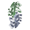
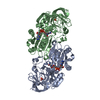
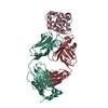
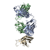
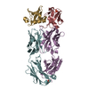
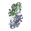
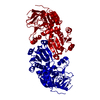
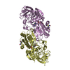
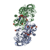
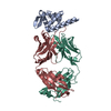
| Title | SARS-CoV-2 Beta spike SD1 in complex with S3H3 Fab | |||||||||
 Map data Map data | ||||||||||
 Sample Sample |
| |||||||||
 Keywords Keywords | SARS-CoV-2 / coronavirus / Beta variant / B.1.351 lineage / spike protein / VIRAL PROTEIN | |||||||||
| Function / homology |  Function and homology information Function and homology informationsymbiont-mediated disruption of host tissue / Maturation of spike protein / Translation of Structural Proteins / Virion Assembly and Release / host cell surface / host extracellular space / viral translation / symbiont-mediated-mediated suppression of host tetherin activity / Induction of Cell-Cell Fusion / structural constituent of virion ...symbiont-mediated disruption of host tissue / Maturation of spike protein / Translation of Structural Proteins / Virion Assembly and Release / host cell surface / host extracellular space / viral translation / symbiont-mediated-mediated suppression of host tetherin activity / Induction of Cell-Cell Fusion / structural constituent of virion / membrane fusion / entry receptor-mediated virion attachment to host cell / Attachment and Entry / host cell endoplasmic reticulum-Golgi intermediate compartment membrane / positive regulation of viral entry into host cell / receptor-mediated virion attachment to host cell / host cell surface receptor binding / symbiont-mediated suppression of host innate immune response / receptor ligand activity / endocytosis involved in viral entry into host cell / fusion of virus membrane with host plasma membrane / fusion of virus membrane with host endosome membrane / viral envelope / symbiont entry into host cell / virion attachment to host cell / SARS-CoV-2 activates/modulates innate and adaptive immune responses / host cell plasma membrane / virion membrane / identical protein binding / membrane / plasma membrane Similarity search - Function | |||||||||
| Biological species |   | |||||||||
| Method | single particle reconstruction / cryo EM / Resolution: 4.3 Å | |||||||||
 Authors Authors | Wang YF / Cong Y | |||||||||
| Funding support |  China, 1 items China, 1 items
| |||||||||
 Citation Citation |  Journal: Emerg Microbes Infect / Year: 2022 Journal: Emerg Microbes Infect / Year: 2022Title: Mapping cross-variant neutralizing sites on the SARS-CoV-2 spike protein. Authors: Shiqi Xu / Yifan Wang / Yanxing Wang / Chao Zhang / Qin Hong / Chenjian Gu / Rong Xu / Tingfeng Wang / Yong Yang / Jinkai Zang / Yu Zhou / Zuyang Li / Qixing Liu / Bingjie Zhou / Lulu Bai / ...Authors: Shiqi Xu / Yifan Wang / Yanxing Wang / Chao Zhang / Qin Hong / Chenjian Gu / Rong Xu / Tingfeng Wang / Yong Yang / Jinkai Zang / Yu Zhou / Zuyang Li / Qixing Liu / Bingjie Zhou / Lulu Bai / Yuanfei Zhu / Qiang Deng / Haikun Wang / Dimitri Lavillette / Gary Wong / Youhua Xie / Yao Cong / Zhong Huang /  Abstract: The emergence of multiple severe acute respiratory syndrome coronavirus 2 (SARS-CoV-2) variants of concern threatens the efficacy of currently approved vaccines and authorized therapeutic monoclonal ...The emergence of multiple severe acute respiratory syndrome coronavirus 2 (SARS-CoV-2) variants of concern threatens the efficacy of currently approved vaccines and authorized therapeutic monoclonal antibodies (MAbs). It is hence important to continue searching for SARS-CoV-2 broadly neutralizing MAbs and defining their epitopes. Here, we isolate 9 neutralizing mouse MAbs raised against the spike protein of a SARS-CoV-2 prototype strain and evaluate their neutralizing potency towards a panel of variants, including B.1.1.7, B.1.351, B.1.617.1, and B.1.617.2. By using a combination of biochemical, virological, and cryo-EM structural analyses, we identify three types of cross-variant neutralizing MAbs, represented by S5D2, S5G2, and S3H3, respectively, and further define their epitopes. S5D2 binds the top lateral edge of the receptor-binding motif within the receptor-binding domain (RBD) with a binding footprint centred around the loop, and efficiently neutralizes all variant pseudoviruses, but the potency against B.1.617.2 was observed to decrease significantly. S5G2 targets the highly conserved RBD core region and exhibits comparable neutralization towards the variant panel. S3H3 binds a previously unreported epitope located within the evolutionarily stable SD1 region and is able to near equally neutralize all of the variants tested. Our work thus defines three distinct cross-variant neutralizing sites on the SARS-CoV-2 spike protein, providing guidance for design and development of broadly effective vaccines and MAb-based therapies. | |||||||||
| History |
|
- Structure visualization
| Movie |
 Movie viewer Movie viewer |
|---|---|
| Structure viewer | EM map:  SurfView SurfView Molmil Molmil Jmol/JSmol Jmol/JSmol |
| Supplemental images |
- Downloads & links
-EMDB archive
| Map data |  emd_32434.map.gz emd_32434.map.gz | 1.2 MB |  EMDB map data format EMDB map data format | |
|---|---|---|---|---|
| Header (meta data) |  emd-32434-v30.xml emd-32434-v30.xml emd-32434.xml emd-32434.xml | 14.9 KB 14.9 KB | Display Display |  EMDB header EMDB header |
| Images |  emd_32434.png emd_32434.png | 122.4 KB | ||
| Filedesc metadata |  emd-32434.cif.gz emd-32434.cif.gz | 6.4 KB | ||
| Archive directory |  http://ftp.pdbj.org/pub/emdb/structures/EMD-32434 http://ftp.pdbj.org/pub/emdb/structures/EMD-32434 ftp://ftp.pdbj.org/pub/emdb/structures/EMD-32434 ftp://ftp.pdbj.org/pub/emdb/structures/EMD-32434 | HTTPS FTP |
-Validation report
| Summary document |  emd_32434_validation.pdf.gz emd_32434_validation.pdf.gz | 369.5 KB | Display |  EMDB validaton report EMDB validaton report |
|---|---|---|---|---|
| Full document |  emd_32434_full_validation.pdf.gz emd_32434_full_validation.pdf.gz | 369.1 KB | Display | |
| Data in XML |  emd_32434_validation.xml.gz emd_32434_validation.xml.gz | 5.3 KB | Display | |
| Data in CIF |  emd_32434_validation.cif.gz emd_32434_validation.cif.gz | 6 KB | Display | |
| Arichive directory |  https://ftp.pdbj.org/pub/emdb/validation_reports/EMD-32434 https://ftp.pdbj.org/pub/emdb/validation_reports/EMD-32434 ftp://ftp.pdbj.org/pub/emdb/validation_reports/EMD-32434 ftp://ftp.pdbj.org/pub/emdb/validation_reports/EMD-32434 | HTTPS FTP |
-Related structure data
| Related structure data |  7wd8MC  7wcrC  7wczC  7wd0C  7wd7C  7wd9C  7wdfC M: atomic model generated by this map C: citing same article ( |
|---|---|
| Similar structure data |
- Links
Links
| EMDB pages |  EMDB (EBI/PDBe) / EMDB (EBI/PDBe) /  EMDataResource EMDataResource |
|---|---|
| Related items in Molecule of the Month |
- Map
Map
| File |  Download / File: emd_32434.map.gz / Format: CCP4 / Size: 11.4 MB / Type: IMAGE STORED AS FLOATING POINT NUMBER (4 BYTES) Download / File: emd_32434.map.gz / Format: CCP4 / Size: 11.4 MB / Type: IMAGE STORED AS FLOATING POINT NUMBER (4 BYTES) | ||||||||||||||||||||||||||||||||||||||||||||||||||||||||||||||||||||
|---|---|---|---|---|---|---|---|---|---|---|---|---|---|---|---|---|---|---|---|---|---|---|---|---|---|---|---|---|---|---|---|---|---|---|---|---|---|---|---|---|---|---|---|---|---|---|---|---|---|---|---|---|---|---|---|---|---|---|---|---|---|---|---|---|---|---|---|---|---|
| Projections & slices | Image control
Images are generated by Spider. | ||||||||||||||||||||||||||||||||||||||||||||||||||||||||||||||||||||
| Voxel size | X=Y=Z: 1.093 Å | ||||||||||||||||||||||||||||||||||||||||||||||||||||||||||||||||||||
| Density |
| ||||||||||||||||||||||||||||||||||||||||||||||||||||||||||||||||||||
| Symmetry | Space group: 1 | ||||||||||||||||||||||||||||||||||||||||||||||||||||||||||||||||||||
| Details | EMDB XML:
CCP4 map header:
| ||||||||||||||||||||||||||||||||||||||||||||||||||||||||||||||||||||
-Supplemental data
- Sample components
-Entire : SARS-CoV-2 Beta spike in complex with S3H3 Fab
| Entire | Name: SARS-CoV-2 Beta spike in complex with S3H3 Fab |
|---|---|
| Components |
|
-Supramolecule #1: SARS-CoV-2 Beta spike in complex with S3H3 Fab
| Supramolecule | Name: SARS-CoV-2 Beta spike in complex with S3H3 Fab / type: complex / ID: 1 / Parent: 0 / Macromolecule list: all |
|---|---|
| Source (natural) | Organism:  |
-Supramolecule #2: SARS-CoV-2 spike protein
| Supramolecule | Name: SARS-CoV-2 spike protein / type: complex / ID: 2 / Parent: 1 / Macromolecule list: #1 |
|---|---|
| Source (natural) | Organism:  |
-Supramolecule #3: Heavy chain of S3H3 Fab
| Supramolecule | Name: Heavy chain of S3H3 Fab / type: complex / ID: 3 / Parent: 1 / Macromolecule list: #2 |
|---|
-Supramolecule #4: Light chain of S3H3 Fab
| Supramolecule | Name: Light chain of S3H3 Fab / type: complex / ID: 4 / Parent: 1 / Macromolecule list: #3 |
|---|---|
| Source (natural) | Organism:  |
-Supramolecule #5: partial spike of SARS-CoV-2 Beta variant
| Supramolecule | Name: partial spike of SARS-CoV-2 Beta variant / type: complex / ID: 5 / Parent: 1 / Macromolecule list: #1 |
|---|---|
| Source (natural) | Organism:  |
-Macromolecule #1: Spike glycoprotein
| Macromolecule | Name: Spike glycoprotein / type: protein_or_peptide / ID: 1 / Number of copies: 2 / Enantiomer: LEVO |
|---|---|
| Source (natural) | Organism:  |
| Molecular weight | Theoretical: 139.650516 KDa |
| Recombinant expression | Organism:  Homo sapiens (human) Homo sapiens (human) |
| Sequence | String: MFVFLVLLPL VSSQCVNFTT RTQLPPAYTN SFTRGVYYPD KVFRSSVLHS TQDLFLPFFS NVTWFHAIHV SGTNGTKRFA NPVLPFNDG VYFASTEKSN IIRGWIFGTT LDSKTQSLLI VNNATNVVIK VCEFQFCNDP FLGVYYHKNN KSWMESEFRV Y SSANNCTF ...String: MFVFLVLLPL VSSQCVNFTT RTQLPPAYTN SFTRGVYYPD KVFRSSVLHS TQDLFLPFFS NVTWFHAIHV SGTNGTKRFA NPVLPFNDG VYFASTEKSN IIRGWIFGTT LDSKTQSLLI VNNATNVVIK VCEFQFCNDP FLGVYYHKNN KSWMESEFRV Y SSANNCTF EYVSQPFLMD LEGKQGNFKN LREFVFKNID GYFKIYSKHT PINLVRGLPQ GFSALEPLVD LPIGINITRF QT LHISYLT PGDSSSGWTA GAAAYYVGYL QPRTFLLKYN ENGTITDAVD CALDPLSETK CTLKSFTVEK GIYQTSNFRV QPT ESIVRF PNITNLCPFG EVFNATRFAS VYAWNRKRIS NCVADYSVLY NSASFSTFKC YGVSPTKLND LCFTNVYADS FVIR GDEVR QIAPGQTGNI ADYNYKLPDD FTGCVIAWNS NNLDSKVGGN YNYLYRLFRK SNLKPFERDI STEIYQAGST PCNGV KGFN CYFPLQSYGF QPTYGVGYQP YRVVVLSFEL LHAPATVCGP KKSTNLVKNK CVNFNFNGLT GTGVLTESNK KFLPFQ QFG RDIADTTDAV RDPQTLEILD ITPCSFGGVS VITPGTNTSN QVAVLYQGVN CTEVPVAIHA DQLTPTWRVY STGSNVF QT RAGCLIGAEH VNNSYECDIP IGAGICASYQ TQTNSPGSAS SVASQSIIAY TMSLGVENSV AYSNNSIAIP TNFTISVT T EILPVSMTKT SVDCTMYICG DSTECSNLLL QYGSFCTQLN RALTGIAVEQ DKNTQEVFAQ VKQIYKTPPI KDFGGFNFS QILPDPSKPS KRSFIEDLLF NKVTLADAGF IKQYGDCLGD IAARDLICAQ KFNGLTVLPP LLTDEMIAQY TSALLAGTIT SGWTFGAGA ALQIPFAMQM AYRFNGIGVT QNVLYENQKL IANQFNSAIG KIQDSLSSTA SALGKLQDVV NQNAQALNTL V KQLSSNFG AISSVLNDIL SRLDPPEAEV QIDRLITGRL QSLQTYVTQQ LIRAAEIRAS ANLAATKMSE CVLGQSKRVD FC GKGYHLM SFPQSAPHGV VFLHVTYVPA QEKNFTTAPA ICHDGKAHFP REGVFVSNGT HWFVTQRNFY EPQIITTDNT FVS GNCDVV IGIVNNTVYD PLQPELDSFK EELDKYFKNH TSPDVDLGDI SGINASVVNI QKEIDRLNEV AKNLNESLID LQEL GKYEQ GSGYIPEAPR DGQAYVRKDG EWVLLSTFLE NLYFQGDYKD DDDKHHHHHH HHH UniProtKB: Spike glycoprotein |
-Macromolecule #2: Heavy chain of S3H3 Fab
| Macromolecule | Name: Heavy chain of S3H3 Fab / type: protein_or_peptide / ID: 2 / Number of copies: 1 / Enantiomer: LEVO |
|---|---|
| Source (natural) | Organism:  |
| Molecular weight | Theoretical: 23.619521 KDa |
| Sequence | String: QVQLQQPGAE LVRPGASVKL SCKASGYSFT RFWMNWVKQR PGQGLEWIGM IHPSDSETRL NQKFKDKATL TVDKSSTTAY MQLSSPTSE DSAVYYCARK DYDYDAWFAY WGQGTLVTVS AAKTTPPSVY PLAPGSAAQT NSMVTLGCLV KGYFPEPVTV T WNSGSLSS ...String: QVQLQQPGAE LVRPGASVKL SCKASGYSFT RFWMNWVKQR PGQGLEWIGM IHPSDSETRL NQKFKDKATL TVDKSSTTAY MQLSSPTSE DSAVYYCARK DYDYDAWFAY WGQGTLVTVS AAKTTPPSVY PLAPGSAAQT NSMVTLGCLV KGYFPEPVTV T WNSGSLSS GVHTFPAVLQ SDLYTLSSSV TVPSSTWPSE TVTCNVAHPA SSTKVDKKI |
-Macromolecule #3: Light chain of S3H3 Fab
| Macromolecule | Name: Light chain of S3H3 Fab / type: protein_or_peptide / ID: 3 / Number of copies: 1 / Enantiomer: LEVO |
|---|---|
| Source (natural) | Organism:  |
| Molecular weight | Theoretical: 23.5401 KDa |
| Sequence | String: DIVLTQSPAS LAVSLGQRAT ISCRASKSVS ASVYSYMHWY QQKPGQPPKL LIYLASSLES GVPARFSGSG SGTDFTLNIH PVEEEDAAT YYCHHSRELP PAFGGGTKLE IKRADAAPTV SIFPPSSEQL TSGGASVVCF LNNFYPKDIN VKWKIDGSER Q NGVLNSWT ...String: DIVLTQSPAS LAVSLGQRAT ISCRASKSVS ASVYSYMHWY QQKPGQPPKL LIYLASSLES GVPARFSGSG SGTDFTLNIH PVEEEDAAT YYCHHSRELP PAFGGGTKLE IKRADAAPTV SIFPPSSEQL TSGGASVVCF LNNFYPKDIN VKWKIDGSER Q NGVLNSWT DQDSKDSTYS MSSTLTLTKD EYERHNSYTC EATHKTSTSP IVKSFNR |
-Experimental details
-Structure determination
| Method | cryo EM |
|---|---|
 Processing Processing | single particle reconstruction |
| Aggregation state | particle |
- Sample preparation
| Buffer | pH: 7.5 |
|---|---|
| Vitrification | Cryogen name: ETHANE |
- Electron microscopy
| Microscope | FEI TITAN KRIOS |
|---|---|
| Image recording | Film or detector model: GATAN K3 (6k x 4k) / Average electron dose: 50.0 e/Å2 |
| Electron beam | Acceleration voltage: 300 kV / Electron source:  FIELD EMISSION GUN FIELD EMISSION GUN |
| Electron optics | Illumination mode: FLOOD BEAM / Imaging mode: BRIGHT FIELD / Nominal defocus max: 2.5 µm / Nominal defocus min: 0.8 µm |
| Experimental equipment |  Model: Titan Krios / Image courtesy: FEI Company |
- Image processing
| Startup model | Type of model: EMDB MAP EMDB ID: |
|---|---|
| Final reconstruction | Resolution.type: BY AUTHOR / Resolution: 4.3 Å / Resolution method: FSC 0.143 CUT-OFF / Number images used: 102262 |
| Initial angle assignment | Type: MAXIMUM LIKELIHOOD |
| Final angle assignment | Type: MAXIMUM LIKELIHOOD |
 Movie
Movie Controller










 Z (Sec.)
 Y (Row.)
 X (Col.)


