 Movie
Movie Controller

[English] 日本語
 Yorodumi
- EMDB-32250: Coxsackievirus B3 at pH7.4 (VP3-234E) incubation with coxsackievi... -
+ Open data

- Basic information
| Entry | Database: EMDB / ID: EMD-32250 | |||||||||
|---|---|---|---|---|---|---|---|---|---|---|
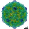
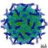
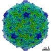
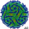
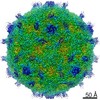
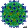
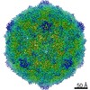
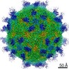
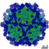
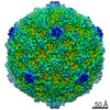
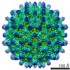
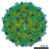
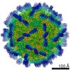
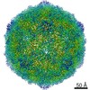
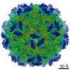
| Title | Coxsackievirus B3 at pH7.4 (VP3-234E) incubation with coxsackievirus and adenovirus receptor for 20min | |||||||||
 Map data Map data | ||||||||||
 Sample Sample |
| |||||||||
 Keywords Keywords | CVB3 / VP3-234E / coxsackievirus and adenovirus receptor / 20min / VIRUS | |||||||||
| Function / homology |  Function and homology information Function and homology informationAV node cell-bundle of His cell adhesion involved in cell communication / cell adhesive protein binding involved in AV node cell-bundle of His cell communication / homotypic cell-cell adhesion / AV node cell to bundle of His cell communication / epithelial structure maintenance / regulation of AV node cell action potential / gamma-delta T cell activation / apicolateral plasma membrane / primordial germ cell migration / connexin binding ...AV node cell-bundle of His cell adhesion involved in cell communication / cell adhesive protein binding involved in AV node cell-bundle of His cell communication / homotypic cell-cell adhesion / AV node cell to bundle of His cell communication / epithelial structure maintenance / regulation of AV node cell action potential / gamma-delta T cell activation / apicolateral plasma membrane / primordial germ cell migration / connexin binding / cell-cell junction organization / transepithelial transport / heterophilic cell-cell adhesion / cardiac muscle cell development / intercalated disc / bicellular tight junction / neutrophil chemotaxis / cell adhesion molecule binding / acrosomal vesicle / Cell surface interactions at the vascular wall / filopodium / adherens junction / PDZ domain binding / neuromuscular junction / mitochondrion organization / beta-catenin binding / integrin binding / Immunoregulatory interactions between a Lymphoid and a non-Lymphoid cell / cell-cell junction / cell junction / heart development / growth cone / virus receptor activity / actin cytoskeleton organization / cell body / defense response to virus / basolateral plasma membrane / neuron projection / membrane raft / signaling receptor binding / protein-containing complex / : / extracellular region / nucleoplasm / identical protein binding / plasma membrane / cytoplasm Similarity search - Function | |||||||||
| Biological species |  Homo sapiens (human) / Homo sapiens (human) /   Coxsackievirus B3 Coxsackievirus B3 | |||||||||
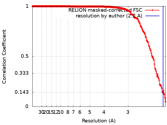
| Method | single particle reconstruction / cryo EM / Resolution: 2.2 Å | |||||||||
 Authors Authors | Wang QL / Liu CC | |||||||||
| Funding support |  China, 1 items China, 1 items
| |||||||||
 Citation Citation | Journal: Proc Natl Acad Sci U S A / Year: 2022 Title: Molecular basis of differential receptor usage for naturally occurring CD55-binding and -nonbinding coxsackievirus B3 strains. Authors: Qingling Wang / Qian Yang / Congcong Liu / Guoqing Wang / Hao Song / Guijun Shang / Ruchao Peng / Xiao Qu / Sheng Liu / Yingzi Cui / Peiyi Wang / Wenbo Xu / Xin Zhao / Jianxun Qi / Mengsu Yang / George F Gao / Abstract: Receptor usage defines cell tropism and contributes to cell entry and infection. Coxsackievirus B (CVB) engages coxsackievirus and adenovirus receptor (CAR), and selectively utilizes the decay- ...Receptor usage defines cell tropism and contributes to cell entry and infection. Coxsackievirus B (CVB) engages coxsackievirus and adenovirus receptor (CAR), and selectively utilizes the decay-accelerating factor (DAF; CD55) to infect cells. However, the differential receptor usage mechanism for CVB remains elusive. This study identified VP3-234 residues (234Q/N/V/D/E) as critical population selection determinants during CVB3 virus evolution, contributing to diverse binding affinities to CD55. Cryoelectron microscopy (cryo-EM) structures of CD55-binding/nonbinding isolates and their complexes with CD55 or CAR were obtained under both neutral and acidic conditions, and the molecular mechanism of VP3-234 residues determining CD55 affinity/specificity for naturally occurring CVB3 strains was elucidated. Structural and biochemical studies in vitro revealed the dynamic entry process of CVB3 and the function of the uncoating receptor CAR with different pH preferences. This work provides detailed insight into the molecular mechanism of CVB infection and contributes to an in-depth understanding of enterovirus attachment receptor usage. | |||||||||
| History |
|
- Structure visualization
| Movie |
Movie viewer |
|---|---|
| Structure viewer | EM map: SurfViewMolmilJmol/JSmol |
| Supplemental images |

- Downloads & links
-EMDB archive
| Map data | emd_32250.map.gz | 59.7 MB | EMDB map data format | |
|---|---|---|---|---|
| Header (meta data) | emd-32250-v30.xmlemd-32250.xml | 21.3 KB 21.3 KB | Display Display | EMDB header |
| FSC (resolution estimation) | emd_32250_fsc.xml | 17.9 KB | Display | FSC data file |
| Images | 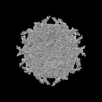 emd_32250.png emd_32250.png | 110.5 KB | ||
| Masks | emd_32250_msk_1.map | 512 MB | Mask map | |
| Filedesc metadata | emd-32250.cif.gz | 6.7 KB | ||
| Archive directory |  http://ftp.pdbj.org/pub/emdb/structures/EMD-32250ftp://ftp.pdbj.org/pub/emdb/structures/EMD-32250 http://ftp.pdbj.org/pub/emdb/structures/EMD-32250ftp://ftp.pdbj.org/pub/emdb/structures/EMD-32250 | HTTPS FTP |
-Related structure data
| Related structure data |  7w14MC  7vxhC  7vxzC  7vy0C 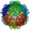 7vy5C  7vy6C 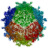 7vykC 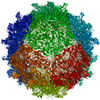 7vylC  7vymC  7w17C M: atomic model generated by this map C: citing same article ( |
|---|---|
| Similar structure data |


-Links
| EMDB pages | EMDB (EBI/PDBe) / EMDataResource |
|---|---|
| Related items in Molecule of the Month |


-Map
| File | Download / File: emd_32250.map.gz / Format: CCP4 / Size: 512 MB / Type: IMAGE STORED AS FLOATING POINT NUMBER (4 BYTES) | ||||||||||||||||||||||||||||||||||||||||||||||||||||||||||||||||||||
|---|---|---|---|---|---|---|---|---|---|---|---|---|---|---|---|---|---|---|---|---|---|---|---|---|---|---|---|---|---|---|---|---|---|---|---|---|---|---|---|---|---|---|---|---|---|---|---|---|---|---|---|---|---|---|---|---|---|---|---|---|---|---|---|---|---|---|---|---|---|
| Projections & slices | Image control
Images are generated by Spider. | ||||||||||||||||||||||||||||||||||||||||||||||||||||||||||||||||||||
| Voxel size | X=Y=Z: 1.08 Å | ||||||||||||||||||||||||||||||||||||||||||||||||||||||||||||||||||||
| Density |
| ||||||||||||||||||||||||||||||||||||||||||||||||||||||||||||||||||||
| Symmetry | Space group: 1 | ||||||||||||||||||||||||||||||||||||||||||||||||||||||||||||||||||||
| Details | EMDB XML:
CCP4 map header:
| ||||||||||||||||||||||||||||||||||||||||||||||||||||||||||||||||||||
 Z (Sec.)
 Y (Row.)
 X (Col.)


-Supplemental data
-Mask #1
| File | emd_32250_msk_1.map | ||||||||||||
|---|---|---|---|---|---|---|---|---|---|---|---|---|---|
| Projections & Slices |
| ||||||||||||
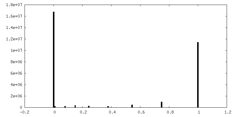
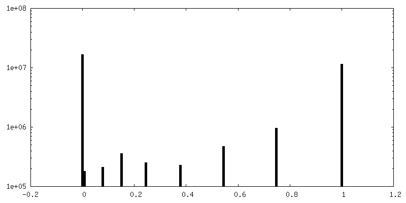
| Density Histograms |


- Sample components
-Entire : Coxsackievirus B3 incubation with coxsackievirus and adenovirus r...
| Entire | Name: Coxsackievirus B3 incubation with coxsackievirus and adenovirus receptor |
|---|---|
| Components |
|
-Supramolecule #1: Coxsackievirus B3 incubation with coxsackievirus and adenovirus r...
| Supramolecule | Name: Coxsackievirus B3 incubation with coxsackievirus and adenovirus receptor type: complex / ID: 1 / Parent: 0 / Macromolecule list: all |
|---|
-Supramolecule #3: receptor
| Supramolecule | Name: receptor / type: complex / ID: 3 / Parent: 1 / Macromolecule list: #5 |
|---|---|
| Source (natural) | Organism: Homo sapiens (human) |
-Supramolecule #2: Coxsackievirus B3
| Supramolecule | Name: Coxsackievirus B3 / type: virus / ID: 2 / Parent: 1 / Macromolecule list: #1-#4 / NCBI-ID: 12072 / Sci species name: Coxsackievirus B3 / Virus type: VIRION / Virus isolate: STRAIN / Virus enveloped: No / Virus empty: No |
|---|
-Macromolecule #1: Capsid protein VP1
| Macromolecule | Name: Capsid protein VP1 / type: protein_or_peptide / ID: 1 / Number of copies: 1 / Enantiomer: LEVO |
|---|---|
| Source (natural) | Organism: Coxsackievirus B3 |
| Molecular weight | Theoretical: 31.561285 KDa |
| Recombinant expression | Organism: Homo sapiens (human) |
| Sequence | String: GPVEDAVTAA IGRVADTVGT GPTNSEAIPA LTAAETGHTS QVVPGDTMQT RHVKNYHSRS ESTVENFLCR SACVYFTEYK NSGSKRYAE WVVTTRQAAQ LRRKLEFFTY IRFDLELTFV ITSTQQPSTT QNQDAQILTH QIMYVPPGGP VPDKVDSYVW Q TSTNPSVF ...String: GPVEDAVTAA IGRVADTVGT GPTNSEAIPA LTAAETGHTS QVVPGDTMQT RHVKNYHSRS ESTVENFLCR SACVYFTEYK NSGSKRYAE WVVTTRQAAQ LRRKLEFFTY IRFDLELTFV ITSTQQPSTT QNQDAQILTH QIMYVPPGGP VPDKVDSYVW Q TSTNPSVF WTEGNAPPRM SIPFLSIGNA YSNFYDGWSD FSRDGVYGIN TLNSMGTLYA RHVNTGGTGP IKSTIRIYFK PK HVKAWIP RPPRLCQYEK AKNVNFQPSG VTTTRQSITA MTNTGAF |
-Macromolecule #2: Capsid protein VP2
| Macromolecule | Name: Capsid protein VP2 / type: protein_or_peptide / ID: 2 / Number of copies: 1 / Enantiomer: LEVO |
|---|---|
| Source (natural) | Organism: Coxsackievirus B3 |
| Molecular weight | Theoretical: 28.85649 KDa |
| Recombinant expression | Organism: Homo sapiens (human) |
| Sequence | String: SPTVEECGYS DRVRSITLGN STITTQECAN VVVGYGVWPD YLKDNEATAE DQPTQPDVAT CRFYTLDSVQ WQKTSPGWWW KLPDALSNL GLFGQNMQYH YLGRTGYTIH VQCNASKFHQ GCLLVVCVPE AEMGCATLDN TPSSAELLGG DAAKEFAGEP I ASGSNKLV ...String: SPTVEECGYS DRVRSITLGN STITTQECAN VVVGYGVWPD YLKDNEATAE DQPTQPDVAT CRFYTLDSVQ WQKTSPGWWW KLPDALSNL GLFGQNMQYH YLGRTGYTIH VQCNASKFHQ GCLLVVCVPE AEMGCATLDN TPSSAELLGG DAAKEFAGEP I ASGSNKLV QRVVYNAGMG IGVGNLTIFP HQWINLRTNN SATIVMPYTN SVPMDNMFRH NNVTLMVIPF VPLDYCPGST TY VPITVTI APMNAEYNGL RLAGHQ |
-Macromolecule #3: Capsid protein VP3
| Macromolecule | Name: Capsid protein VP3 / type: protein_or_peptide / ID: 3 / Number of copies: 1 / Enantiomer: LEVO |
|---|---|
| Source (natural) | Organism: Coxsackievirus B3 |
| Molecular weight | Theoretical: 26.154695 KDa |
| Recombinant expression | Organism: Homo sapiens (human) |
| Sequence | String: GLPTMNTPGS CQFLTSDDFQ SPSAMPQYDV TPEMRIPGEV KNLMEIAEVD SVVPVQNVGE KVNSMEAYQI PVRSNEGSGT QVFGFPLQP GYSSVFSRTL LGEILNYYTH WSGSIKLTFM FCGSAMATGK FLLAYSPLGA GAPTKRVDAM LGTHVVWDVG L QSSCVLCI ...String: GLPTMNTPGS CQFLTSDDFQ SPSAMPQYDV TPEMRIPGEV KNLMEIAEVD SVVPVQNVGE KVNSMEAYQI PVRSNEGSGT QVFGFPLQP GYSSVFSRTL LGEILNYYTH WSGSIKLTFM FCGSAMATGK FLLAYSPLGA GAPTKRVDAM LGTHVVWDVG L QSSCVLCI PWISQTHYRY VASDECTAGG FITCWYQTNI VVPADAQSSC YIMCFVSACN DFSVRLLKDT PFISQENFFQ |
-Macromolecule #4: Capsid protein VP4
| Macromolecule | Name: Capsid protein VP4 / type: protein_or_peptide / ID: 4 / Number of copies: 1 / Enantiomer: LEVO |
|---|---|
| Source (natural) | Organism: Coxsackievirus B3 |
| Molecular weight | Theoretical: 7.306014 KDa |
| Recombinant expression | Organism: Homo sapiens (human) |
| Sequence | String: GAQVSTQKTG AHETGLNASG NSIIHYTNIN YYKDAASNSA TRQDFAQDPG KFTEPVKDIM IKSLPALN |
-Macromolecule #5: Coxsackievirus and adenovirus receptor
| Macromolecule | Name: Coxsackievirus and adenovirus receptor / type: protein_or_peptide / ID: 5 / Number of copies: 1 / Enantiomer: LEVO |
|---|---|
| Source (natural) | Organism: Homo sapiens (human) |
| Molecular weight | Theoretical: 24.277525 KDa |
| Recombinant expression | Organism: Homo sapiens (human) |
| Sequence | String: MSITTPEEMI EKAKGETAYL PCKFTLSPED QGPLDIEWLI SPADNQKVDQ VIILYSGDKI YDDYYPDLKG RVHFTSNDLK SGDASINVT NLQLSDIGTY QCKVKKAPGV ANKKIHLVVL VKPSGARCYV DGSEEIGSDF KIKCEPKEGS LPLQYEWQKL S DSQKMPTS ...String: MSITTPEEMI EKAKGETAYL PCKFTLSPED QGPLDIEWLI SPADNQKVDQ VIILYSGDKI YDDYYPDLKG RVHFTSNDLK SGDASINVT NLQLSDIGTY QCKVKKAPGV ANKKIHLVVL VKPSGARCYV DGSEEIGSDF KIKCEPKEGS LPLQYEWQKL S DSQKMPTS WLAEMTSSVI SVKNASSEYS GTYSCTVRNR VGSDQCLLRL NVVPPSNKAL E UniProtKB: Coxsackievirus and adenovirus receptor |
-Experimental details
-Structure determination
| Method | cryo EM |
|---|---|
 Processing Processing | single particle reconstruction |
| Aggregation state | particle |
-Sample preparation
| Buffer | pH: 7.4 |
|---|---|
| Grid | Model: PELCO Ultrathin Carbon with Lacey Carbon / Support film - Material: CARBON / Support film - topology: CONTINUOUS |
| Vitrification | Cryogen name: ETHANE / Chamber humidity: 100 % / Chamber temperature: 277 K / Instrument: FEI VITROBOT MARK IV |
- Electron microscopy
| Microscope | FEI TITAN KRIOS |
|---|---|
| Temperature | Min: 70.0 K / Max: 70.0 K |
| Image recording | Film or detector model: FEI FALCON III (4k x 4k) / Detector mode: COUNTING / Digitization - Dimensions - Width: 4096 pixel / Digitization - Dimensions - Height: 4096 pixel / Average exposure time: 1.0 sec. / Average electron dose: 40.0 e/Å2 |
| Electron beam | Acceleration voltage: 300 kV / Electron source:  FIELD EMISSION GUN FIELD EMISSION GUN |
| Electron optics | C2 aperture diameter: 70.0 µm / Calibrated defocus max: 5.0 µm / Calibrated defocus min: 1.8 µm / Illumination mode: FLOOD BEAM / Imaging mode: BRIGHT FIELD / Cs: 2.7 mm / Nominal defocus max: 2.5 µm / Nominal defocus min: 1.5 µm / Nominal magnification: 75000 |
| Sample stage | Specimen holder model: FEI TITAN KRIOS AUTOGRID HOLDER / Cooling holder cryogen: NITROGEN |
| Experimental equipment |  Model: Titan Krios / Image courtesy: FEI Company |
+Image processing
-Atomic model buiding 1
| Initial model |
| ||||||||||
|---|---|---|---|---|---|---|---|---|---|---|---|
| Refinement | Space: REAL / Protocol: RIGID BODY FIT | ||||||||||
| Output model | PDB-7w14: |
