Movie
Movie Controller

[English] 日本語
 Yorodumi
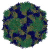
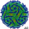
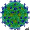
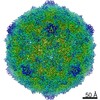
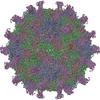
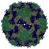
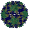
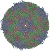
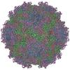
- PDB-7vyk: Coxsackievirus B3 at pH7.4 (VP3-234Q) incubation with coxsackievi... -
+ Open data

- Basic information
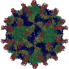
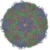
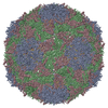
| Entry | Database: PDB / ID: 7vyk | ||||||
|---|---|---|---|---|---|---|---|
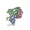
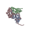
| Title | Coxsackievirus B3 at pH7.4 (VP3-234Q) incubation with coxsackievirus and adenovirus receptor for 10min | ||||||
 Components Components |
| ||||||
 Keywords Keywords | VIRUS / CVB3 | ||||||
| Function / homology |  Function and homology information Function and homology informationAV node cell-bundle of His cell adhesion involved in cell communication / cell adhesive protein binding involved in AV node cell-bundle of His cell communication / symbiont-mediated perturbation of host transcription / homotypic cell-cell adhesion / AV node cell to bundle of His cell communication / epithelial structure maintenance / regulation of AV node cell action potential / primordial germ cell migration / gamma-delta T cell activation / apicolateral plasma membrane ...AV node cell-bundle of His cell adhesion involved in cell communication / cell adhesive protein binding involved in AV node cell-bundle of His cell communication / symbiont-mediated perturbation of host transcription / homotypic cell-cell adhesion / AV node cell to bundle of His cell communication / epithelial structure maintenance / regulation of AV node cell action potential / primordial germ cell migration / gamma-delta T cell activation / apicolateral plasma membrane / cell-cell junction organization / connexin binding / transepithelial transport / cardiac muscle cell development / heterophilic cell-cell adhesion / symbiont-mediated suppression of host cytoplasmic pattern recognition receptor signaling pathway via inhibition of RIG-I activity / intercalated disc / bicellular tight junction / symbiont-mediated suppression of host cytoplasmic pattern recognition receptor signaling pathway via inhibition of MDA-5 activity / neutrophil chemotaxis / symbiont-mediated suppression of host cytoplasmic pattern recognition receptor signaling pathway via inhibition of MAVS activity / cell adhesion molecule binding / acrosomal vesicle / Cell surface interactions at the vascular wall / picornain 2A / filopodium / symbiont-mediated suppression of host mRNA export from nucleus / adherens junction / PDZ domain binding / mitochondrion organization / neuromuscular junction / symbiont genome entry into host cell via pore formation in plasma membrane / picornain 3C / T=pseudo3 icosahedral viral capsid / host cell cytoplasmic vesicle membrane / beta-catenin binding / integrin binding / Immunoregulatory interactions between a Lymphoid and a non-Lymphoid cell / cell junction / cell-cell junction / ribonucleoside triphosphate phosphatase activity / heart development / nucleoside-triphosphate phosphatase / growth cone / virus receptor activity / channel activity / actin cytoskeleton organization / cell body / monoatomic ion transmembrane transport / defense response to virus / symbiont-mediated suppression of host NF-kappaB cascade / basolateral plasma membrane / DNA replication / RNA helicase activity / neuron projection / endocytosis involved in viral entry into host cell / membrane raft / signaling receptor binding / symbiont-mediated activation of host autophagy / RNA-directed RNA polymerase / cysteine-type endopeptidase activity / viral RNA genome replication / RNA-directed RNA polymerase activity / virion attachment to host cell / host cell nucleus / structural molecule activity / DNA-templated transcription / protein-containing complex / proteolysis / : / RNA binding / extracellular region / zinc ion binding / ATP binding / identical protein binding / plasma membrane / cytoplasm Similarity search - Function | ||||||
| Biological species |   Coxsackievirus B3 Coxsackievirus B3 Homo sapiens (human) Homo sapiens (human) | ||||||
| Method | ELECTRON MICROSCOPY / single particle reconstruction / cryo EM / Resolution: 2.79 Å | ||||||
 Authors Authors | Wang, Q.L. / Liu, C.C. | ||||||
| Funding support |  China, 1items China, 1items
| ||||||
 Citation Citation | Journal: Proc Natl Acad Sci U S A / Year: 2022 Title: Molecular basis of differential receptor usage for naturally occurring CD55-binding and -nonbinding coxsackievirus B3 strains. Authors: Qingling Wang / Qian Yang / Congcong Liu / Guoqing Wang / Hao Song / Guijun Shang / Ruchao Peng / Xiao Qu / Sheng Liu / Yingzi Cui / Peiyi Wang / Wenbo Xu / Xin Zhao / Jianxun Qi / Mengsu Yang / George F Gao / Abstract: Receptor usage defines cell tropism and contributes to cell entry and infection. Coxsackievirus B (CVB) engages coxsackievirus and adenovirus receptor (CAR), and selectively utilizes the decay- ...Receptor usage defines cell tropism and contributes to cell entry and infection. Coxsackievirus B (CVB) engages coxsackievirus and adenovirus receptor (CAR), and selectively utilizes the decay-accelerating factor (DAF; CD55) to infect cells. However, the differential receptor usage mechanism for CVB remains elusive. This study identified VP3-234 residues (234Q/N/V/D/E) as critical population selection determinants during CVB3 virus evolution, contributing to diverse binding affinities to CD55. Cryoelectron microscopy (cryo-EM) structures of CD55-binding/nonbinding isolates and their complexes with CD55 or CAR were obtained under both neutral and acidic conditions, and the molecular mechanism of VP3-234 residues determining CD55 affinity/specificity for naturally occurring CVB3 strains was elucidated. Structural and biochemical studies in vitro revealed the dynamic entry process of CVB3 and the function of the uncoating receptor CAR with different pH preferences. This work provides detailed insight into the molecular mechanism of CVB infection and contributes to an in-depth understanding of enterovirus attachment receptor usage. | ||||||
| History |
|
- Structure visualization
| Movie |
Movie viewer |
|---|---|
| Structure viewer | Molecule: MolmilJmol/JSmol |
- Downloads & links
-Download
| PDBx/mmCIF format | 7vyk.cif.gz | 185.5 KB | Display | PDBx/mmCIF format |
|---|---|---|---|---|
| PDB format | pdb7vyk.ent.gz | 142.3 KB | Display | PDB format |
| PDBx/mmJSON format | 7vyk.json.gz | Tree view | PDBx/mmJSON format | |
| Others |  Other downloads Other downloads |
-Validation report
| Arichive directory | https://data.pdbj.org/pub/pdb/validation_reports/vy/7vykftp://data.pdbj.org/pub/pdb/validation_reports/vy/7vyk | HTTPS FTP |
|---|
-Related structure data
| Related structure data | 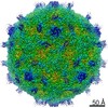 32207MC  7vxhC 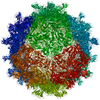 7vxzC  7vy0C 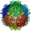 7vy5C  7vy6C 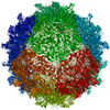 7vylC  7vymC  7w14C  7w17C C: citing same article ( M: map data used to model this data |
|---|---|
| Similar structure data |


-Links
 PDBj
PDBj


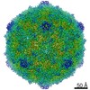
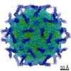
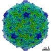
- Assembly
| Deposited unit | 
|
|---|---|
| 1 | x 60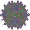
|
| 2 |
|
| 3 | x 5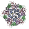
|
| 4 | x 6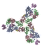
|
| 5 | 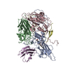
|
| Symmetry | Point symmetry: (Schoenflies symbol: I (icosahedral)) |
-Components
-Capsid protein ... , 4 types, 4 molecules ABCD
| #1: Protein | Mass: 30053.660 Da / Num. of mol.: 1 Source method: isolated from a genetically manipulated source Source: (gene. exp.) Coxsackievirus B3 / Cell line (production host): HEK293 / Production host: Homo sapiens (human) / References: UniProt: P03313 |
|---|---|
| #2: Protein | Mass: 28822.475 Da / Num. of mol.: 1 Source method: isolated from a genetically manipulated source Source: (gene. exp.) Coxsackievirus B3 / Cell line (production host): HEK293 / Production host: Homo sapiens (human) / References: UniProt: P03313 |
| #3: Protein | Mass: 26227.725 Da / Num. of mol.: 1 Source method: isolated from a genetically manipulated source Source: (gene. exp.) Coxsackievirus B3 / Cell line (production host): HEK293 / Production host: Homo sapiens (human) / References: UniProt: P03313 |
| #4: Protein | Mass: 7480.235 Da / Num. of mol.: 1 Source method: isolated from a genetically manipulated source Source: (gene. exp.) Coxsackievirus B3 / Cell line (production host): HEK293 / Production host: Homo sapiens (human) / References: UniProt: P03313 |
-Protein / Non-polymers , 2 types, 2 molecules E
| #5: Protein | Mass: 25106.408 Da / Num. of mol.: 1 Source method: isolated from a genetically manipulated source Source: (gene. exp.) Homo sapiens (human) / Gene: CXADR, CAR / Cell line (production host): HEK293 / Production host: Homo sapiens (human) / References: UniProt: P78310 |
|---|---|
| #6: Chemical | ChemComp-PLM /  Mass: 256.424 Da / Num. of mol.: 1 / Source method: obtained synthetically / Formula: C16H32O2 / Feature type: SUBJECT OF INVESTIGATION Mass: 256.424 Da / Num. of mol.: 1 / Source method: obtained synthetically / Formula: C16H32O2 / Feature type: SUBJECT OF INVESTIGATION |
-Details
| Has ligand of interest | Y |
|---|---|
| Has protein modification | Y |
| Sequence details | AUTHORS STATE THAT THE GENEBANK ACCESSION NUMBER IS KJ025083 for this capsid protein. |
-Experimental details
-Experiment
| Experiment | Method: ELECTRON MICROSCOPY |
|---|---|
| EM experiment | Aggregation state: PARTICLE / 3D reconstruction method: single particle reconstruction |
- Sample preparation
| Component |
| ||||||||||||||||||||||||
|---|---|---|---|---|---|---|---|---|---|---|---|---|---|---|---|---|---|---|---|---|---|---|---|---|---|
| Molecular weight | Experimental value: NO | ||||||||||||||||||||||||
| Source (natural) |
| ||||||||||||||||||||||||
| Source (recombinant) |
| ||||||||||||||||||||||||
| Details of virus | Empty: NO / Enveloped: NO / Isolate: STRAIN / Type: VIRION | ||||||||||||||||||||||||
| Natural host | Organism: Homo sapiens | ||||||||||||||||||||||||
| Buffer solution | pH: 7.4 | ||||||||||||||||||||||||
| Specimen | Embedding applied: NO / Shadowing applied: NO / Staining applied: NO / Vitrification applied: YES | ||||||||||||||||||||||||
| Specimen support | Grid type: PELCO Ultrathin Carbon with Lacey Carbon | ||||||||||||||||||||||||
| Vitrification | Instrument: FEI VITROBOT MARK IV / Cryogen name: ETHANE / Humidity: 100 % / Chamber temperature: 277 K |
- Electron microscopy imaging
| Experimental equipment |  Model: Titan Krios / Image courtesy: FEI Company |
|---|---|
| Microscopy | Model: FEI TITAN KRIOS |
| Electron gun | Electron source:  FIELD EMISSION GUN / Accelerating voltage: 300 kV / Illumination mode: FLOOD BEAM FIELD EMISSION GUN / Accelerating voltage: 300 kV / Illumination mode: FLOOD BEAM |
| Electron lens | Mode: BRIGHT FIELD / Nominal magnification: 75000 X / Nominal defocus max: 2500 nm / Nominal defocus min: 1500 nm / Calibrated defocus min: 1800 nm / Calibrated defocus max: 5000 nm / Cs: 2.7 mm / C2 aperture diameter: 70 µm / Alignment procedure: COMA FREE |
| Specimen holder | Cryogen: NITROGEN / Specimen holder model: FEI TITAN KRIOS AUTOGRID HOLDER / Temperature (max): 70 K / Temperature (min): 70 K |
| Image recording | Average exposure time: 1 sec. / Electron dose: 40 e/Å2 / Detector mode: COUNTING / Film or detector model: FEI FALCON III (4k x 4k) |
| Image scans | Width: 4096 / Height: 4096 |
- Processing
| EM software |
| ||||||||||||||||||||||||||||||||||||||||||||||||||
|---|---|---|---|---|---|---|---|---|---|---|---|---|---|---|---|---|---|---|---|---|---|---|---|---|---|---|---|---|---|---|---|---|---|---|---|---|---|---|---|---|---|---|---|---|---|---|---|---|---|---|---|
| CTF correction | Type: PHASE FLIPPING AND AMPLITUDE CORRECTION | ||||||||||||||||||||||||||||||||||||||||||||||||||
| Particle selection | Num. of particles selected: 158446 | ||||||||||||||||||||||||||||||||||||||||||||||||||
| 3D reconstruction | Resolution: 2.79 Å / Resolution method: FSC 0.143 CUT-OFF / Num. of particles: 35532 / Symmetry type: POINT | ||||||||||||||||||||||||||||||||||||||||||||||||||
| Atomic model building | Protocol: RIGID BODY FIT / Space: REAL | ||||||||||||||||||||||||||||||||||||||||||||||||||
| Atomic model building | 3D fitting-ID: 1 / Accession code: 1COV / Initial refinement model-ID: 1 / PDB-ID: 1COV / Source name: PDB / Type: experimental model
|