Movie
Movie Controller


+ Open data

- Basic information
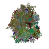
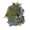
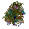
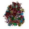
| Entry | Database: EMDB / ID: EMD-3221 | |||||||||
|---|---|---|---|---|---|---|---|---|---|---|
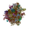
| Title | Mammalian 80S HCV-IRES complex, Classical | |||||||||
 Map data Map data | Reconstruction of ribosome complex | |||||||||
 Sample Sample |
| |||||||||
 Keywords Keywords | ribosome / translation initiation / Hepatitis C Virus internal ribosome entry site | |||||||||
| Function / homology |  Function and homology information Function and homology informationnegative regulation of endoplasmic reticulum unfolded protein response / oxidized pyrimidine DNA binding / response to TNF agonist / positive regulation of base-excision repair / positive regulation of respiratory burst involved in inflammatory response / positive regulation of gastrulation / positive regulation of intrinsic apoptotic signaling pathway in response to DNA damage / protein tyrosine kinase inhibitor activity / positive regulation of ubiquitin-protein transferase activity / IRE1-RACK1-PP2A complex ...negative regulation of endoplasmic reticulum unfolded protein response / oxidized pyrimidine DNA binding / response to TNF agonist / positive regulation of base-excision repair / positive regulation of respiratory burst involved in inflammatory response / positive regulation of gastrulation / positive regulation of intrinsic apoptotic signaling pathway in response to DNA damage / protein tyrosine kinase inhibitor activity / positive regulation of ubiquitin-protein transferase activity / IRE1-RACK1-PP2A complex / positive regulation of Golgi to plasma membrane protein transport / nucleolus organization / positive regulation of DNA-templated transcription initiation / TNFR1-mediated ceramide production / negative regulation of RNA splicing / neural crest cell differentiation / ZNF598 and the Ribosome-associated Quality Trigger (RQT) complex dissociate a ribosome stalled on a no-go mRNA / supercoiled DNA binding / negative regulation of DNA repair / PELO:HBS1L and ABCE1 dissociate a ribosome on a non-stop mRNA / cysteine-type endopeptidase activator activity involved in apoptotic process / oxidized purine DNA binding / NF-kappaB complex / cytoplasmic translational initiation / rRNA modification in the nucleus and cytosol / negative regulation of intrinsic apoptotic signaling pathway in response to hydrogen peroxide / erythrocyte homeostasis / regulation of establishment of cell polarity / negative regulation of phagocytosis / negative regulation of bicellular tight junction assembly / ubiquitin-like protein conjugating enzyme binding / cytoplasmic side of rough endoplasmic reticulum membrane / Formation of the ternary complex, and subsequently, the 43S complex / pigmentation / ion channel inhibitor activity / protein kinase A binding / laminin receptor activity / Ribosomal scanning and start codon recognition / positive regulation of mitochondrial depolarization / Translation initiation complex formation / negative regulation of Wnt signaling pathway / fibroblast growth factor binding / Protein hydroxylation / BH3 domain binding / negative regulation of translational frameshifting / TOR signaling / regulation of adenylate cyclase-activating G protein-coupled receptor signaling pathway / monocyte chemotaxis / mTORC1-mediated signalling / SARS-CoV-1 modulates host translation machinery / regulation of cell division / iron-sulfur cluster binding / positive regulation of GTPase activity / cellular response to ethanol / Peptide chain elongation / Selenocysteine synthesis / negative regulation of protein binding / Formation of a pool of free 40S subunits / positive regulation of intrinsic apoptotic signaling pathway by p53 class mediator / Eukaryotic Translation Termination / protein serine/threonine kinase inhibitor activity / negative regulation of respiratory burst involved in inflammatory response / SRP-dependent cotranslational protein targeting to membrane / Response of EIF2AK4 (GCN2) to amino acid deficiency / ubiquitin ligase inhibitor activity / Viral mRNA Translation / endonucleolytic cleavage to generate mature 3'-end of SSU-rRNA from (SSU-rRNA, 5.8S rRNA, LSU-rRNA) / Nonsense Mediated Decay (NMD) independent of the Exon Junction Complex (EJC) / positive regulation of signal transduction by p53 class mediator / negative regulation of ubiquitin-dependent protein catabolic process / GTP hydrolysis and joining of the 60S ribosomal subunit / L13a-mediated translational silencing of Ceruloplasmin expression / Major pathway of rRNA processing in the nucleolus and cytosol / regulation of translational fidelity / positive regulation of microtubule polymerization / phagocytic cup / Nonsense Mediated Decay (NMD) enhanced by the Exon Junction Complex (EJC) / spindle assembly / cellular response to leukemia inhibitory factor / positive regulation of intrinsic apoptotic signaling pathway / Protein methylation / endonucleolytic cleavage in ITS1 to separate SSU-rRNA from 5.8S rRNA and LSU-rRNA from tricistronic rRNA transcript (SSU-rRNA, 5.8S rRNA, LSU-rRNA) / translation regulator activity / Nuclear events stimulated by ALK signaling in cancer / gastrulation / rough endoplasmic reticulum / ribosomal small subunit export from nucleus / Amplification of signal from unattached kinetochores via a MAD2 inhibitory signal / laminin binding / DNA-(apurinic or apyrimidinic site) endonuclease activity / positive regulation of cell cycle / rescue of stalled cytosolic ribosome / signaling adaptor activity / Maturation of protein E / negative regulation of protein ubiquitination / Maturation of protein E / MDM2/MDM4 family protein binding / ER Quality Control Compartment (ERQC) / Mitotic Prometaphase / Myoclonic epilepsy of Lafora Similarity search - Function | |||||||||
| Biological species |   Hepatitis C virus Hepatitis C virus | |||||||||
| Method | single particle reconstruction / cryo EM / Resolution: 3.9 Å | |||||||||
 Authors Authors | Yamamoto H / Collier M / Loerke J / Ismer J / Schmidt A / Hilal T / Sprink T / Yamamoto K / Mielke T / Burger J ...Yamamoto H / Collier M / Loerke J / Ismer J / Schmidt A / Hilal T / Sprink T / Yamamoto K / Mielke T / Burger J / Shaikh TR / Dabrowski M / Hildebrand PW / Scheerer P / Spahn CMT | |||||||||
 Citation Citation | Journal: EMBO J / Year: 2015 Title: Molecular architecture of the ribosome-bound Hepatitis C Virus internal ribosomal entry site RNA. Authors: Hiroshi Yamamoto / Marianne Collier / Justus Loerke / Jochen Ismer / Andrea Schmidt / Tarek Hilal / Thiemo Sprink / Kaori Yamamoto / Thorsten Mielke / Jörg Bürger / Tanvir R Shaikh / ...Authors: Hiroshi Yamamoto / Marianne Collier / Justus Loerke / Jochen Ismer / Andrea Schmidt / Tarek Hilal / Thiemo Sprink / Kaori Yamamoto / Thorsten Mielke / Jörg Bürger / Tanvir R Shaikh / Marylena Dabrowski / Peter W Hildebrand / Patrick Scheerer / Christian M T Spahn /   Abstract: Internal ribosomal entry sites (IRESs) are structured cis-acting RNAs that drive an alternative, cap-independent translation initiation pathway. They are used by many viruses to hijack the ...Internal ribosomal entry sites (IRESs) are structured cis-acting RNAs that drive an alternative, cap-independent translation initiation pathway. They are used by many viruses to hijack the translational machinery of the host cell. IRESs facilitate translation initiation by recruiting and actively manipulating the eukaryotic ribosome using only a subset of canonical initiation factor and IRES transacting factors. Here we present cryo-EM reconstructions of the ribosome 80S- and 40S-bound Hepatitis C Virus (HCV) IRES. The presence of four subpopulations for the 80S•HCV IRES complex reveals dynamic conformational modes of the complex. At a global resolution of 3.9 Å for the most stable complex, a derived atomic model reveals a complex fold of the IRES RNA and molecular details of its interaction with the ribosome. The comparison of obtained structures explains how a modular architecture facilitates mRNA loading and tRNA binding to the P-site. This information provides the structural foundation for understanding the mechanism of HCV IRES RNA-driven translation initiation. | |||||||||
| History |
|
- Structure visualization
| Movie |
Movie viewer |
|---|---|
| Structure viewer | EM map: SurfViewMolmilJmol/JSmol |
| Supplemental images |

- Downloads & links
-EMDB archive
| Map data | emd_3221.map.gz | 11.9 MB | EMDB map data format | |
|---|---|---|---|---|
| Header (meta data) | emd-3221-v30.xmlemd-3221.xml | 11 KB 11 KB | Display Display | EMDB header |
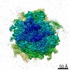
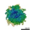
| Images |  EMD-1-3221.png EMD-1-3221.png | 227.8 KB | ||
| Archive directory |  http://ftp.pdbj.org/pub/emdb/structures/EMD-3221ftp://ftp.pdbj.org/pub/emdb/structures/EMD-3221 http://ftp.pdbj.org/pub/emdb/structures/EMD-3221ftp://ftp.pdbj.org/pub/emdb/structures/EMD-3221 | HTTPS FTP |
-Related structure data
| Related structure data | 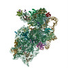 5flxMC 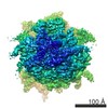 3223C 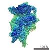 3224C 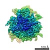 3225C 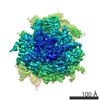 3226C M: atomic model generated by this map C: citing same article ( |
|---|---|
| Similar structure data |

-Links
| EMDB pages | EMDB (EBI/PDBe) / EMDataResource |
|---|---|
| Related items in Molecule of the Month |


-Map
| File | Download / File: emd_3221.map.gz / Format: CCP4 / Size: 204.4 MB / Type: IMAGE STORED AS FLOATING POINT NUMBER (4 BYTES) | ||||||||||||||||||||||||||||||||||||||||||||||||||||||||||||||||||||
|---|---|---|---|---|---|---|---|---|---|---|---|---|---|---|---|---|---|---|---|---|---|---|---|---|---|---|---|---|---|---|---|---|---|---|---|---|---|---|---|---|---|---|---|---|---|---|---|---|---|---|---|---|---|---|---|---|---|---|---|---|---|---|---|---|---|---|---|---|---|
| Annotation | Reconstruction of ribosome complex | ||||||||||||||||||||||||||||||||||||||||||||||||||||||||||||||||||||
| Projections & slices | Image control
Images are generated by Spider. | ||||||||||||||||||||||||||||||||||||||||||||||||||||||||||||||||||||
| Voxel size | X=Y=Z: 1.07 Å | ||||||||||||||||||||||||||||||||||||||||||||||||||||||||||||||||||||
| Density |
| ||||||||||||||||||||||||||||||||||||||||||||||||||||||||||||||||||||
| Symmetry | Space group: 1 | ||||||||||||||||||||||||||||||||||||||||||||||||||||||||||||||||||||
| Details | EMDB XML:
CCP4 map header:
| ||||||||||||||||||||||||||||||||||||||||||||||||||||||||||||||||||||
 Z (Sec.)
 X (Row.)
 Y (Col.)


-Supplemental data
- Sample components
-Entire : Mammalian 80S-HCV-IRES complex, classical
| Entire | Name: Mammalian 80S-HCV-IRES complex, classical |
|---|---|
| Components |
|
-Supramolecule #1000: Mammalian 80S-HCV-IRES complex, classical
| Supramolecule | Name: Mammalian 80S-HCV-IRES complex, classical / type: sample / ID: 1000 / Number unique components: 2 |
|---|---|
| Molecular weight | Theoretical: 4.6 MDa |
-Supramolecule #1: 80S ribosome
| Supramolecule | Name: 80S ribosome / type: complex / ID: 1 / Recombinant expression: No / Ribosome-details: ribosome-eukaryote: ALL |
|---|---|
| Source (natural) | Organism: |
| Molecular weight | Theoretical: 4.5 MDa |
-Macromolecule #1: HCV-IRES
| Macromolecule | Name: HCV-IRES / type: rna / ID: 1 / Classification: OTHER / Structure: DOUBLE HELIX / Synthetic?: Yes |
|---|---|
| Source (natural) | Organism: Hepatitis C virus / synonym: HCV |
| Molecular weight | Theoretical: 162 KDa |
| Sequence | String: GCCAGCCCCC UGAUGGGGGC GACACUCCAC CAUGAAUCAC UCCCCUGUGA GGAACUACUG UCUUCACGCA GAAAGCGUCU AGCCAUGGCG UUAGUAUGAG UGUCGUGCAG CCUCCAGGAC CCCCCCUCCC GGGAGAGCCA UAGUGGUCUG CGGAACCGGU GAGUACACCG ...String: GCCAGCCCCC UGAUGGGGGC GACACUCCAC CAUGAAUCAC UCCCCUGUGA GGAACUACUG UCUUCACGCA GAAAGCGUCU AGCCAUGGCG UUAGUAUGAG UGUCGUGCAG CCUCCAGGAC CCCCCCUCCC GGGAGAGCCA UAGUGGUCUG CGGAACCGGU GAGUACACCG GAAUUGCCAG GACGACCGGG UCCUUUCUUG GAUAAACCCG CUCAAUGCCU GGAGAUUUGG GCGUGCCCCC GCAAGACUGC UAGCCGAGUA GUGUUGGGUC GCGAAAGGCC UUGUGGUACU GCCUGAUAGG GUGCUUGCGA GUGCCCCGGG AGGUCUCGUA GACCGUGCAC CAUGAGCACG AAUCCUAAAC CUCAAAGAAA AACCAAACGU AACACCAACC GUCGCCCACA GGACGUCAAG UUCCCGGGUG GCGGUCUAGA CGCCGAGAUC AGAAAUCCCU CUCUCGGAUC GCAUUUGGAC UUCUGCCUUC GGGCACCACG GUCGGAUCCG AAUU |
-Experimental details
-Structure determination
| Method | cryo EM |
|---|---|
 Processing Processing | single particle reconstruction |
| Aggregation state | particle |
-Sample preparation
| Concentration | 0.15 mg/mL |
|---|---|
| Buffer | pH: 7.6 Details: 20mM Tris-HCl, 7.5mM MgCl2, 100mM KCl, 0.2mM spermidine, 2mM DTT |
| Grid | Details: Quantifoil R3-3 Cu 300 mesh with 2 nm carbon support film |
| Vitrification | Cryogen name: ETHANE / Chamber humidity: 100 % / Chamber temperature: 93 K / Instrument: FEI VITROBOT MARK I / Method: blot for 2-4 seconds before plunging |
- Electron microscopy
| Microscope | FEI TITAN KRIOS |
|---|---|
| Date | Oct 16, 2014 |
| Image recording | Category: CCD / Film or detector model: FEI FALCON II (4k x 4k) / Number real images: 7707 / Average electron dose: 20 e/Å2 / Details: Automated data collection on using EPU |
| Electron beam | Acceleration voltage: 300 kV / Electron source:  FIELD EMISSION GUN FIELD EMISSION GUN |
| Electron optics | Calibrated magnification: 130293 / Illumination mode: FLOOD BEAM / Imaging mode: BRIGHT FIELD / Cs: 2.7 mm / Nominal defocus max: 4.0 µm / Nominal defocus min: 1.0 µm / Nominal magnification: 75000 |
| Sample stage | Specimen holder model: FEI TITAN KRIOS AUTOGRID HOLDER |
| Experimental equipment |  Model: Titan Krios / Image courtesy: FEI Company |
-Image processing
| CTF correction | Details: CTFFIND3 |
|---|---|
| Final reconstruction | Applied symmetry - Point group: C1 (asymmetric) / Resolution.type: BY AUTHOR / Resolution: 3.9 Å / Resolution method: OTHER / Software - Name: spider, sparx Details: To avoid overfitting, the data was refined in a resolution-limited scheme using SPIDER. A final local refinement and the final reconstruction were calculated in Sparx. Number images used: 171820 |