 Movie
Movie Controller

[English] 日本語
 Yorodumi
- EMDB-32092: Cryo-EM structure of amyloid fibril formed by FUS low complexity ... -
+ Open data

- Basic information
| Entry | Database: EMDB / ID: EMD-32092 | |||||||||
|---|---|---|---|---|---|---|---|---|---|---|
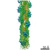
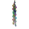
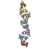
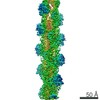
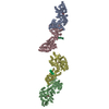
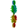
| Title | Cryo-EM structure of amyloid fibril formed by FUS low complexity domain | |||||||||
 Map data Map data | ||||||||||
 Sample Sample |
| |||||||||
 Keywords Keywords | amyloid fibril / PROTEIN FIBRIL | |||||||||
| Function / homology |  Function and homology information Function and homology informationmembraneless organelle assembly / mRNA stabilization / regulation of RNA splicing / Processing of Capped Intron-Containing Pre-mRNA / postsynaptic cytosol / positive regulation of double-strand break repair via homologous recombination / presynaptic cytosol / mRNA Splicing - Major Pathway / RNA splicing / transcription coregulator activity ...membraneless organelle assembly / mRNA stabilization / regulation of RNA splicing / Processing of Capped Intron-Containing Pre-mRNA / postsynaptic cytosol / positive regulation of double-strand break repair via homologous recombination / presynaptic cytosol / mRNA Splicing - Major Pathway / RNA splicing / transcription coregulator activity / mRNA 3'-UTR binding / molecular condensate scaffold activity / protein homooligomerization / GABA-ergic synapse / amyloid fibril formation / transcription coactivator activity / chromatin binding / regulation of transcription by RNA polymerase II / regulation of DNA-templated transcription / glutamatergic synapse / DNA binding / RNA binding / zinc ion binding / nucleoplasm / identical protein binding / nucleus Similarity search - Function | |||||||||
| Biological species |   Aequorea victoria (jellyfish) / Aequorea victoria (jellyfish) /  Homo sapiens (human) Homo sapiens (human) | |||||||||
| Method | helical reconstruction / cryo EM / Resolution: 2.9 Å | |||||||||
 Authors Authors | Sun YP / Xia WC | |||||||||
| Funding support | 1 items
| |||||||||
 Citation Citation | Journal: iScience / Year: 2022 Title: Molecular structure of an amyloid fibril formed by FUS low-complexity domain. Authors: Yunpeng Sun / Shenqing Zhang / Jiaojiao Hu / Youqi Tao / Wencheng Xia / Jinge Gu / Yichen Li / Qin Cao / Dan Li / Cong Liu /  Abstract: FUS is a multifunctional nuclear protein which undergoes liquid-liquid phase separation in response to stress and DNA damage. Dysregulation of FUS dynamic phase separation leads to formation of ...FUS is a multifunctional nuclear protein which undergoes liquid-liquid phase separation in response to stress and DNA damage. Dysregulation of FUS dynamic phase separation leads to formation of pathological fibril closely associated with neurodegenerative diseases such as amyotrophic lateral sclerosis and frontotemporal dementia. In this study, we determined the cryo-EM structure of a cytotoxic fibril formed by the low-complexity (LC) domain of FUS at 2.9 Å resolution. The fibril structure exhibits a new and extensive serpentine fold consisting of three motifs incorporating together via a Tyr triad. FUS LC employs 91 residues to form an enlarged and stable fibril core via hydrophilic interaction and hydrogen bonds, which is distinct from most of previously determined fibrils commonly stabilized by hydrophobic interaction. Our work reveals the structural basis underlying formation of a cytotoxic and thermostable fibril of FUS LC and sheds light on understanding the liquid-to-solid phase transition of FUS in disease. | |||||||||
| History |
|
- Structure visualization
| Movie |
Movie viewer |
|---|---|
| Structure viewer | EM map: SurfViewMolmilJmol/JSmol |
| Supplemental images |

- Downloads & links
-EMDB archive
| Map data | emd_32092.map.gz | 59.8 MB | EMDB map data format | |
|---|---|---|---|---|
| Header (meta data) | emd-32092-v30.xmlemd-32092.xml | 9.3 KB 9.3 KB | Display Display | EMDB header |
| FSC (resolution estimation) | emd_32092_fsc.xml | 9.1 KB | Display | FSC data file |
| Images | 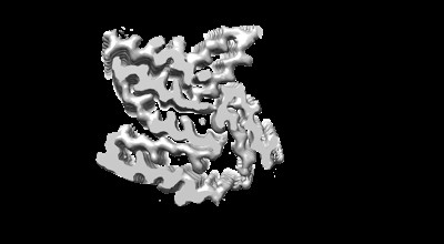 emd_32092.png emd_32092.png | 33.1 KB | ||
| Filedesc metadata | emd-32092.cif.gz | 5.1 KB | ||
| Archive directory |  http://ftp.pdbj.org/pub/emdb/structures/EMD-32092ftp://ftp.pdbj.org/pub/emdb/structures/EMD-32092 http://ftp.pdbj.org/pub/emdb/structures/EMD-32092ftp://ftp.pdbj.org/pub/emdb/structures/EMD-32092 | HTTPS FTP |
-Related structure data
| Related structure data | 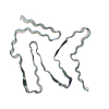 7vqqMC M: atomic model generated by this map C: citing same article ( |
|---|---|
| Similar structure data |


-Links
| EMDB pages | EMDB (EBI/PDBe) / EMDataResource |
|---|---|
| Related items in Molecule of the Month |


-Map
| File | Download / File: emd_32092.map.gz / Format: CCP4 / Size: 64 MB / Type: IMAGE STORED AS FLOATING POINT NUMBER (4 BYTES) | ||||||||||||||||||||||||||||||||||||||||||||||||||||||||||||||||||||
|---|---|---|---|---|---|---|---|---|---|---|---|---|---|---|---|---|---|---|---|---|---|---|---|---|---|---|---|---|---|---|---|---|---|---|---|---|---|---|---|---|---|---|---|---|---|---|---|---|---|---|---|---|---|---|---|---|---|---|---|---|---|---|---|---|---|---|---|---|---|
| Projections & slices | Image control
Images are generated by Spider. | ||||||||||||||||||||||||||||||||||||||||||||||||||||||||||||||||||||
| Voxel size | X=Y=Z: 1.1 Å | ||||||||||||||||||||||||||||||||||||||||||||||||||||||||||||||||||||
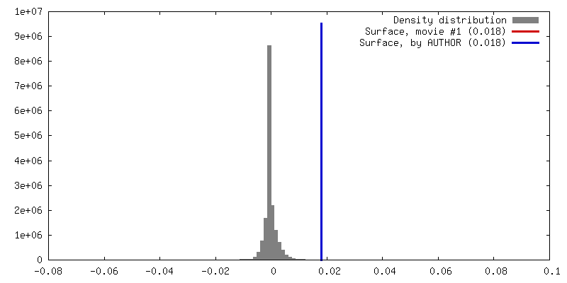
| Density |
| ||||||||||||||||||||||||||||||||||||||||||||||||||||||||||||||||||||
| Symmetry | Space group: 1 | ||||||||||||||||||||||||||||||||||||||||||||||||||||||||||||||||||||
| Details | EMDB XML:
CCP4 map header:
| ||||||||||||||||||||||||||||||||||||||||||||||||||||||||||||||||||||
 Z (Sec.)
 Y (Row.)
 X (Col.)


-Supplemental data
- Sample components
-Entire : Amyloid fibril formed by FUS low complexity domain
| Entire | Name: Amyloid fibril formed by FUS low complexity domain |
|---|---|
| Components |
|
-Supramolecule #1: Amyloid fibril formed by FUS low complexity domain
| Supramolecule | Name: Amyloid fibril formed by FUS low complexity domain / type: organelle_or_cellular_component / ID: 1 / Parent: 0 / Macromolecule list: all |
|---|---|
| Source (natural) | Organism: Aequorea victoria (jellyfish) |
-Macromolecule #1: fusion protein of mCerulean and FUS LCD
| Macromolecule | Name: fusion protein of mCerulean and FUS LCD / type: protein_or_peptide / ID: 1 Details: 6His-tagged mCerulean:(FPbase ID: J2JWA, Link: https://www.fpbase.org/protein/mcerulean/) Number of copies: 3 / Enantiomer: LEVO |
|---|---|
| Source (natural) | Organism: Homo sapiens (human) |
| Molecular weight | Theoretical: 51.948352 KDa |
| Recombinant expression | Organism:  |
| Sequence | String: MSYYHHHHHH DYDIPTTENL YFQGAMVSKG EELFTGVVPI LVELDGDVNG HKFSVSGEGE GDATYGKLTL KFICTTGKLP VPWPTLVTT LTWGVQCFAR YPDHMKQHDF FKSAMPEGYV QERTIFFKDD GNYKTRAEVK FEGDTLVNRI ELKGIDFKED G NILGHKLE ...String: MSYYHHHHHH DYDIPTTENL YFQGAMVSKG EELFTGVVPI LVELDGDVNG HKFSVSGEGE GDATYGKLTL KFICTTGKLP VPWPTLVTT LTWGVQCFAR YPDHMKQHDF FKSAMPEGYV QERTIFFKDD GNYKTRAEVK FEGDTLVNRI ELKGIDFKED G NILGHKLE YNAISDNVYI TADKQKNGIK ANFKIRHNIE DGSVQLADHY QQNTPIGDGP VLLPDNHYLS TQSKLSKDPN EK RDHMVLL EFVTAAGITL GMDELYKAGT MDPASNDYTQ QATQSYGAYP TQPGQGYSQQ SSQPYGQQSY SGYSQSTDTS GYG QSSYSS YGQSQNTGYG TQSTPQGYGS TGGYGSSQSS QSSYGQQSSY PGYGQQPAPS STSGSYGSSS QSSSYGQPQS GSYS QQPSY GGQQQSYGQQ QSYNPPQGYG QQNQYNSSSG GGGGGGGGGN YGQDQSSMSS GGGSGGGYGN QDQSGGGGSG GYGQQ DRG UniProtKB: RNA-binding protein FUS |
-Experimental details
-Structure determination
| Method | cryo EM |
|---|---|
 Processing Processing | helical reconstruction |
| Aggregation state | filament |
-Sample preparation
| Buffer | pH: 7.5 |
|---|---|
| Vitrification | Cryogen name: ETHANE |
- Electron microscopy
| Microscope | FEI TITAN KRIOS |
|---|---|
| Image recording | Film or detector model: GATAN K2 SUMMIT (4k x 4k) / Average electron dose: 40.0 e/Å2 |
| Electron beam | Acceleration voltage: 300 kV / Electron source:  FIELD EMISSION GUN FIELD EMISSION GUN |
| Electron optics | Illumination mode: FLOOD BEAM / Imaging mode: BRIGHT FIELD |
| Experimental equipment |  Model: Titan Krios / Image courtesy: FEI Company |
-Image processing
| Final reconstruction | Applied symmetry - Helical parameters - Δz: 5.0 Å Applied symmetry - Helical parameters - Δ&Phi: -1.44 ° Applied symmetry - Helical parameters - Axial symmetry: C1 (asymmetric) Resolution.type: BY AUTHOR / Resolution: 2.9 Å / Resolution method: FSC 0.143 CUT-OFF / Number images used: 171163 |
|---|---|
| Startup model | Type of model: NONE |
| Final angle assignment | Type: NOT APPLICABLE |
| FSC plot (resolution estimation) | 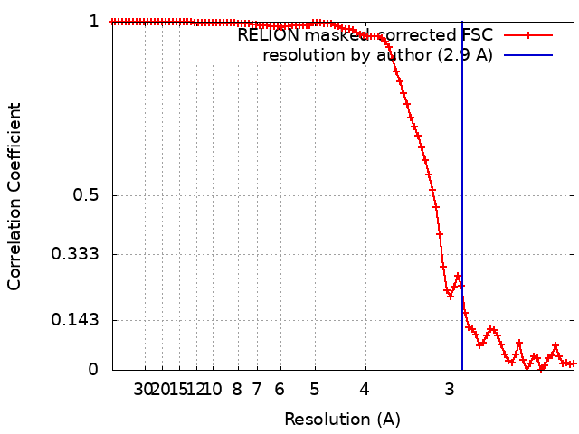 |
