 Movie
Movie Controller

[English] 日本語
 Yorodumi
- PDB-7vqq: Cryo-EM structure of amyloid fibril formed by FUS low complexity ... -
+ Open data

- Basic information
| Entry | Database: PDB / ID: 7vqq | ||||||
|---|---|---|---|---|---|---|---|
| Title | Cryo-EM structure of amyloid fibril formed by FUS low complexity domain | ||||||
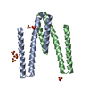
 Components Components | fusion protein of mCerulean and FUS LCD | ||||||
 Keywords Keywords | PROTEIN FIBRIL / amyloid fibril | ||||||
| Function / homology |  Function and homology information Function and homology informationmembraneless organelle assembly / mRNA stabilization / regulation of RNA splicing / Processing of Capped Intron-Containing Pre-mRNA / postsynaptic cytosol / positive regulation of double-strand break repair via homologous recombination / presynaptic cytosol / mRNA Splicing - Major Pathway / RNA splicing / transcription coregulator activity ...membraneless organelle assembly / mRNA stabilization / regulation of RNA splicing / Processing of Capped Intron-Containing Pre-mRNA / postsynaptic cytosol / positive regulation of double-strand break repair via homologous recombination / presynaptic cytosol / mRNA Splicing - Major Pathway / RNA splicing / transcription coregulator activity / mRNA 3'-UTR binding / molecular condensate scaffold activity / protein homooligomerization / GABA-ergic synapse / amyloid fibril formation / transcription coactivator activity / chromatin binding / regulation of transcription by RNA polymerase II / regulation of DNA-templated transcription / glutamatergic synapse / DNA binding / RNA binding / zinc ion binding / nucleoplasm / identical protein binding / nucleus Similarity search - Function | ||||||
| Biological species |   Aequorea victoria (jellyfish) Aequorea victoria (jellyfish) Homo sapiens (human) Homo sapiens (human) | ||||||
| Method | ELECTRON MICROSCOPY / helical reconstruction / cryo EM / Resolution: 2.9 Å | ||||||
 Authors Authors | Sun, Y.P. / Xia, W.C. / Liu, C. | ||||||
| Funding support | 1items
| ||||||
 Citation Citation | Journal: iScience / Year: 2022 Title: Molecular structure of an amyloid fibril formed by FUS low-complexity domain. Authors: Yunpeng Sun / Shenqing Zhang / Jiaojiao Hu / Youqi Tao / Wencheng Xia / Jinge Gu / Yichen Li / Qin Cao / Dan Li / Cong Liu /  Abstract: FUS is a multifunctional nuclear protein which undergoes liquid-liquid phase separation in response to stress and DNA damage. Dysregulation of FUS dynamic phase separation leads to formation of ...FUS is a multifunctional nuclear protein which undergoes liquid-liquid phase separation in response to stress and DNA damage. Dysregulation of FUS dynamic phase separation leads to formation of pathological fibril closely associated with neurodegenerative diseases such as amyotrophic lateral sclerosis and frontotemporal dementia. In this study, we determined the cryo-EM structure of a cytotoxic fibril formed by the low-complexity (LC) domain of FUS at 2.9 Å resolution. The fibril structure exhibits a new and extensive serpentine fold consisting of three motifs incorporating together via a Tyr triad. FUS LC employs 91 residues to form an enlarged and stable fibril core via hydrophilic interaction and hydrogen bonds, which is distinct from most of previously determined fibrils commonly stabilized by hydrophobic interaction. Our work reveals the structural basis underlying formation of a cytotoxic and thermostable fibril of FUS LC and sheds light on understanding the liquid-to-solid phase transition of FUS in disease. | ||||||
| History |
|
- Structure visualization
| Movie |
Movie viewer |
|---|---|
| Structure viewer | Molecule: MolmilJmol/JSmol |
- Downloads & links
-Download
| PDBx/mmCIF format | 7vqq.cif.gz | 71.8 KB | Display | PDBx/mmCIF format |
|---|---|---|---|---|
| PDB format | pdb7vqq.ent.gz | 43.2 KB | Display | PDB format |
| PDBx/mmJSON format | 7vqq.json.gz | Tree view | PDBx/mmJSON format | |
| Others |  Other downloads Other downloads |
-Validation report
| Arichive directory | https://data.pdbj.org/pub/pdb/validation_reports/vq/7vqqftp://data.pdbj.org/pub/pdb/validation_reports/vq/7vqq | HTTPS FTP |
|---|
-Related structure data
| Related structure data | 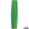 32092MC M: map data used to model this data C: citing same article ( |
|---|---|
| Similar structure data |
-Links
 PDBj
PDBj


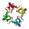
- Assembly
| Deposited unit | 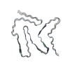
|
|---|---|
| 1 |
|
-Components
| #1: Protein | Mass: 51948.352 Da / Num. of mol.: 3 Source method: isolated from a genetically manipulated source Details: 6His-tagged mCerulean:(FPbase ID: J2JWA, Link: https://www.fpbase.org/protein/mcerulean/) Source: (gene. exp.) Aequorea victoria (jellyfish), (gene. exp.) Homo sapiens (human)Gene: FUS, TLS / Production host:  |
|---|
-Experimental details
-Experiment
| Experiment | Method: ELECTRON MICROSCOPY |
|---|---|
| EM experiment | Aggregation state: FILAMENT / 3D reconstruction method: helical reconstruction |
- Sample preparation
| Component | Name: Amyloid fibril formed by FUS low complexity domain / Type: ORGANELLE OR CELLULAR COMPONENT / Entity ID: all / Source: RECOMBINANT | ||||||||||||
|---|---|---|---|---|---|---|---|---|---|---|---|---|---|
| Molecular weight | Experimental value: NO | ||||||||||||
| Source (natural) |
| ||||||||||||
| Source (recombinant) |
| ||||||||||||
| Buffer solution | pH: 7.5 | ||||||||||||
| Specimen | Embedding applied: NO / Shadowing applied: NO / Staining applied: NO / Vitrification applied: YES | ||||||||||||
| Vitrification | Cryogen name: ETHANE |
- Electron microscopy imaging
| Experimental equipment |  Model: Titan Krios / Image courtesy: FEI Company |
|---|---|
| Microscopy | Model: FEI TITAN KRIOS |
| Electron gun | Electron source:  FIELD EMISSION GUN / Accelerating voltage: 300 kV / Illumination mode: FLOOD BEAM FIELD EMISSION GUN / Accelerating voltage: 300 kV / Illumination mode: FLOOD BEAM |
| Electron lens | Mode: BRIGHT FIELD |
| Image recording | Electron dose: 40 e/Å2 / Film or detector model: GATAN K2 SUMMIT (4k x 4k) |
- Processing
| CTF correction | Type: NONE |
|---|---|
| Helical symmerty | Angular rotation/subunit: -1.44 ° / Axial rise/subunit: 5 Å / Axial symmetry: C1 |
| 3D reconstruction | Resolution: 2.9 Å / Resolution method: FSC 0.143 CUT-OFF / Num. of particles: 171163 / Symmetry type: HELICAL |
