 Movie
Movie Controller

[English] 日本語
 Yorodumi
- EMDB-32003: Structural insights into the membrane microdomain organization by... -
+ Open data

- Basic information
| Entry | Database: EMDB / ID: EMD-32003 | |||||||||
|---|---|---|---|---|---|---|---|---|---|---|
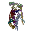
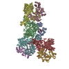
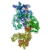
| Title | Structural insights into the membrane microdomain organization by SPFH family proteins | |||||||||
 Map data Map data | ||||||||||
 Sample Sample |
| |||||||||
 Keywords Keywords | membrane microdomain organization / MEMBRANE PROTEIN | |||||||||
| Function / homology |  Function and homology information Function and homology informationHydrolases; Acting on peptide bonds (peptidases); Metalloendopeptidases / ATP-dependent peptidase activity / membrane => GO:0016020 / protein catabolic process / metalloendopeptidase activity / peptidase activity / cell division / proteolysis / zinc ion binding / ATP binding ...Hydrolases; Acting on peptide bonds (peptidases); Metalloendopeptidases / ATP-dependent peptidase activity / membrane => GO:0016020 / protein catabolic process / metalloendopeptidase activity / peptidase activity / cell division / proteolysis / zinc ion binding / ATP binding / membrane / plasma membrane Similarity search - Function | |||||||||
| Biological species |  | |||||||||
| Method | single particle reconstruction / cryo EM / Resolution: 3.27 Å | |||||||||
 Authors Authors | Ma CY / Wang CK | |||||||||
| Funding support | 1 items
| |||||||||
 Citation Citation | Journal: Cell Res / Year: 2022 Title: Structural insights into the membrane microdomain organization by SPFH family proteins. Authors: Chengying Ma / Chengkun Wang / Dingyi Luo / Lu Yan / Wenxian Yang / Ningning Li / Ning Gao /  Abstract: The lateral segregation of membrane constituents into functional microdomains, conceptually known as lipid raft, is a universal organization principle for cellular membranes in both prokaryotes and ...The lateral segregation of membrane constituents into functional microdomains, conceptually known as lipid raft, is a universal organization principle for cellular membranes in both prokaryotes and eukaryotes. The widespread Stomatin, Prohibitin, Flotillin, and HflK/C (SPFH) family proteins are enriched in functional membrane microdomains at various subcellular locations, and therefore were hypothesized to play a scaffolding role in microdomain formation. In addition, many SPFH proteins are also implicated in highly specific processes occurring on the membrane. However, none of these functions is understood at the molecular level. Here we report the structure of a supramolecular complex that is isolated from bacterial membrane microdomains and contains two SPFH proteins (HflK and HflC) and a membrane-anchored AAA+ protease FtsH. HflK and HflC form a circular 24-mer assembly, featuring a laterally segregated membrane microdomain (20 nm in diameter) bordered by transmembrane domains of HflK/C and a completely sealed periplasmic vault. Four FtsH hexamers are embedded inside this microdomain through interactions with the inner surface of the vault. These observations provide a mechanistic explanation for the role of HflK/C and their mitochondrial homologs prohibitins in regulating membrane-bound AAA+ proteases, and suggest a general model for the organization and functionalization of membrane microdomains by SPFH proteins. | |||||||||
| History |
|
- Structure visualization
| Movie |
Movie viewer |
|---|---|
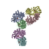
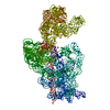
| Structure viewer | EM map: SurfViewMolmilJmol/JSmol |
| Supplemental images |
- Downloads & links
-EMDB archive
| Map data | emd_32003.map.gz | 4.8 MB | EMDB map data format | |
|---|---|---|---|---|
| Header (meta data) | emd-32003-v30.xmlemd-32003.xml | 13.6 KB 13.6 KB | Display Display | EMDB header |
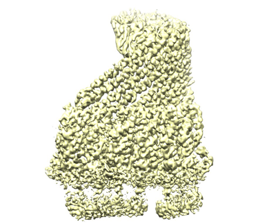
| Images |  emd_32003.png emd_32003.png | 138.4 KB | ||
| Filedesc metadata | emd-32003.cif.gz | 5.7 KB | ||
| Archive directory |  http://ftp.pdbj.org/pub/emdb/structures/EMD-32003ftp://ftp.pdbj.org/pub/emdb/structures/EMD-32003 http://ftp.pdbj.org/pub/emdb/structures/EMD-32003ftp://ftp.pdbj.org/pub/emdb/structures/EMD-32003 | HTTPS FTP |
-Related structure data
| Related structure data |  7vhqMC  7vhpC M: atomic model generated by this map C: citing same article ( |
|---|---|
| Similar structure data |


-Links
| EMDB pages | EMDB (EBI/PDBe) / EMDataResource |
|---|---|
| Related items in Molecule of the Month |


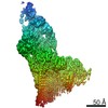
-Map
| File | Download / File: emd_32003.map.gz / Format: CCP4 / Size: 244.1 MB / Type: IMAGE STORED AS FLOATING POINT NUMBER (4 BYTES) | ||||||||||||||||||||||||||||||||||||||||||||||||||||||||||||
|---|---|---|---|---|---|---|---|---|---|---|---|---|---|---|---|---|---|---|---|---|---|---|---|---|---|---|---|---|---|---|---|---|---|---|---|---|---|---|---|---|---|---|---|---|---|---|---|---|---|---|---|---|---|---|---|---|---|---|---|---|---|
| Projections & slices | Image control
Images are generated by Spider. | ||||||||||||||||||||||||||||||||||||||||||||||||||||||||||||
| Voxel size | X=Y=Z: 1.057 Å | ||||||||||||||||||||||||||||||||||||||||||||||||||||||||||||
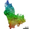
| Density |
| ||||||||||||||||||||||||||||||||||||||||||||||||||||||||||||
| Symmetry | Space group: 1 | ||||||||||||||||||||||||||||||||||||||||||||||||||||||||||||
| Details | EMDB XML:
CCP4 map header:
| ||||||||||||||||||||||||||||||||||||||||||||||||||||||||||||
 Z (Sec.)
 Y (Row.)
 X (Col.)


-Supplemental data
- Sample components
-Entire : KCF complex
| Entire | Name: KCF complex |
|---|---|
| Components |
|
-Supramolecule #1: KCF complex
| Supramolecule | Name: KCF complex / type: complex / ID: 1 / Parent: 0 / Macromolecule list: all |
|---|---|
| Source (natural) | Organism: |
-Macromolecule #1: ATP-dependent zinc metalloprotease FtsH
| Macromolecule | Name: ATP-dependent zinc metalloprotease FtsH / type: protein_or_peptide / ID: 1 / Number of copies: 6 / Enantiomer: LEVO EC number: Hydrolases; Acting on peptide bonds (peptidases); Metalloendopeptidases |
|---|---|
| Source (natural) | Organism: |
| Molecular weight | Theoretical: 7.345267 KDa |
| Recombinant expression | Organism: |
| Sequence | String: RKVDYSTFLQ EVNNDQVREA RINGREINVT KKDSNRYTTY IPVQDPKLLD NLLTKNVKVV GEP UniProtKB: ATP-dependent zinc metalloprotease FtsH |
-Macromolecule #2: Protein HflK
| Macromolecule | Name: Protein HflK / type: protein_or_peptide / ID: 2 / Number of copies: 3 / Enantiomer: LEVO |
|---|---|
| Source (natural) | Organism: |
| Molecular weight | Theoretical: 30.164129 KDa |
| Recombinant expression | Organism: |
| Sequence | String: RVVTIAAAAI VIIWAASGFY TIKEAERGVV TRFGKFSHLV EPGLNWKPTF IDEVKPVNVE AVRELAASGV MLTSDENVVR VEMNVQYRV TNPEKYLYSV TSPDDSLRQA TDSALRGVIG KYTMDRILTE GRTVIRSDTQ RELEETIRPY DMGITLLDVN F QAARPPEE ...String: RVVTIAAAAI VIIWAASGFY TIKEAERGVV TRFGKFSHLV EPGLNWKPTF IDEVKPVNVE AVRELAASGV MLTSDENVVR VEMNVQYRV TNPEKYLYSV TSPDDSLRQA TDSALRGVIG KYTMDRILTE GRTVIRSDTQ RELEETIRPY DMGITLLDVN F QAARPPEE VKAAFDDAIA ARENEQQYIR EAEAYTNEVQ PRANGQAQRI LEEARAYKAQ TILEAQGEVA RFAKLLPEYK AA PEITRER LYIETMEKVL GNTRKVLVND UniProtKB: Protein HflK |
-Macromolecule #3: Modulator of FtsH protease HflC
| Macromolecule | Name: Modulator of FtsH protease HflC / type: protein_or_peptide / ID: 3 / Number of copies: 3 / Enantiomer: LEVO |
|---|---|
| Source (natural) | Organism: |
| Molecular weight | Theoretical: 37.176293 KDa |
| Recombinant expression | Organism: |
| Sequence | String: MRKSVIAIII IVLVVLYMSV FVVKEGERGI TLRFGKVLRD DDNKPLVYEP GLHFKIPFIE TVKMLDARIQ TMDNQADRFV TKEKKDLIV DSYIKWRISD FSRYYLATGG GDISQAEVLL KRKFSDRLRS EIGRLDVKDI VTDSRGRLTL EVRDALNSGS A GTEDEVTT ...String: MRKSVIAIII IVLVVLYMSV FVVKEGERGI TLRFGKVLRD DDNKPLVYEP GLHFKIPFIE TVKMLDARIQ TMDNQADRFV TKEKKDLIV DSYIKWRISD FSRYYLATGG GDISQAEVLL KRKFSDRLRS EIGRLDVKDI VTDSRGRLTL EVRDALNSGS A GTEDEVTT SAADNAIAEA AERVTAETKG KVPVINPNSM AALGIEVVDV RIKQINLPTE VSEAIYNRMR AEREAVARRH RS QGQEEAE KLRATADYEV TRTLAEAERQ GRIMRGEGDA EAAKLFADAF SKDPDFYAFI RSLRAYENSF SGNQDVMVMS PDS DFFRYM KTP UniProtKB: Modulator of FtsH protease HflC |
-Experimental details
-Structure determination
| Method | cryo EM |
|---|---|
 Processing Processing | single particle reconstruction |
| Aggregation state | particle |
-Sample preparation
| Buffer | pH: 7.4 |
|---|---|
| Vitrification | Cryogen name: ETHANE |
- Electron microscopy
| Microscope | FEI TITAN KRIOS |
|---|---|
| Image recording | Film or detector model: GATAN K2 SUMMIT (4k x 4k) / Average electron dose: 60.0 e/Å2 |
| Electron beam | Acceleration voltage: 300 kV / Electron source:  FIELD EMISSION GUN FIELD EMISSION GUN |
| Electron optics | Illumination mode: FLOOD BEAM / Imaging mode: BRIGHT FIELD |
| Experimental equipment |  Model: Titan Krios / Image courtesy: FEI Company |
